Original Fritz Kahn Posters and Key Booklet, Sotheby's Vintage Posters Auction, May 13


Morbid Anatomy reader Gotthold is a long time collector of Fritz Kahn books and posters. He is currently selling two of his original posters (as pictured above) along with a "key booklet" as part of Sotheby's May 13 Vintage Posters Auction.

I asked Gotthold to tell me and the Morbid Anatomy readership a bit about this special collection he is actioning off in the hopes of helping it find a proper and loving home; here is his response:

Dear Morbid Anatomy readers:

I have been a keen reader of this blog since I discovered it about a year ago when searching for information on anatomical posters I bought for use in an art project.

My personal artistic fascination with death, pornography, science and religion has taken me on a strange and fascinating journey over the past year through the cavernous bookshop cellars of Vienna, the seedy sex shops of London’s Soho, and the wonderful Morbid Anatomy blog in search of new materials and ideas. In my search for materials to use for my work, I spend a seemingly senseless amount of time and money looking for rare, obscure, and interesting materials to use and take inspiration from. It was on one of these escapades when visiting Vienna that I first stumbled upon the wonderful works of Fritz Kahn whose unique mechanical anatomy illustrations have earned much attention on this very blog (recent posts here, here, and here).

Since this initial discovery, I have managed to amass an extensive collection of Fritz Kahn's books, all featuring his wonderful illustrations, and have also had the luck to acquire a few original posters, including the famed ‘Der Mensch als Industriepalast’ or 'Man as Industrial Palace' of 1926 as seen above, top; you can found out more about that piece here.

Conducting more commercially oriented research around these works, I stumbled upon Morbid Anatomy for the first time to read a post on a Christies ‘Anatomy as Art’ auction in New York where this poster sold for some $3,500. The financially conscious side of myself forced me to reluctantly get in touch with Christies in London regarding a sale. I was informed by their experts there was no specialist auction coming up anytime soon but that I could still consign the poster to a ‘Vintage Posters’ auction in May. I chose to sell the two posters and a ‘key’ booklet together as a lot; I still believe this is extremely unique, given that the key booklet acts as an index to the numerical and alphabetical indicators on the poster without which it is difficult to fully comprehend the intended meaning of the illustrations.

The marketing around this auction has been weak, and there isn’t much explanation of the uniqueness of the key booklet or even an image of the second poster in the lot (as seen above, bottom). When I looked at the other posters for sale at this the auction I realized that my item is out of place and I doubt that it will strike the right chord with the bidders.

I have still however decided to proceed with the auction, not in the least because I need the proceeds of this sale to help further my artistic pursuits. I therefore implore anyone who knows relevant collectors to spread the word about the auction, and encourage anyone who’s interested to bid on these items as they are impeccable (the nice thing about Christies auctions is that anyone can place bids from anywhere in the world online). You can see the lot on the auction website by clicking here.

So please, any and all of you medical art aficionados out there, check out (and bid on!) Gotthold's Sotheby's lot on May 13th; you can find out more about the lot by clicking here and more about the auction by clicking here. And yes, online/remote bidding is very much a possibility! Also, please feel free to forward this post to any interested parties!

If you are interested in learning more about Fritz Kahn and seeing more of his incredible work, I highly recommend the beautiful, lavishly illustrated book Fritz Kahn: Man Machine / Maschine Mensch, which comes complete with a frame-worthy poster-sized reproduction of ‘Der Mensch als Industriepalast’ ('Man as Industrial Palace'). Good stuff!

Tonight!!! "Experimenting with Death: An Introduction to Terror Management Theory," Lecture, Observatory


Tonight! Michael Johns on all things Terror Management Theory! 8:00! Observatory!

Full details follow. Hope to see you there!

Experimenting with Death: An Introduction to Terror Management Theory
An Illustrated Lecture by Michael Johns, Former Assistant Professor of Psychology at the University of Wyoming
Date: Thursday, May 6
Time: 8:00 PM
Admission: $5
Presented by Morbid Anatomy

In his Pulitzer Prize-winning book, Denial of Death, cultural anthropologist Ernest Becker attempted to develop a unified theory of human behavior. He argued that it was the human capacity to grasp and contemplate our own mortality–and our need to suppress this knowledge–that was at the root of human culture and behavior, from genocide to altruism, religion to philosophy. Terror Management Theory (TMT) is a psychological theory directly based on Becker’s work, developed by a group of social psychologists interested in testing Becker’s assertions about death as a core motivator of human behavior. Over the last 25 years, psychologists in the North America, Europe and the Middle East have conducted hundreds of studies to test hypothesis derived from Becker’s work and the Terror Management Theory it inspired. This body of research compellingly supports Becker’s thesis and reveals the ways in which mortality salience influences behaviors ranging from aggression and stereotyping to creativity and sexuality. Using segments from the documentary “Flight from Death: The Quest for Immortality,” this lecture will introduce Terror Management Theory and discuss the often clever experiments that have been conducted to test its tenets.

Michael Johns is a social psychologist and works as a research scientist in the NYC Department of Health. He has published numerous research articles and book chapters on a variety of topics, including Terror Management Theory. Before moving to Brooklyn, Mike was an Assistant Professor of Psychology at the University of Wyoming.

You can find out more about this presentation here. For more on Ernest Becker's wonderful book Denial of Death, click here; for more on the film "Flight From Death - The Quest for Immortality," click here. You can get directions to Observatory--which is next door to the Morbid Anatomy Library--by clicking here. You can find out more about Observatory here, join our mailing list by clicking here, and join us on Facebook by clicking here.

The Taxidermy of Mr. Walter Potter and his Museum of Curiosities, Melissa Milgrom


Melissa Milgrom--author of Still Life: Adventures in Taxidermy and panelist at the recent Congress for Curious People--has just published a nice article about that undisputed king of Victorian anthropomorhic taxidermy, animal artist and museologoist Walter Potter; following is a brief excerpt:

Athletic toads? Rats gambling in a dollhouse of decadence? How about bespectacled gentlemen lobsters?

No, this isn’t Wes Anderson’s sequel to Fantastic Mr. Fox, but the work of English Victorian taxidermist Mr. Walter Potter. Potter was famous for his over-the-top anthropomorphic scenes—kittens at the tea table; guinea pigs playing cricket—which were displayed in his Museum of Curiosities from 1861 until 2003 when his wondrous collection was sold in a contentious auction, which I attended in Cornwall.

One of England’s oldest private museums, Potter’s belonged to the era of the amateur nature lover when museums were spirited jumbles, not the sober typologies they would become post-Darwin. Potter’s verged on the freakish: random, cluttered, crammed to the rafters with curios and oddities, weird accumulations and creatures that were stuffed, pickled, dissected, and deformed. And I was lucky, though it filled me with sadness, to wander through Potter’s crooked corridors on its very last day...

Had Potter attended the Great Expo (very likely) he would have seen among the taxidermy displays a comic depiction of Goethe’s fable Reinecke the Fox reenacted with semi-human foxes. Sounds childlike—and it was in the best, most passionate way—but in the days before irony anthropomorphism was a form of endearment (imagine Beatrix Potter, no relation). More so, the facial expressions were expertly manipulated, raising the taxidermic bar and inspiring followers.

Known as the Grotesque School, “mirth-provoking” characters were the equivalent of a blockbuster movie. Queen Victoria herself stopped to linger and laugh at a frog shaving another frog. And taxidermists began transforming all sorts of animals into tiny humans: crows playing violin, frogs doing the cancan, squirrels as Romeo. None were as ambitious as Mr. Walker Potter...

You can read the full article on the Wonders and Marvels blog by clicking here. You can find out more about Milgrom's Still Life--which contains a nice discussion of Potter and his work--by clicking here. If the life and work of Walter Potter is of interest, I also highly highly recommend that you check out the wonderful, lavishly-illustrated Walter Potter and his Museum of Curious Taxidermy, written by Congress for Curious People lecturer Pat Morris; you can do so by clicking here or by visiting Observatory (more on that here).

All images are of Walter Potter's work and are drawn from the wonderful Ravishing Beasts blog; you can see them in context by clicking here.

Axillary Node Dissection, Sentinel Lymph Node Biopsy May Pose More Harm Than Good In Patients With Pure DCIS

Medscape (5/3, Ellis; registration required) reported that "two commonly used procedures -- axillary node dissection and sentinel lymph node biopsy -- might pose more harm than good to patients with pure ductal carcinoma in situ (DCIS)," according to a study presented at the American Society of Breast Surgeons 11th Annual Meeting. One of the study's authors said that "her findings indicate that not only do these procedures and their subsequent findings not affect survival 'in any way,' the procedure itself carries some morbidity -- albeit low."

"Experimenting with Death: An Introduction to Terror Management Theory," Lecture, Observatory, Thursday May 6


This Thursday, May 6, join Morbid Anatomy and Michael Johns at Observatory for a night of all things Terror Management Theory! Full details follow; This will be a very good night and I hope very much to see you there!

Experimenting with Death: An Introduction to Terror Management Theory
An Illustrated Lecture by Michael Johns, Former Assistant Professor of Psychology at the University of Wyoming
Date: Thursday, May 6
Time: 8:00 PM
Admission: $5
Presented by Morbid Anatomy

In his Pulitzer Prize-winning book, Denial of Death, cultural anthropologist Ernest Becker attempted to develop a unified theory of human behavior. He argued that it was the human capacity to grasp and contemplate our own mortality–and our need to suppress this knowledge–that was at the root of human culture and behavior, from genocide to altruism, religion to philosophy. Terror Management Theory (TMT) is a psychological theory directly based on Becker’s work, developed by a group of social psychologists interested in testing Becker’s assertions about death as a core motivator of human behavior. Over the last 25 years, psychologists in the North America, Europe and the Middle East have conducted hundreds of studies to test hypothesis derived from Becker’s work and the Terror Management Theory it inspired. This body of research compellingly supports Becker’s thesis and reveals the ways in which mortality salience influences behaviors ranging from aggression and stereotyping to creativity and sexuality. Using segments from the documentary “Flight from Death: The Quest for Immortality,” this lecture will introduce Terror Management Theory and discuss the often clever experiments that have been conducted to test its tenets.

Michael Johns is a social psychologist and works as a research scientist in the NYC Department of Health. He has published numerous research articles and book chapters on a variety of topics, including Terror Management Theory. Before moving to Brooklyn, Mike was an Assistant Professor of Psychology at the University of Wyoming.

You can find out more about this presentation here. For more on Ernest Becker's wonderful book Denial of Death, click here; for more on the film "Flight From Death - The Quest for Immortality," click here. You can get directions to Observatory--which is next door to the Morbid Anatomy Library--by clicking here. You can find out more about Observatory here, join our mailing list by clicking here, and join us on Facebook by clicking here.

Head of Discovery and Engagement, Wellcome Library, Employment Opportunity


To quote the new call for applications for "Head of Discovery and Engagement at the Wellcome Library," "The Wellcome Library is the one of the world's great cultural treasures: a unique and irreplaceable collection, which documents medicine and its role in society, past and present." The Wellcome Library also happens to be one of my favorite places in the world, and the newly created position of "Head of Discovery and Engagement" seems like a potentially pretty darn great job.

The closing date for applications is May 10th; full job description and details follow:

Head of Discovery and Engagement
Wellcome Library
Closing Date: 5/10/2010
Salary: £50 000 - £60 000

Job Details
The Wellcome Trust is a global charity dedicated to achieving extraordinary improvements in human and animal health. We support the brightest minds in biomedical research and the medical humanities.

The Wellcome Library is the one of the world's great cultural treasures: a unique and irreplaceable collection, which documents medicine and its role in society, past and present. As Head of Discovery and Engagement, you will play a pivotal role in making these outstanding collections accessible, a key part of an ambitious strategy to transform the Wellcome Library. This will include revolutionising our web presence and reading-room services to meet the needs of existing and new audiences and developing the Library's role as not only a world-class research resource, but also as part of Wellcome Collection, one of London's most exciting cultural destinations.

A passionate advocate for our collections, you will lead the Library's outreach, communication and marketing activities and, by developing our understanding of users and their needs, ensure we have a robust framework for evaluating our success. As a key member of the senior management team, reporting to the Head of Library, you will need to demonstrate: significant experience in a public/user focused role in a cultural environment; a commitment to audience development and engagement programmes; a proven understanding of commissioning audience research and evaluation; a good knowledge of social media and web technologies and experience of creating/commissioning web content; previous staff management experience and an ability to manage budgets/resources; excellent written and verbal communication skills across a broad range of stakeholders; a demonstrable ability to contribute creatively and enthusiastically at a strategic level. In addition a strong interest in the history of health, medicine or science would be advantageous.

For more information on the Wellcome Library and the transformation strategy, please visit: http://library.wellcome.ac.uk For more information on this role or a job description and to apply online visit: http://www.wellcome.ac.uk/jobs Alternatively write to: HR, Wellcome Trust, 215 Euston Road, London, NW1 2BE. Please send a CV (including salary details) and covering letter explaining how you meet the criteria and what you feel you can bring to this role.

You can find out more by clicking here. To find out more about the astounding Wellcome Library, click here.

Image: The Wellcome Library via Himetop and drawn from chrisjohnbeckett's Flickr photostream.

Will Pathology PACS eliminate injuries or cause different ones?

I wonder who is more susceptible to work-related injuries.  Pathologists or radiologists?  Is anyone aware of any data as that below for radiology for pathology?  Will pathology PACS eliminate stress and strain from microscope use or simply create other injuries?

Radiologists may be prone to work-related injuries

By Erik L. Ridley
AuntMinnie staff writer

Thanks to a lack of attention paid to developing comfortable and ergonomic work spaces, radiologists appear susceptible to work-related musculoskeletal symptoms, according to research presented Monday at the American Roentgen Ray Society (ARRS) meeting in San Diego.


Researchers from Massachusetts General Hospital (MGH) in Boston found that only 7.2% of surveyed radiologists did not have any work-related musculoskeletal symptoms. Investments in training and education seem required to address this health risk to radiologists, according to the study team.

"It's an important problem that affects every radiologist, regardless of your level of training, what specialty you're in, your location, and [whether you're in] academic or private practice," said Dr. Anand Prabhakar, a clinical fellow in abdominal imaging. "We need to do something about it before it becomes very debilitating to people."

Following anecdotal reports that many radiologists suffer from work-related musculoskeletal symptoms, the MGH research team sought to determine the prevalence of these symptoms in radiologists at a large tertiary-care hospital. They also wanted to investigate the effects of several workstation ergonomic factors and work habits on these symptoms, according to Prabhakar.

They administered a written questionnaire to 28 randomly selected radiologists (17 male, 11 female; age range = 36-50 years) from various divisions of a single radiology department. All of these radiologists primarily utilized PACS and speech recognition for interpretation, but also occasionally viewed hard-copy images, according to the researchers.

The questionnaire gathered information such as demographics, work-related musculoskeletal symptoms, work habits, and workstation design. Of the respondents, 50% had worked on the same system for longer than five years. In addition, 96% had two to three monitors at their workstation.

Only 7.2% were symptom-free, and 70% had seen a physician for work-related musculoskeletal symptoms. The researchers also found that 75% had changed their work schedule in response to symptoms.

Prevalence of musculoskeletal symptoms included:

  • Neck pain in 42.8%
  • Lower back pain in 39.2%
  • Headache in 32.1%
  • Shoulder pain in 32.1%
  • Wrist pain in 7.4%

The MGH researchers also discovered that three radiologists (17%) never adjusted their chairs; all had three or more symptoms of musculoskeletal pain. Only 32.3% of respondents always adjust their chairs, while 42.8% said they sometimes did.

As for adjusting their monitors, 60.7% never did. Elbow rests were never used by 53.5% of respondents, of which 46% had shoulder pain. Only 50% had a regular exercise program.

However, the researchers did not find any relationship between those who had more adaptive responses overall and those who had fewer symptoms.

Prabhakar attributed the prevalence of musculoskeletal problems to a combination of factors, including the piecemeal approach of putting together radiology work spaces in the PACS era.

"For example, the PACS station is not purchased at the same time [from] the same vendor; it's not integrated with, say, the desk, telephone, mouse, keyboard, and dictation system," he told AuntMinnie.com. "There really has not been any interest in somebody putting it all together and really taking into account radiologist comfort."

Cost also can be a factor, Prabhakar said. Training and education are often not provided on how to set up work spaces to be more comfortable and ergonomic.

In another notable survey finding, radiologists expressed strong preferences for having more tools for combating repetitive stress injury, some of which are very low cost, he said. For example, approximately 70% of respondents thought a wireless mouse would be helpful and approximately 60% said a wireless keyboard would be helpful. And although it would be more expensive than a wireless keyboard or wireless mouse, more than 80% of respondents wanted a desk with an adjustable height.

"If we can somehow test whether those small things could help people, then I think it's a small investment in the long-term health of radiologists," he said. "I suspect, and I haven't proven it, that if radiologists are more comfortable, then they're going to work more efficiently. And that will increase productivity, which I think is the measure that administrators will look at before they invest in it."

As for specific steps radiologists can take to improve their work-space environment, Prabhakar suggests examining recommendations produced by the U.S. Department of Labor's Occupational Safety and Health Administration (OSHA). Those guidelines, which are available online as an e-tool, provide a range of tips for creating safe and comfortable workstations.

"Ultimately, I hope that this project inspires other people to take control of their own workspace, maybe inspire industry or academic institutions to address the problem, and hopefully future research will prove that this may improve our productivity," he said.

Future research efforts will evaluate the impact of training and education on the symptoms of work-related musculoskeletal symptoms, Prabhakar said.

Kabinett des Grotesken ("Cabinet of the Grotesque"), Berliner Medizinhistorisches Museum der Charité, Spiegel Online


My friend, German journalist Michael Kneissler, just sent me a link to an article and an amazing short film celebrating the world famous Berliner Medizinhistorisches Museum der Charité on its 300th birthday, prompted by a new exhibition at the museum entitled "Charité--300 years of medicine in Berlin."

Following is an excerpt from the article--found on Spiegel Online and entitled "Kabinett des Grotesken" ("Cabinet of the Grotesque")--via a sloppy Google Translation:

Human malformations, surgical instruments, the Dildo-box of a sex researcher: The Collection of the Berlin Charité shows the dazzling variety of medical research. To mark its 300th anniversary Clinic presents highlights from the world famous now its archive.

Hands upset, steal: impossible. In the showcases the treasures of the Lord Virchow are safe. Very safe. And yet the guards sneak past every now and again. Ready to intervene immediately. They know that the temptation is to press for the issue "Charité - 300 Years of Medicine in Berlin" on the trigger...

Brains, livers, lungs, testes, ovaries removed - from the different and peaceful perished miserably, preserved in jars for viewing, Educate and quenching. An exhibition of the Interior, without taboos. Even human fetuses are also included. One with legs fused together, one with eyes grown together in the middle of the forehead. A Cyclops. Unreal and yet real.

Virchow himself called this collection - eagerly gathered for medical students and the public in order to warn of an unhealthy lifestyle - his "favorite child", for some visitors to the house if these preparations now the favorite image design: "Krass," it escapes some...

This dazzling looking exhibition is on view at the Berlin-based Medizinhistorisches Museum der Charité until February 2011; very much hope to see it before it comes down!

You can read the whole article and watch the wonderful video walk-through on the same page (just click the play button!) by clicking here. You can find out more about the museum in English by clicking here. Image above is drawn from the video.

Thanks so much to Michael Kneissler for sending this along!

Announcement for Upcoming Aperio Digital Pathology Seminars in Chicago and Cleveland

Join Us for the Chicago and Cleveland Digital Pathology Seminars

CHICAGO
May 13, 2010
Hyatt Regency
on the Riverwalk

CLEVELAND
May 14, 2010
Hyatt Regency Cleveland

Aperio is pleased to announce the first-annual Chicago and Cleveland Digital Pathology Seminars.  The seminar is complimentary and meals will be provided.

Meet and network with fellow clinicians, pathologists, histologists, biostatisticians, researchers, informaticians, and biologists to share ideas about implementing digital pathology in drug development, academic, and research environments.

SAMPLE SEMINAR TOPICS:

Marc Friedman, PhD, Aperio
Hands-On Breakfast Workshop: Slide Scanning 101

Scott Spear, Aperio
Spectrum Plus and Data Management

Peter Gann, MD, ScD, University of Illinois at Chicago
Aging, the Architecture of Normal Breast Tissue, and Breast Cancer Risk

Christopher Tully, Aperio
Image Analysis 101: Tools for Analyzing Histology

Gerard Gagne, Abbott Laboratories
Bringing the Divide - Using Digital Pathology to Guide Ultrastructural Pathology Evaluations

Bill Standwill, HistoRx
AQUA® Technology for Precise Biomarker Expression Measurement in Tissue

Frank Voelker, DVM, DACVP, Flagship Biosciences
Measuring Fat and Vacuolar Space Using
Digital Pathology

Jared Schwartz, MD, PhD, Aperio
Overcoming Psychological Barriers to Adoption of Digital Pathology and Other Novel Technologies

Ossama (Sam) Tawfik, MD, PhD, Kansas University Medical Center
Interactive Multi-Disciplinary Web Conferencing for Radiology/Pathology Correlation of Breast Biopsies: How Integrated Breast Cancer Diagnostics are Improving Patient Care

Bruce Dunn, MD, Milwaukee VA Medical Center
Primary Diagnostic Telepathology

RSVP TODAY TO RESERVE YOUR SEAT!
To RSVP for this seminar, contact Nicole Siska, Aperio Events Team, at 760.539.1193 or nsiska@aperio.com.

Squamous cell carcinoma and Epstein Bar

You did not mention ear insuflation. I thought you would since the ears are so close to the tongue. Is there a reason that the ear insuflations would not do the trick for this cancer of the tongue?

Also she lives 15 minutes away from me. Is there a way I could ozonate water for her and the ozone would hold for the day till she drank it? I once heard that you can do something with magnets to keep the water ozonated for a little while. I do not remember all the information on it.

Also you mentioned rinsing her mouth with ozonated water. How much ozonated water should she drink?

We are in total agreement that her body is run down.

thanks a lot Read more...


Immunice for Immune Support

Tomorrow Night at Observatory! "Three Unique Medical Museums in Northern Italy," Lecture by Marie Dauenheimer


Just a quick reminder: tomorrow night at Observatory! Marie Dauenheimer--the curator of the "Anatomical Art: Dissection to Illustration" exhibition discussed in this recent post--will be on hand at Observatory to deliver an illustrated lecture that "will survey the collections of three unique and often over-looked anatomical museums in Northern Italy." You can read a full description here. Full event details follow; hope very much to see you there!

Three Unique Medical Museums in Northern Italy
An illustrated presentation by Marie Dauenheimer of the Vesalius Trust
Date: May 1, 2010
Time: 8:00 P.M.
Admission: $5
Presented by Morbid Anatomy

Tonight’s visual presentation by Marie Dauenheimer will survey the collections of three unique and often over-looked anatomical museums in Northern Italy which Dauenheimer toured as part of last years Vesalius Trust “Art and Anatomy Tour.” First, the University of Florence Museum of Pathological Anatomy, famous for its collection of wax pathological models created in the 19th century, including an amazing life size leper; then The Museum of Human Anatomy in Bologna featuring the work of famed wax modeling team of Anna Morandi Manzolini and her husband Giovanni Manzolini, whose life size wax models inspired Clement Susini and the wax-modeling workshop in Florence (see image above); and lastly the fascinating University of Pavia Museum of Anatomy, which houses the beautiful 18th century frescoed dissection theater, where anatomist Antonio Scarpa. So join us tonight for wine, fellowship, and a virtual and very visual tour of some of the finest and most fascinating medical museums of Italy!

Marie Dauenheimer is a Board Certified Medical Illustrator working in the Washington, DC Metropolitan area. She specializes in creating medical illustrations and animations for educational materials, including posters, brochures, books, websites and interactive media. Since 1997 Marie has organized and led numerous “Art and Anatomy Tours” throughout Europe for the Vesalius Trust. Past tours have explored anatomical museums, rare book collections and dissection theatres in Italy, The Netherlands, Belgium, France, Scotland and England. In addition to illustrating Marie teaches drawing, life drawing and human and animal anatomy at the Art Institute of Washington. Part of Marie’s anatomy class involves study and drawing from cadavers in the Anatomy Lab at Howard University College of Medicine in Washington, DC (for more on that, see this recent post).

You can find out more about this presentation here. You can get directions to Observatory--which is next door to the Morbid Anatomy Library--by clicking here. You can find out more about Observatory here, join our mailing list by clicking here, and join us on Facebook by clicking here. To learn more about Marie's "Anatomical Art: Dissection to Illustration" exhibition, click here. For more on the Vesalius Trust, click here.

Image: Self-portrait of wax modeller Anna Morandi Manzolini dissecting a human brain, Bologna, c. 1760; Via Scienza a Due Voci

Prevention of Acupuncture Infection Needs More Focus

(HealthDay News) -- More needs to be done to prevent a rising tide of infections related to acupuncture, researchers from the University of Hong Kong say.

In a commentary, published online March 19 in BMJ, Patrick Woo and his colleagues stressed that "to prevent infections transmitted by acupuncture, infection control measures should be implemented, such as use of disposable needles, skin disinfection procedures, and aseptic techniques. Stricter regulation and accreditation requirements are also needed."

Five percent to 10 percent of acupuncture patients who develop certain kinds of bacterial infections go on to experience serious complications, the authors pointed out. These can include joint deterioration, flesh-eating disease and even paralysis and organ failure. Read more...


Kama Yogi for Optimal Male Sexual Performance!

"Three Unique Medical Museums in Northern Italy," Lecture by Marie Dauenheimer, Observatory, Saturday May 1


This Saturday night, Marie Dauenheimer--the curator of the "Anatomical Art: Dissection to Illustration" exhibition discussed in yesterday's post--will be on hand at Observatory to deliver an illustrated lecture that "will survey the collections of three unique and often over-looked anatomical museums in Northern Italy." One of the museums discussed will be The Museum of Human Anatomy in Bologna, which houses--among other works--an incredible wax self-portrait of Anna Morandi Manzolini dissecting a brain (c. 1760 ; see above). The other two musems she will discuss will be the fantastic and difficult-to-access University of Florence Museum of Pathological Anatomy and the University of Pavia Museum of Anatomy.

Marie--who also leads tours of medical museums for the Vesalius Trust (as discussed in this recent post)--is an excellent speaker; her lecture on Italian Wax Anatomical Models in European Collections, which she gave about a year ago, was beloved by all, and we are exceptionally pleased to be hosting her again!

Full details follow; hope very much to see you there!

Three Unique Medical Museums in Northern Italy
An illustrated presentation by Marie Dauenheimer of the Vesalius Trust
Date: May 1, 2010
Time: 8:00 P.M.
Admission: $5
Presented by Morbid Anatomy

Tonight’s visual presentation by Marie Dauenheimer will survey the collections of three unique and often over-looked anatomical museums in Northern Italy which Dauenheimer toured as part of last years Vesalius Trust “Art and Anatomy Tour.” First, the University of Florence Museum of Pathological Anatomy, famous for its collection of wax pathological models created in the 19th century, including an amazing life size leper; then The Museum of Human Anatomy in Bologna featuring the work of famed wax modeling team of Anna Morandi Manzolini and her husband Giovanni Manzolini, whose life size wax models inspired Clement Susini and the wax-modeling workshop in Florence (see image above); and lastly the fascinating University of Pavia Museum of Anatomy, which houses the beautiful 18th century frescoed dissection theater, where anatomist Antonio Scarpa. So join us tonight for wine, fellowship, and a virtual and very visual tour of some of the finest and most fascinating medical museums of Italy!

Marie Dauenheimer is a Board Certified Medical Illustrator working in the Washington, DC Metropolitan area. She specializes in creating medical illustrations and animations for educational materials, including posters, brochures, books, websites and interactive media. Since 1997 Marie has organized and led numerous “Art and Anatomy Tours” throughout Europe for the Vesalius Trust. Past tours have explored anatomical museums, rare book collections and dissection theatres in Italy, The Netherlands, Belgium, France, Scotland and England. In addition to illustrating Marie teaches drawing, life drawing and human and animal anatomy at the Art Institute of Washington. Part of Marie’s anatomy class involves study and drawing from cadavers in the Anatomy Lab at Howard University College of Medicine in Washington, DC (for more on that, see this recent post).

You can find out more about this presentation here. You can get directions to Observatory--which is next door to the Morbid Anatomy Library--by clicking here. You can find out more about Observatory here, join our mailing list by clicking here, and join us on Facebook by clicking here. To learn more about Marie's "Anatomical Art: Dissection to Illustration" exhibition, click here. For more on the Vesalius Trust, click here.

Image: Self-portrait of wax modeller Anna Morandi Manzolini dissecting a human brain, Bologna, c. 1760; Via Scienza a Due Voci

ASCP 2010 Annual Meeting Digital Pathology Course Offerings

The upcoming ASCP Annual Meeting will feature several sesssions discussing Pathology 2.0, digital pathology, including telepathology and image analysis and personalized healthcare. 

Looks like an excellent faculty for these sessions.  The meeting will take place in San Francisco between October 27-31, 2010. 

A257 Harnessing Web 2.0 to Improve Consultation and Communication

Thursday, October 28 9:30 am - 11:30 am

Get an overview of the power of Web 2.0 to link laboratory practitioners across the globe. Discussions will include a big-picture view of how the internet is changing communication among professionals, how digital pathology can facilitate consultations, and much more. Interactive lectures will offer you an opportunity to connect with select web sites and participate in a Q&A session. You will leave with the ability to:

Establish communication networks with colleagues around the world.
Understand how digital image capture/sharing will transform practice in the next decade.
Access web sites that offer specialized content in pathology and laboratory medicine.

Moderator:
William E. Schreiber, MD, FASCP, University of British Columbia

Faculty:
Dean Giustini, MLS, MEd, University of British Columbia
Michael Feldman, MD, FASCP, University of Pennsylvania

Credits: 2.0 CME

SP88 Hauling Pathology and Pathologist into the 21st Century – Telepathology and Image Analysis

Thursday, October 28 9:30 am - 11:30 am

Review telepathology for patient care, including new systems, software, government regulations, and a real-world two-year experience with a successful telepathology deployment. You will also benefit from a multifaceted view of image analysis, including a brief overview, current commonly used applications, potential research applications, and possible future developments in image analysis. You will leave with the ability to:

Recognize the challenges for so-called low- and high-end telepathology systems.
Appreciate the utility of image analysis for patient care and how these solutions can generate revenue for pathology departments.
Appreciate how images analysis can be applied to daily practice and research.

Moderator:
N. Volkan Adsay, MD, FASCP, Emory University

Faculty:
Alexis B. Carter, MD, FASCP, Emory University
Alton B. "Brad" Farris, III, MD, FASCP, Emory University

Credits: 2.0 CME

SP89 Expanding the Pathologists’ Toolbox in the Era of Personalized Healthcare – Next-Generation Pathology Technology

Thursday, October 28 3:00 pm - 5:00 pm

Hear about new information regarding several technologies – including virtual slide imaging/image analysis, circulating tumor cells, and quantum dot multiplexing – with the potential to form the basis of future high-value medical diagnostics performed and interpreted by pathologists. The faculty will discuss the need for research-grade discovery technologies to be adapted rapidly into diagnostic products that are not only analytically accurate, but also fit into routine clinical practice. You will leave with the ability to:

Recognize the relevance of the Personalized Healthcare (PHC) model for pathologists.
Understand the basic principles underlying key technologies that may play an important role in the future of pathology.

Moderator:
Jared Schwartz, MD, FASCP, Aperio Technologies, Inc.
Gary Pestano, PhD, Ventana Medical Systems
Mara Aspinall, MBA, On-Q-ity

Credits: 3.0 CME

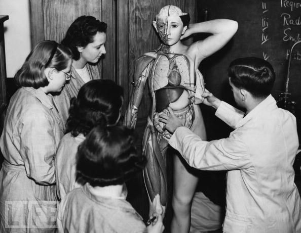
"Excellent Old-School Science Models," Life Magazine Photo Gallery


The images you see above--and the captions below--are drawn from a really fantastic Life Magazine online photo gallery entitled "Excellent Old-School Science Models." You can see the entire gallery of 29 images--well worth your perusal!--by clicking here.

Captions top to bottom, as supplied by the gallery:

  1. Isn't She Lovely: Trainee nurses examine a model of a human body to learn anatomy, Gerry Cranham, Oct. 7, 1938
  2. Behind It All: A technician works on life-like models for use in science and health lectures at the Cologne Health Museum in Germany, Ralph Crane, Feb 01, 1955
  3. Going Deep: A technician at the Cologne Health Museum gets into his work, Ralph Crane, Feb 01, 1955
  4. The Egg Factory: An exhibit illustrates the biology of the chicken at the World Poultry Exhibition at the Crystal Palace exhibition hall in London, Fox Photos, Jul 28, 1930
  5. Universal: A girl scout leans in to take a closer look at an enclosed model of the solar system, circa 1920s, George Eastman House, Jan 01, 1920

Found via Morbid Anatomy Library intern Amber Duntley's Facebook feed. Thanks, Amber!

ATA 2010 Meeting Announcement

To zero in on the latest telemedicine, telehealth, ehealth, mobile applications, and advanced remote medical technology, sign up now for the 15th American Telemedicine Association Annual International Meeting and Exposition to take place at the Henry B. Gonzalez Convention Center in San Antonio, Texas, on May 16-18 2010.

In recent months, the industry has seen huge changes in both national and state telehealth policies, National Health Reform, billions spent on broadband and health IT, new FCC broadband activities, and states mandating telemedicine coverage. The next year will be equally critical for the industry.

To address the many changes and issues involved, Aneesh Chopra, Federal Chief Technology Officer of the U.S. and John P. Howe, III, MD, President and CEO, Project Hope will be the featured speakers at the opening plenary session on Tuesday May 18th.

If you use telecommunications solutions for healthcare, the ATA Annual Meeting is the single most important event of the year to keep you up-to-date on the latest developments in your field. ATA 2010 will provide insightful keynote speakers, peer-reviewed presentations, executive roundtables, professional networking events, daily breakout sessions, and pre-meeting certificate courses.

Educational concurrent sessions will feature 72 peer-reviewed sessions and discussions on clinical services, telemental health, pediatric telehealth, remote monitoring, consumer and mobile health, emergency and disaster response, operations, business and finance, regulatory issues, and reimbursement issues.

For a preliminary program and further details, go to http://www.americantelemed.org.

World’s Smallest, Lightest Telemedicine Microscope

via MedGadget

Researchers at The California NanoSystems Institute at UCLA (CNSI) have developed a tiny telemedicine microscope for imaging blood samples or other fluids, testing water quality or other public health need in resource-limited settings.

From the press release:

Slightly wider than a US quarter and weighing just 46 grams, the lensless microscope is a self-contained imaging device. Using LUCAS (Lensless Ultra-wide-field Cell Monitoring Array) technology, it generates holographic images of microparticles or cells by employing a light-emitting diode to (LED) illuminate the objects and a digital sensor array to capture their images.

Samples are loaded using a small chip that can be filled with saliva or a blood smear for health monitoring. With blood smears, the lensless microscope is capable of accurately identifying cells and particles, including red blood cells, white blood cells and platelets. The technology has the potential to help monitor diseases like malaria, HIV and tuberculosis.

The microscope is fairly robust with few moving parts and a large aperture. Images can be uploaded via a direct USB connection to a smart phone or other device, and sent to a hospital.

More information: UCLA engineer invents world's smallest, lightest telemedicine microscope