To Eat More Fruit, Picture a Fruit Salad

(HealthDay News) -- Creating a healthy eating action plan and visualizing yourself carrying it out may help improve the way you eat, researchers suggest.

"Telling people to just change the way they eat doesn't work; we've known that for a long time," study author Barbel Knauper, an associate professor of psychology at McGill University in Montreal, said in a university news release.

"But research has shown that if people make a concrete plan about what they are going to do, they are better at acting on their intentions. What we've done that's new is to add visualization techniques to the action plan," she explained.

Her study included 177 students who were asked to set the goal of eating more fruit for a week. All of the students ate more fruit during that time. However, those who made a concrete plan, wrote it down and also visualized how they were going to carry out their plan (i.e. when, where and how they would buy, prepare and eat fruit) increased their fruit consumption twice as much as those who didn't plan or visualize. Read more...

AyurGold for Healthy Blood

Chocolate compounds fight high cholesterol

Chocolate has received a lot of attention for being a treasure trove of nutritional goodness. Polyphenols in cacao beans are linked to promoting heart, brain, and liver health, which has sparked renewed interest in chocolate as a medicinal food. And a new study adds to the growing list of benefits, showing that chocolate polyphenols also help to lower bad cholesterol.

Published in the journal Diabetic Medicine, the study tested the effects of polyphenol-rich chocolate in a group of 12 volunteers with type-2 diabetes. After 16 weeks, the researchers from Hull University in the U.K. discovered that the polyphenols helped lower participants' bad cholesterol levels while raising good cholesterol levels.

"Chocolate with a high cocoa content should be included in the diet of individuals with type-2 diabetes as part of a sensible, balanced approach to diet and lifestyle," said professor Steve Akin, author of the study.Read more...

AyurGold for Healthy Blood

PathXL in the Classroom

"Students have been delighted with its (PathXL’s) ease of use and its 24 hour, 365 day accessibility." Dr. Stephen McCullough, Head of Anatomy, Queens University Belfast

i-Path’s PathXL software continues to provide an innovative approach to classroom learning. A number of leading educational centres in the UK and further afield are recognising the benefits that PathXL can bring to medical education.

Queen's University, Belfast (link) is an institution with a world-class academic reputation and is a member of the Russell Group of leading UK universities. Conventional microscopy was proving to be ineffective in teaching the approximately 2300 students in the School of Medicine, Dentistry and Biomedical Sciences; it was time consuming, difficult to establish standards in teaching or examining and unpopular among students.

With the help of i-Path, Queen’s introduced a virtual microscopy solution. By using PathXL, virtual slides could be viewed on-line and used in the same way as a conventional microscope by large numbers of students or trainees over a computer network, thus avoiding the necessity for them to be at a particular venue at a set time to attend a teaching or training session.

PathXL has helped enhance the learning experience of students who can practice histology in a digital environment in both the classroom and at home. ?PathXL facilitates group learning as it enables students to discuss interesting slides on large computer monitors by viewing them digitally. In addition, with classroom control software, tutors can control single or multiple classroom monitors and communicate with individual students, with groups or the entire classroom from the front of the class.

"This versatile system has allowed us to construct practical classes that can interactively link a range of study material including virtual slides, micrographs, x-rays, anatomical specimens and lecture material." Dr. Stephen McCullough, Head of Anatomy, QUB

---------------------------------------------------------------------------------------------------------------

The University of Glasgow, School of Veterinary Medicine (link) has also chosen PathXL to help transform its learning environment.

In 2010 a new course in veterinary pathology was introduced for the undergraduate veterinary students, focussing on developing their enthusiasm for pathology in both practical, clinical and research contexts.

PathXL now provides the students with access to histopathological slides, including cytological smears. This system has been used to enhance laboratory-based microscopy classes at the University of Glasgow by enabling students to view, at their convenience, labelled slides in their area of study.

"Research has shown that veterinary students are comfortable with this type of computer-based study of pathology, committing larger periods of time to study of the course material than they are able to in microscope-based classes alone"  Dr. Pamela Johnston, University of Glasgow, Faculty of Veterinary Medicine

PathXL has also been used in institutions such as Michigan State University (Veterinary Online Atlas), Nottingham University Hospital (Education Portal) and the Pathological Society (Educational Portal).


PathXL – Key Benefits For StudentsEnhanced learning experience

  • Ease of use
  • Freedom from optical stress
  • Better image quality
  • Time saving

Path XL – Key Benefits For Administrators

  • Ability to construct course-specific modules
  • Versatility
  • Practical classes can include a range of resources
  • Slides available to everyone at the same time
  • Cost savings

i-Path has a unique team of scientists and software developers that can offer advice and consultancy on a wide range of issues relating to virtual microscopy.

For further information please visit http://www.i-path.co.uk or contact us at contact@i-path.co.uk.


New Digital Pathology Vendor – Welcome to DigiPath, Inc.

A new digital pathology vendor hits the market today.  Affordable and innovative solutions to meet both clinical and research/biopharma needs will be required for DigiPath in an increasingly competitive landscape, particularly for hospitals - both community-based groups and academic medical centers to be successful.  Check out their new website at http://www.digipath.biz for more information.

DigiPath DigiPath, Inc.™ today announced its entry into the digital pathology market. DigiPath provides the next generation of affordable, innovative, and reliable digital pathology solutions. DigiPath’s solutions will support community pathology practices, hospitals, academic medical centers, reference laboratories, biopharma organizations, and life science research institutions.

“The medical community is looking for more affordable, innovative, and reliable digital pathology solutions to assist in enhancing patient care,” commented Keith Kaplan, M.D., Editor, Digital Pathology Blog™ at http://www.tissuepathology.com. “DigiPath seems well positioned to help our community practices expand digital pathology adoption.”

“In June 2010, we surveyed over 400 CLIA™ certified labs concerning digital pathology adoption”, stated Jondavid Klipp, Publisher, Laboratory Economics™. “55% of the respondents said that digital pathology was too expensive. Affordability is an important concern to pathology laboratories.” 

“The DigiPath team brings a combined 60 year experience within the digital pathology field”, said Eric Stoppenhagen, Chief Executive Officer. “Our team pioneered digital pathology by implementing over 500 installations worldwide. During the coming years, DigiPath will be providing affordable, innovative, and reliable digital pathology solutions.”

About DigiPath, Inc.

DigiPath, Inc. provides the next generation of affordable, innovative, and reliable digital pathology solutions. DigiPath’s advisors bring over 60 years combined expertise in pioneering digital pathology, implementing over 500 installations at community pathology practices, hospitals, academic medical centers, reference laboratories, biopharma organizations, and life science research institutions worldwide.

Visit http://www.digipath.biz monthly to see new solutions emerge during 2011.


SAFE HARBOR STATEMENT

This Press Release may contain certain forward-looking statements within the meaning of the Securities Litigation Reform Act of 1995. DigiPath has tried, whenever possible, to identify these forward-looking statements using words such as "anticipates," "believes," "estimates," "expects," "plans," "intends," "potential" and similar expressions. These statements reflect DigiPath's current beliefs and are based upon information currently available to it. Accordingly, such forward-looking statements involve known and unknown risks, uncertainties and other factors which could cause DigiPath's actual results, performance or achievements to differ materially from those expressed in or implied by such statements. DigiPath undertakes no obligation to update or provide advice in the event of any change, addition or alteration to the information catered in this Press Release including such forward-looking statements.


Leica Microsystems Launches SCN400 F Combined Fluorescence and Brightfield Slide Scanner

Leica continues to innovate in short order considering how long they have been in the digital pathology market I think this is very quick to launch a florescent scanner.

Leica is now offering a combined brightfield and fluorescent scanner and image management application that may be attractive to the research world as well as large clinical laboratories.  See link below to preview some of their images.  

Welcome your feedback on the blog about image quality, much like Facebook on Leica's iPad application.

Multi-channel fluorescent scanning for Digital Pathology

Wetzlar, Germany. Leica Microsystems announces the release of the Leica SCN400 F combined fluo­rescent and brightfield digital slide scanner. The SCN400 F brings together Leica’s award winning brightfield slide scanning capabilities and multi-channel fluorescence imaging in a single platform, providing versatility to researchers and core laboratories utilizing Digital Pathology.

By employing two separate illumination and scanning methods, the Leica SCN400 F ensures excellent brightfield scans and crisp multi-channel fluorescent images from one device. Scanned slides are readily re­viewed online through the SlidePath Digital Image Hub web-viewer, incorporating the ability to turn on and off separate channels with the click of a button to examine each marker individually.

To view a sample fluorescent image, please visit our Virtual Slide Gallery at http://www.leica-microsystems.com/products/digital-pathology.

Dr. Donal O’Shea, Head of Digital Pathology in Leica Microsystems says, “The Leica SCN400 F is a significant breakthrough for researchers looking for an affordable solution for scanning brightfield and fluorescent slides. With the ever growing adoption of immunofluorescent staining in research to study multi-marker co-expression in oncology and neuroscience, we see the SCN400 F as a valuable tool for core laboratories, bringing increased efficiency and quality to research practices.”

The Leica SCN400 F is the newest scanning solution in the Total Digital Pathology portfolio, demon­strating Leica’s drive and innovation in this market. Leica Microsystems’ broad range of interoperable Digital Pathology components provides customers with the flexibility to choose the solution that is right for their needs.

_ _ _ _ _ _ _ _ _ _ _ _ _ _ _

Leica Microsystems is a world leader in microscopes and scientific instruments. Founded as a family business in the nineteenth century, the company’s history was marked by unparalleled innovation on its way to becoming a global enterprise.

Its historically close cooperation with the scientific community is the key to Leica Microsystems’ tradition of innovation, which draws on users’ ideas and creates solutions tailored to their requirements. At the global level, Leica Microsystems is organized in four divisions, all of which are among the leaders in their respective fields: the Life Science Division, Industry Division, Biosystems Division and Medical Division.

Leica Microsystems’ Biosystems Division, also known as Leica Biosystems, offers histopathology labo­ratories the most extensive product range with appropriate products for each work step in histology and for a high level of productivity in the working processes of the entire laboratory.

The company is represented in over 100 countries with 12 manufacturing facilities in 7 countries, sales and service organizations in 19 countries and an international network of dealers. The company is head­quartered in Wetzlar, Germany.


"The Secret Museum: Collections as Muse," Artist Talk and Artifacts from the Stores, Natural History Museum, London, Thursday July 7


Hi all! Will be giving a free and open-to-the-public artist's talk augmented by artifacts drawn from the stores at the London Natural History Museum this Thursday at 2:30. Full details follow; hope very much to see you there!

The Secret Museum: Collections as Muse with artist Joanna Ebenstein
Artist Talk with Artifacts from the Stores
Museum of Natural History, London, Attenborough Studio
Thursday July 7
2:30 PM

Ancient wood and glass cases, elaborate labels from centuries past, rows of dusty bell jars, atmospherically decayed specimens, the unintentionally surreal and sublime vistas of the Museum backstage... these are the kinds of things that intrigue and inspire New York artist Joanna Ebenstein. Today, join her for a look at some of her artwork engaging with these themes. Also on view will be a variety of rarely seen artifacts specially drawn for this talk from the Natural History Museum's extensive and astounding stores.

More here.

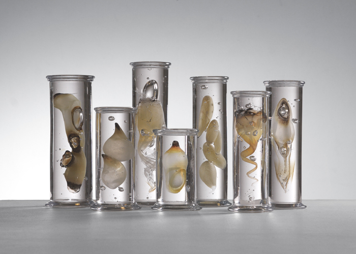
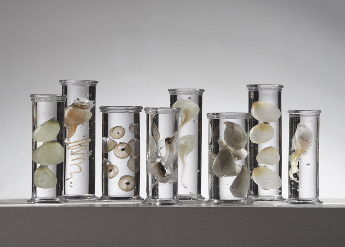
The Biologically-Inspired Glass Work of Danish Artist Steffan Dam


Wow. I have just come across the exquisite, biologically-inspired glass work of Danish artist Steffan Dam. The work--organized into series titled "Flower Blocks," "Specimen Blocks," "Fossils" and "Marine Biology--reminds me quite a bit of the work of revered natural history artists Ernst Heackel and Leopold and Rudolf Blaschka.

Above are just a few of my favorite pieces; I highly encourage you to visit the website to see the full collection by clicking here; you can also download a PDF catalog of his work by clicking here.

Via Wunderkammer.

Cabinet Cards / Storydress II, Albumen Print Photographs of Life-size Paper Mache and Plaster Sculpture, Christine Elfman, 2008

Cabinet Cards / Storydress II

albumen prints from wet-plate collodion negatives
4.25 x 6.5 inches, series of 5 mounted on cabinet cards
6.5 x 8.5 inches, series of 10 framed
2008

Storydress II is a series of photographs of a life-size paper mache and plaster sculpture. The dress is made of paper mache stories that I recorded of my great-grandmother’s autobiographical reminiscences. Each photograph contains legible words. The sculpture was photographed with the wet-plate collodion negative process, printed on handmade gold-toned Albumen paper, and burnished onto antique Cabinet Card mounts. For exhibition the cabinet card photographs are displayed using an antique wooden Graphoscope (magnifying device) and shelf.

Finding unknown relatives in my family photograph collection, and noticing old photographs of anonymous people in antique stores, I was taken by how many people were forgotten regardless of photography’s intention to “Secure the shadow, ‘ere the substance fades away.” The older the picture, the more forlorn the subject appeared to me. Holding their image, I was impressed with their absence. Storydress II tries to show this underlying subject of photographic portraiture. The 19th century cabinet card is turned inside out, revealing the presence of absence in a medium characterized by rigid detail and anonymity. The figure of reminiscence, cast in plaster, parallels the poetic immobility of the head clamp, used in early photography to prevent movement during long exposures, aptly defined by Barthes as “the corset of my imaginary existence”. The life size cast figure wears a paper mache dress made of family stories: recorded, torn up, and glued back together again. The tedious processes involved in making both the subject and photograph are offerings to time’s taking.

I really, really love this piece--which uses as its base a life-size paper mache and plaster sculpture!--and encourage you to visit Christine Elfman's website and click on "view close up here" to appreciate it fully. Or click on image to see a pleasing larger version.

Via Foxes in Breeches.

Gay Men More Likely to Have Had Cancer

(HealthDay News) -- A new study finds that homosexual men are twice as likely as other males to have been diagnosed with and then survive a cancer, shining a light on the unique medical risks that gay people may face.

It's not the first time that researchers have noted differences in health risks linked to sexual orientation. Gay men, of course, are at higher risk of becoming infected with HIV, while lesbians may be more likely than heterosexual women to get breast cancer. Both gay men and lesbians have higher rates of tobacco use than the general population, and research has shown that lesbians drink more and are more prone to obesity than other women.

The new study adds to existing knowledge, but "there's a painful dearth of data about lesbian, gay, bisexual and transgender health in general," noted Liz Margolies, executive director of the National LGBT Cancer Network, who's familiar with the new research. Read more...

Joint Mender for Joint Care

Humorous pathology videos from University of Florida Department of Pathology

A loyal reader put me onto DRdoubleB over at YouTube, a pathology resident at the University of Florida who put together some high quality music videos with a pathology theme.  

Check out "Dynamite Case" about the trials and tribulations of daily sign-out, grossing and trying to go to meetings, "I Got It" for nailing a sarcoma diagnosis & "I Like Path That Way" which has more attending input on the video. Pretty sure my attendings in residency would either not have supported these videos or appeared in them. 

Looks like a fun department with a talented resident and supporting cast.


Metamark Genetics Partners with Definiens on Tissue-Based Cancer Diagnostics

Partnership forged to tackle tumor heterogeneity

Cambridge, MA and Munich, Germany – June 30, 2011 – Metamark Genetics, a privately held oncology molecular diagnostic company, and Definiens, the leading Health Image Intelligence™ company, today announced a development and commercialization partnership in which Metamark will combine its proprietary multiplex protein detection platform with image analysis technology developed jointly with Definiens to deliver innovative prognostic and predictive assays for early-stage cancers.

"Definiens is a clear world leader in quantitative digital pathology and in the analysis of biomarkers in tissue image data,” said Eric Devroe, Ph.D, Vice President of Business & Strategy Development for Metamark. “We see great synergy in combining Definiens’ technology and expertise with Metamark’s unique understanding of tumor heterogeneity and insight into the molecular drivers regulating tumor formation and aggression. We believe that this partnership will enable us to co-develop and implement unique and important image analysis tools."

Metamark’s quantitative diagnostic approach can identify and account for small regions of molecularly aggressive cancer cells within an otherwise indolent tumor. The company is developing a portfolio of prognostic and predictive assays with the ability to address the "heterogeneous" nature of tumors not only between one patient and the next, but also within an individual patient’s tumor.

Definiens has made ground-breaking achievements in the field of biomarker development based on its highly robust and scalable image analysis framework that enables scientists and clinicians to rapidly extract comprehensive biomarker and morphology expression profiles and to identify a relevant subset of descriptors by correlating them against patient outcomes.

"Definiens is pleased to partner with Metamark Genetics, and to incorporate our image and data analysis software technology into Metamark’s pioneering Oncology diagnostic offerings," said Thomas Colarusso, Vice President of Sales & Operations for Definiens Inc. "The capability to provide detailed readouts from tissue analysis is opening up new gateways for developing diagnostic assays as we enter the age of personalized medicine. We believe the Definiens Developer XD and Tissue Studio software will enable Metamark to classify tumor heterogeneity of multiplexed tissue stains and derive prognostic and predictive assays."

About Metamark Genetics, Inc.
Metamark is a privately held oncology company focused on the development of function-based prognostic assays for early staged cancers. The MetamarkDx™ Prognostic Assays under development are based on Metamark’s proprietary Prognosis Determinants™, genes discovered through leading edge cancer research and demonstrated to play a causal role in promoting tumor progression and spread. For further information, please visit the company’s website at  http://www.metamarkgenetics.com.

About Definiens
Definiens is a leading Health Image Intelligence™ company that develops software solutions for biomedical image analysis, data mining and clinical decision support. The company’s software analyzes images from cell-based assays, whole tissue slides and full body scans and allows users to correlate this information with data derived from other sources, supporting better decisions in research, diagnostics and therapy. By automating analysis workflows and generating new knowledge, Definiens provides pharmaceutical and biotechnology companies, research institutions, clinical service organizations and medical professionals with deeper insights, faster results and better decision support. Harnessing the power of image intelligence, Definiens supports personalized medicine and aims to significantly improve the quality of patients’ lives. Definiens is headquartered in Munich, Germany, and has offices throughout the United States. Further information is available at http://www.definiens.com.

Prepare Your Lab for a Medicare RAC Audit: Essential Do’s and Don’ts to Reduce Compliance Risk and Avoid Penalties

Pre-emptive steps to avoid fines, overpayments and even jail time

LIVE EVENT JULY 20

Jane Pine WoodTom Rehwaldjustin clark

YOUR PRESENTERS:

Jane Pine Wood, Attorney, McDonald Hopkins, LLC

Tom Rehwald, CPA, Chief Financial Officer, InCyte Pathology, PS

Justin M. Clark (Moderator), Director of Operations, THE DARK REPORT

LIVE Event July 20 – Register NOW!

Recovery Audit Contractors Program (RAC) is the new Medicare program that sends independent auditors to provider facilities, including clinical labs and pathology groups. These auditors are out to recover an estimated $24 billion a year resulting from abuse, fraud, and reimbursement problems that have plagued the system for years.

How ready is your lab is for a RAC audit? Probably not very, which leaves you vulnerable when the first letter arrives from an auditor. That’s because the Centers for Medicare and Medicaid Services (CMS) have designated four independent companies to conduct RAC audits, and they’ll have surprising powers to view your source documentation.

So what steps can you take now to prepare your lab for a RAC audit? What are your rights during an audit? And how can you effectively challenge and appeal the results of a RAC audit?

Get answers to these questions and more when you register to attend the latest Dark Report and DarkDaily.com audio conference on Wednesday, July 20, 2011. Listen as two experts help you prepare your lab for an RAC audit-and how to minimize its impact.

CMS admits that most overpayments are caused by errors and omissions rather than fraud or abuse-many the result of incorrect coding or insufficient documentation. Still, it’s important to take steps now to fully understand the impact of a RAC audit, and what you can do to ensure that proper procedures are in place to reduce risk and limit your lab’s exposure.

In 90 minutes, you’ll get advice on surviving a RAC audit from the following two experts:

First, Jane Pine Wood, a health-law attorney who specializes in regulatory and compliance issues, provides critical details about the RAC audit program: what it is, how audits are performed, what areas are at risk of being audited, and more. She’ll walk you through the process, starting the moment your lab is notified it’s about to be audited. Using case-study examples, Ms. Wood will explain the procedures your lab should follow when responding to a RAC audit.

Our second speaker, Tom Rehwald, CFO at InCyte Pathology LLC, will share what happened when a RAC audit was conducted at his lab. He’ll provide the practical insights and lessons-learned that come from taking part in an actual audit. Find out how you can stay one step ahead of the auditors. Get a better understanding of the process, including the potential pitfalls you’ll need to watch out for when responding to the results of your audit.

In sports, they say the best defense is a good offense and that definitely applies when it comes to RAC audits. Taking steps now to identify and resolve potential problems before you’re audited can substantially reduce your risk of the auditors finding anything wrong.

Register today
 and get the best, most up-to-date information on how to prepare your lab for a RAC audit. Find out how to make risk assessment of your lab’s coding, billing and collections processes an ongoing part of your compliance activities. And how improving them can help you avoid future problems.

Finally, the results of an audit aren’t set in stone so don’t be afraid to challenge them, say our experts. One Florida lab appealed overpayments found during their RAC audit and was able to overturn almost 25% of them. In this session, you’ll find out how to decide whether an appeal makes sense, and if so, what steps your lab can take to contest the results of an audit.

This is a terrific opportunity for you and your management team to come up to speed on preparing for a RAC audit. Find out how to resolve noncompliance and overpayment issues before you’re hit with an audit.

And don’t forget: You can have everyone on your lab team participate with you. For just one registration, you can all listen, learn, and get personalized answers to questions about your lab’s unique needs. Register now to guarantee your participation.

THE DARK REPORT AUDIO CONFERENCE AT A GLANCE


DATE:
 Wednesday, July 20, 2011

TIME: 1 p.m. EDT; 12 p.m. CDT; 11 a.m. MDT; 10 a.m. PDT

PLACE: Your telephone or speakerphone

COST: $195 per dial-in site (unlimited attendance per site) through 7/8/11; $245 thereafter

TO REGISTER NOW: Click here or call 1-800-560-6363 toll-free


For one low price—just $195 (through 7/8/11; $245 thereafter)—you and your entire team can take part in this fast-paced, insightful audio conference. Best of all, you’ll be able to connect personally with our speakers when we open up the phone lines for live Q&A.

Here’s just some of what you’ll learn during this in-depth 90-minute audio conference:


o The difference between a redetermination letter and reconsideration request, and what could happen if you mix up the two.

o The do’s and don’ts of crafting a redetermination letter to correct possible errors in your audit.

o Key steps for meeting compliance recommendations.

o The documentation that best supports the claims you submit to Medicare.

o How to prepare for an audit if you use a third-party billing service.

o How your lab can use the RAC experience to better educate staff and avoid future compliance and overpayment issues.


o The parallel strategies for attacking a RAC audit, including political action, that your lab should consider implementing

…and much more!


How to Register Now:

1. Online
2. Call toll free: 800-560-6363.

Your audio conference registration includes:

  • A site license to attend the conference (invite as many people as you can fit around your speakerphone at no extra charge)
  • A downloadable PowerPoint presentations from our speaker
  • A full transcript emailed to you soon after the conference
  • The opportunity to connect directly with the speaker during the audience Q&A session

Register Now!  Or for more information, call us toll-free at 800-560-6363

Distinguished Faculty: 

Jane Pine Wood is a member of McDonald Hopkins, LLC which specializes in health law, including regulatory, compliance, and contractual matters, as well as Medicaid and Medicare reimbursement issues. Ms. Wood represents physicians, hospitals, clinical and anatomic laboratories, imaging centers, home health agencies, mental health providers, clinics, independent practice associations and integrated delivery systems in corporate, regulatory, reimbursement, contractual and other areas. She received her J.D. degree in 1987 from Vanderbilt University School of Law and is admitted to practice in Massachusetts, Ohio and Tennessee.

Tom Rehwald, CPA
, has been Chief Financial Officer for InCyte Pathology, PS since 2007. He has been involved in healthcare finance and administration for more than 26 years, with experience that runs the full spectrum of a healthcare systems continuum of care. Prior to his current position, he was the CFO at Spokane Ear, Nose, & Throat Clinic and was CFO at Pathology Associates Medical Laboratories. He also held several positions at Providence Health Care/Sacred Heart Medical Center, including Director of Patient Accounts. He graduated Summa Cum Laude from San Diego State University with a Bachelor of Science degree in business administration with an emphasis in accounting.

ACCENT® Continuing Education Credit
The American Association of Clinical Chemistry (AACC) designates this program for a maximum of 1.5 ACCENT® credit hours towards the AACC Clinical Chemist’s Recognition Award. AACC is an approved provider of continuing education for clinical laboratory scientists in the states of California, Florida, Louisiana, Montana, Nevada, North Dakota, Rhode Island, and West Virginia.

 

Leica Microsystems Launches Free Digital Pathology App for iPad and iPhone

Have not downloaded the app yet and tried it out but wanted to share this news on the blog.

Wetzlar, Germany. Leica Microsystems announces the release of the SlidePath Gateway application for iPad and iPhone, which is the latest addition to the Total Digital Pathology portfolio. SlidePath Gateway is a Digital Pathology viewing application with built-in educational content from leading institutes and pathologists. The free application is available now for download from the Apple App store.

IPad SlidePath Gateway

SlidePath Gateway provides access to high quality whole slide images captured on the Leica SCN400 slide scanner, with expert review findings and content provided by contributing bodies including:

  • ASCP CheckPath Anatomical Pathology EQA Program (2006)
  • Cerviva Cervical Cytology EQA Pilot (2009)
  • NHS Breast Screening Pathology EQA Program (2010)
  • UK National Urologic Pathology EQA Program (2009)
  • UKNEQAS HER2 Breast Interpretive EQA Pilo (2011)

Dr. Donal O’Shea, Head of Digital Pathology in Leica Microsystems says, “The release of the SlidePath Gateway App displays Leica Microsystems’ commitment to develop and expand its Digital Pathology portfolio. With this App we are giving everyone the opportunity to try Digital Pathology, access high resolution whole slide imagesand expert opinions, through a highly intuitive user interface. With the rapid adoption and acceptance of iPads and other tablet devices in hospitals and clinical settings, we see SlidePath Gateway paving the way for future adoption of Digital Pathology in routine use.”

SlidePath Gateway for the iPad puts Digital Pathology at your fingertips. Users who install the application can browse folders of images from each of the External Quality Assessment (EQA) programs, select individual cases and view, zoom and navigate around the slides. Each case includes expert pathologist assessments, which can be viewed with the slides, giving increased insight into the samples. They can also see a sample set of Leica Biosystems’ Novocastra antibody range.

Existing users of Leica Microsystems’ Digital Pathology management solution, SlidePath Digital Image Hub (DIH), who usually access their system via a standard internet browser, can now securely connect to their own DIH library of images and review them through the application.

Building on a strong reputation for providing high quality, easy-to-use systems, SlidePath Gateway demonstrates Leica’s drive to deliver cutting edge, flexible tools to the Digital Pathology market.

---------------------------------------------------------------------------------------------------------------------------

Leica Microsystems is a world leader in microscopes and scientific instruments. Founded as a family business in the nineteenth century, the company’s history was marked by unparalleled innovation on its way to becoming a global enterprise.

Its historically close cooperation with the scientific community is the key to Leica Microsystems’ tradition of innovation, which draws on users’ ideas and creates solutions tailored to their requirements. At the global level, Leica Microsystems is organized in four divisions, all of which are among the leaders in their respective fields: the Life Science Division, Industry Division, Biosystems Division and Medical Division.

Leica Microsystems’ Biosystems Division, also known as Leica Biosystems, offers histopathology laboratories the most extensive product range with appropriate products for each work step in histology and for a high level of productivity in the working processes of the entire laboratory.

The company is represented in over 100 countries with 12 manufacturing facilities in 7 countries, sales and service organizations in 19 countries and an international network of dealers. The company is headquartered in Wetzlar, Germany.


From Votives to Venuses: A Brief History of the Human Anatomical Model, July 5th, The Science Museum, London


Hi All! Next Tuesday, July 5th, I will be giving a lecture at London's Science Museum in the museum's lecture theatre. The lecture if free and open to the public.

Full details follow; If you are free and in the neighborhood, why not stop by?

From Votives to Venuses: A Brief History of the Human Anatomical Model
An Illustrated Lecture by Joanna Ebenstein of The Morbid Anatomy Library
Date: 5th July
Time: 4 PM - 5 PM
The Science Museum’s Lecture Theatre
Exhibition Road, South Kensington, London SW7 2DD (Map here)
Admission: Free and open to the public

In the 1690s, surgeon Guillaume Desnoues commissioned wax-worker Gaetano Zumbo to create a life-sized wax copy of one of his most important--and sadly deteriorating--human dissections. This partnership launched a long tradition of collaboration between artists and medical practitioners in the creation of extraordinary and uncannily lifelike anatomical models intended to preserve important anatomical preparations in perpetuity and to instruct and incite wonder in medical students and laypersons alike. Today, join artist and independent researcher Joanna Ebenstein for a lavishly illustrated walk through the world of these fascinating artifacts that are equal parts art and science and which flicker enticingly on the edges of relic and specimen, mysticism and medicine, life and death. This talk will discuss the histories of these objects and their makers as well as their uses and contexts of display; introduce you to many of the amazing museums that house these artifacts; and consider the ways in which these objects relate to a long tradition of religious, allegorical, and artistic approaches to mortality, supplication, and the quest for bodily immortalization.

To download a PDF with more on this and other associated talks, click here. You can find out more about The Science Museum by clicking here. For more information, please contact Selina Pang, Curatorial Coordinator at CuratorialServices@sciencemuseum.org.uk.

Image: "The Slashed Beauty," full-length anatomical model in the Josephinum Collection, Vienna, Austria; Wax model with human hair in rosewood and Venetian glass case; Workshop of Clemente Susini of Florence, 1781-1786 Photographed by Joanna Ebenstein

Field Trip, Anyone? or, a Day of Brains in Jars, Old Libraries, and Underground Crypts in New Haven, Connecticut


Anyone fancy a chartered bus trip to view the legendary Cushing Collection (pictured above), an underground crypt, and a couple of libraries thrown in for good measure? Yeah; me too! Hope very much to see you there.

FIELD TRIP: Day of Brains in Jars, Old Libraries, and Underground Crypts in New Haven, Connecticut
A chartered bus field trip to New Haven, Connecticut with guided tours of The Cushing Brain Collection, The Institute Library, and The Center Church Crypt and an unguided visit to the Beinecke Rare Book and Manuscript Library
Date: Saturday, July 16th
Time: 10:00 AM- 7 PM
Admission: $60
Presented by Morbid Anatomy

*** 28 Person Limit; MUST RSVP to morbidanatomy [at] gmail.com

On Saturday, July 16th join Observatory and Morbid Anatomy for a special field trip to New Haven, Connecticut. Our first stop will be the amazing Cushing Collection, with its over 500 human brains in glass jars and haunting pre- and post-operative photographs amassed by "father of modern neurosurgery" Dr. Harvey Cushing. We will be introduced to this collection-- newly open to the public--via a guided tour by Terry Dagradi, curator of the collection. Our next stop will be the historic and lovely Institute Library (founded 1826), Connecticut's oldest living independent literary institution and one of the last remaining membership libraries in North America, where director Will Baker will give us a tour followed by an opportunity for unguided exploration and lunch. Next, we will be treated to a special after-hours tour of the Center Church Crypt, an underground cemetery featuring 137 grave stones of New Haven's founders and earliest citizens going back to 1687. Our final stop will be an unguided visit to the incredible Beinecke Rare Book and Manuscript Library before hopping on the bus for our return home.

Trip Details: The $60 event cost of this event includes round trip transportation on a special chartered bus from Observatory to New Haven and back again as well as tour costs. Please bring your lunch, which we will have an opportunity to eat at our second stop. The bus will pick up and drop off in front of the 543 Union Street (at Nevins Street) entrance to Observatory. Pick up is 10:00 AM sharp and drop off approximately 7:00 PM depending on traffic.

There is a 28 person limit for this trip, so please RSVP to morbidanatomy [at] gmail.com if interested.

Images: Of and from The Cushing Collection as featured in The New York Times.

Pathology publisher goes digital

Sage Scope Adds Deeper Look For Sage Journals Online Users

SAGE is adding a new feature to its SAGE Journals Online offerings with SAGE scope, a tool that provides readers with interactive slide imagery as supplements to articles posted on SAGE Journals Online. The digital pathology scanning and hosting will be provided by Flagship Biosciences, a digital pathology services firm, and powered by SlidePath’s Digital Image Hub software.

This new addition will allow readers to view the pathology slides not as a flat image, but as if they were seeing it under their own light microscope. The ultra-high resolution slide with the proper digitization software can pan, zoom, and focus slides while adding specific application functionality unique to each user.  Researchers are able to drill down to 20x magnification with rich detail and high resolution, giving them full access to all the data the author or authors saw when they took the sample.

“SAGE scope will help bridge the gap between just a picture and the ability to interact with a sample as the researcher did,” said Ron Epstein, SAGE Director, STM Journals. “This added feature allows SAGE journal readers to discover the information more closely to the way the author intended.”

SAGE is on of the first academic publishers to create an active digital database for pathology, and sees further potential for use in its medical journals.

“We’re excited to meet the needs of the market with this addition and know that not only will our authors see an added value, but those who utilize SAGE journals as well,” said Epstein.

SAGE scope can be seen now in selected SAGE Journals Online. To view the digital files please go to http://sagescope.flagshipbio.com. Select VetPath, Volume 48, Issue 1.


Webinar from Aperio: An Integrated Digital Pathology Workflow for Digital Scanning, Image Management, and Image Analysis

Wednesday, July 6, 2011

8:00 am Pacific Time, 11:00 am Eastern Time
(4:00 pm London GMT / 10:00 am Mexico City Standard Time) 


Presenters:
Dr. Florian Leiss, Definiens Trainer and Marketing Specialist
Priya Vaidyanathan, Ph.D, Image Analysis Specialist, Aperio
 

In this 60-minute complimentary webinar, you will learn about the new solutions available as a result of the partnership between Aperio and Definiens. 

Topics include:

  • Integrating image aquisition, management, and analysis workflow 
  • Spectrum database management software for managing digital slides
  • Live demo of Definiens' Tissue Studio for Her2 analysis in breast cancel tissue sections

To register for the 8 am EDT session, click here.https://aperio.webex.com/aperio/onstage/g.php?d=768950587&t=a

You may also register by visiting the Aperio Events page.

You will receive a confirmation email upon registration with a web link that will lead you to the online event. Simply visit the link at the assigned time. The session is secure and easy to access. If you have any questions or would like additional information, please contact the Aperio Events Team at events@aperio.com or 760.539.1192. 


InView Training Through Simulation in Thames Histopathology Training School

 “i-Path’s InView is a useful additional learning resource which complements the traditional methods of teaching” Dr Sarah Hill, Joint Chair, Thames Histopathology Training School.

The Thames Histopathology Training School is the largest in the UK with over 100 trainee posts in the program.  Training is undertaken in 12 training centres across England.

InView is an online platform aimed at training pathologists and biomedical scientists in diagnostics cytology and histology. InView uses a completely innovative approach to education in diagnostic practice.

“Our trainees find InView very useful. It helps them to bridge the gap between learning from the text books and double headed reporting of real cases with consultants” Dr Sarah Hill, Joint Chair, Thames Histopathology Training School.

InView supplements traditional learning methods with virtual case material, integrating on-line education into pathology training, by combining digital images, e-learning tools and streaming video used to deliver virtual case material in pathology.

Aperio Webinar Announcement: GLP Compliance in Digital Pathology – June 30

Thursday, June 30, 2011

2 Sessions to choose from: 

7:00 am Pacific Time, 10:00 am Eastern Time 
(3:00 pm London GMT / 9:00 am Mexico City Standard Time) 

 
 OR


4:00 pm Pacific Time, 7:00 pm Eastern Time
(8:00 am Japan Time / 9:00 am Australia EST July 1)  

Presenter:
Curtis Adams, Ph.D., Sr. Life Sciences Product Manager, Aperio 

In this 60-minute complimentary webinar, we will present an overview of how digital pathology can help meet GLP regulations. 

Topics include:

  • Research areas requiring GLP compliance
  • Requirements for GLP validation
  • IQ/OQ/PQ
  • Advantages of being GLP verified
  • In house testing vs. vendor services for GLP validation

To register for the 7am PDT session,  click here.https://aperio.webex.com/aperio/onstage/g.php?t=a&d=486968685

To register for the 4pm PDT session, click here.https://aperio.webex.com/aperio/onstage/g.php?t=a&d=485389991

You may also register by visiting the Aperio Events page.

You will receive a confirmation email upon registration with a web link that will lead you to the online event. Simply visit the link at the assigned time. The session is secure and easy to access. If you have any questions or would like additional information, please contact the Aperio Events Team at events@aperio.comor 760.539.1192.