FDA Releases Draft Of Mobile Health App Guidelines

The Washington Post (7/19, Overly) in its Capital Business column reported that the Food and Drug Administration has "proposed its first-ever regulations of the burgeoning mobile health industry" Tuesday, concentrating on a "segment of applications that officials described as being akin to medical devices." FDA policy adviser Bakul Patel, who helped develop the guidelines, "said the agency sought to balance the potential for innovation with patient safety in the rapidly changing mobile space." The guidelines suggest "three types of applications should require the FDA's sign off: a mobile application that acts as an accessory to a regulated medical device, turns a mobile gadget into such a device or makes suggestions regarding a patient's diagnosis or treatment."


How Should Personalized Medicine & Digital Pathology Work Together?

Courtesy of the Digital Pathology Association.

I think this year's Pathology Visions program is going to be excellent with more news about the program to follow shortly.  If you have attended before, I think you will find the topics for this year's speakers very engaging, current and practical.  If you haven't been before, this may be one program to include among your meetings this Fall.

World-renown pathologist, Dr. Mahul B. Amin, will discuss this and more at the 2011 Pathology Visions Conference. Dr. Amin's keynote address will also focus on the role of the pathologist in contemporary health care and discuss the contributions, strengths and limitations of traditional microscopy.

(PRWEB) July 19, 2011 

How does personalized medicine fit into the ever-evolving world of digital pathology? Mahul B. Amin, M.D. will share his perspective on the topic and more at the 2011 Digital Pathology Association’s Pathology Visions Conference. The 7th annual international conference will be held Oct. 30 – Nov. 2, 2011 in San Diego, CA at the Manchester Grand Hyatt.

Dr. Amin’s keynote address promises to shed light on how personalized medicine relates to the discipline of pathology. Dr. Amin currently serves as the Chairman and Professor of the Department of Pathology and Laboratory Medicine at Cedars-Sinai Medical Center. His talk will also review the role of the pathologist in contemporary health care and discuss the contributions, strengths and limitations of traditional microscopy. Topics many in the field of digital pathology will find intriguing.

Logo_dpa “We are honored to have Dr. Amin, a recognized pathology leader, as the keynote speaker at this year’s Pathology Visions Conference,” said Dirk Soenksen, President of the Digital Pathology Association and Chair of the Pathology Visions Program Committee. “The focus of Dr. Amin’s keynote address is particularly relevant, since digital pathology is a catalyst in providing pathologists with the tools and information required to be successful in the era of personalized medicine.”

The Pathology Visions Conference will also showcase more than 35 additional distinguished speakers who will share their breadth of digital pathology knowledge in the clinical, research and education arenas.

Additionally, the Pathology Visions Conference will feature dozens of cutting-edge industry workshops, an extensive exhibit hall filled with the latest product solutions, access to white paper sessions, round-table discussions, scientific poster sessions, and networking events.

The Digital Pathology Association’s annual meeting is the premiere conference dedicated solely to the field of digital pathology. Conference attendees travel from around the world to take part in this yearly must-attend event for anyone working with, or interested in, digital pathology. The Pathology Visions Conference supports the mission of the Digital Pathology Association to facilitate education and awareness of digital pathology applications in health care.

To learn more and to register for the 2011 Pathology Vision’s Conference visit http://digitalpathologyassociation.org/pathology-visions-conference.


FDA seeks comments on proposed policy for companion diagnostics

(Submitted by Steve Potts, CEO of Flagship Biosciences, a digital pathology services company). 

On July 12, 2011, the FDA released a new draft guidance on the development and review of companion diagnostics (CDx). I would encourage anyone working in anatomic pathology to review the draft, as it will have widespread impact on digital pathology companies, antibody providers, and anatomic pathologists in both private practices and large reference laboratories. The guidance was also discussed in Dr. Elizabeth Mansfield's presentation available at the Molecular Diagnostics for Cancer Drug Development June meeting in Boston. 

The most important point can be summarized by one equation:

Failure/lack of test approval = no therapeutic product approval

If this doesn't make get your attention, nothing will. An approved drug can cost from $200M to $1.2B to develop (wide variability in costs is largely due to indication type, you can make your own calculations). Let's recast the equation in terms of the cost of failure for the companion diagnostic and the cost of failure for the therapeutic. A good example is HER2 IHC, which brings in $50M in total global annual sales divided across a handful of chemistry and image analysis players. Let's assume the next big companion diagnostic is almost this big -- and the firm has only four other competitors. So $10M in lost sales per year, perhaps spread over 10 years, and perhaps $10M spent on development. Total cost of failure for the CDx -- $100M. 

Now let's look at the failure cost of the therapeutic due to CDx failure, continuing with the HER2 example. Herceptin had total ten-year sales from 1998 to 2008 of $7.7B, and has climbed higher the last two years. Let's assume only 10 year sales, meaning total cost of failure is lost development costs of $1B and lost ten-year sales of $7.7B.  

$100M =? 7.7B                    Lost Sales Cost
$10M =? $1B                       Lost Development Costs 

One can see the imbalance clearly between the diagnostics and therapeutics industry. Having worked in both diagnostics and therapeutics, the only thing the two industries have in common is biology.

Clearly every pharmaceutical executive will re-examine carefully all the risks involved in their companion diagnostics programs, as CDx competency becomes a determining factor in success or failure for pharma. Does their translational companion diagnostic team have experience launching diagnostic products? Do they have experience in regulatory filings of 510k and PMAs? Is there any part of the diagnostic technology that is not already well proven and widely used in clinical laboratory settings? Has their CDx internal pharma team or external CDx team actually worked in clinical laboratory settings?  

In the next several posts, we will look at the specifics in the CDx policy, and then what impacts we might predict for IHC and FISH/CISH based anatomic pathology testing. 

How Internet-Based Lab Tests Access Helps Clinical Laboratories Serve Un-Insured and Under-Insured Patients

FREE Special Edition White Paper

download your report now!

Download Your FREE Special Report Today!
Simply Complete the Form Below


How Internet-Based Lab Tests Access Helps Clinical Laboratories Serve Un-Insured and Under-Insured Patients

Internet-based companies that provide consumers with access to lab tests provide a significant and highly valued service for patients, particularly for those patients who are uninsured and underinsured.

The companies also provide services for patients who have high deductible health insurance plans and for any patients looking to keep the cost of lab tests low. Typically, patients who are fully insured do not need to the services of Internet-based companies that provide consumers with access to lab tests because their lab tests are paid in full by third-party insurance  company or these patients are required to pay only a small co-pay or coinsurance at the time of service.

The Dark Report is happy to offer our readers a chance to download our recently published FREE White Paper “How Internet-Based Lab Tests Access Helps Clinical Laboratories Serve Un-Insured and Under-Insured Patients” at absolutely no charge. This free download will provide readers with a detailed explanation on how these services work and the benefits these companies provide to the uninsured, the underinsured, and those with high-deductible health insurance. This report also will outline the value of transparency in healthcare pricing and how price transparency helps physicians in Ohio provide better patient care for the uninsured and underinsured.


download your report now!

—

Among other topics, this FREE White Paper specifically addresses:

  1. Current State of Uninsured Patients in US
  2. The Pros and Cons for Health Insurers
  3. Transparency in Laboratory Test Pricing
  4. Why Lab Test Firms Can Improve Patient Care

For more about internet-based laboratory test access for un-insured, please CLICK HERE

download your report now!

Table of Contents

Introduction — Page 3

Chapter 1.
Current State of Uninsured/Under-Insured Patients in the US. — Page 5

Chapter 2.
Transparency in Medical/Medicare Laboratory Test Pricing — Page 7

Chapter 3.
Pros and Cons for Health Insurers — Page 11

Chapter 4.
Case Study: Creating Transparency in Today’s Laboratory Test Pricing — Page 14

Chapter 5.
Why Internet Lab Test Firms are Important for Improving Patient Care — Page 17

Chapter 6.
Conclusion — Page 19

Appendices

A-1 About Joseph Burns — Page 22
A-2
About PrePaidLab.com — Page 23
A-3 About DARK DAILY— Page 24
A-4 About The Dark Intelligence Group, Inc., and The Dark Report— Page 25
A-5 About Executive War College on Laboratory and Pathology Management— Page 26
A-6 About Robert Michel — Page 28  

Terms of Use — Page 32

download your report now!

The 51 Preserved Dogs of Castle Bitov, Czech Republic


They can sit and stay – and are excellent at playing dead – but this room full of obedient dogs will never go walkies again. The odd-ball collection of 51 stuffed dogs is the star attraction at the picturesque Castle Bitov in the Czech Republic.

The castle’s last owner, the ever-so-slightly eccentric Baron Georg Haas, was an animal lover – to say the least. He was the proud owner of thousands of animals – including a lioness called Mietzi-Mausi, with whom it is said he enjoyed sharing lunch every day.

But his favourite style of four-legged friend was the humble canine, and he eventually had more than 200 in the castle grounds. It means the castle might well have been the hardest building to sneak into in the 1940s – certainly the hardest to walk around without looking down.

When the playful pets passed away, the baron buried the majority of them – their final resting places can still be seen in several cemeteries in the castle grounds, each with a wooden cross and small metal plate bearing their name.

But, for a select few, the baron had loftier plans – and the handiwork of the local taxidermist is still being enjoyed today. It’s clear that the baron did not play favourites. Spaniels, terriers, poodles, boxers – hounds of every shape and size – are included in the collection...

Georg Haas was as eccentric an aristocrat as they come. But he was also ahead of his time, designing a magnificent zoo for his animals, with terrariums, bird cages, and various paddocks that he filled with exotic creatures from around the world.

--"The perfectly preserved pooches of Castle Bitov," The Daily Mail, July 19th 2011

You can read the whole entire story--from which the above images were drawn--by clicking here. Click on images to see much larger, more detailed version.

Thanks to Eleanor Crook for bringing this to my attention!

Zoe Beloff at The Great Coney Island Spectacularium, Opening Reception, This Friday, July 22


Opening reception for Zoe Beloff's "Four Hysterical Dramas" at The Great Coney Island Spectacularium
Date: This Friday, July 22nd
Time: 7-10 PM
Where: The Coney Island Museum, 1208 Surf Avenue
Admission: Free

This Friday, Morbid Anatomy and The Great Coney Island Spectacularium cordially invite you to an opening reception to celebrate the launch of our short-term exhibition of Zoe Beloff's installation "Four Hysterical Dramas" This exhibition will be on view at The Spectacularium from July 22nd until August 20th.

More on the exhibition following; hope to see you there!

Four Hysterical Dramas
Beloff will present four miniature theaters housing depictions of actual hysterics filmed by doctors in Belgium, Romania, and the United States. Updating a Victorian stage trick called "Pepper's Ghost", Beloff has transformed these patients into ghostly figures performing an endless loop of madness within the space of each diorama. Beloff was inspired by several remarkable developments at the end of the 19th century: the discovery of the unconscious by psychotherapists, doctors' emerging practice of filming their hysterical patients with motion picture cameras, and the public's fascination with madness which manifested itself in the emotive, hysterical behavior of actors in Parisian cabarets.

You can find out more about the event here and more about Zoe and her work here.

MikroScan Achieves Groundbreaking Footprint in Desktop Whole Slide Scanner for Digital Pathology

Courtesy of the San Francisco Chronicle via PRWeb

Vista, CA (PRWEB) July 22, 2011

Whole slide imaging (WSI) manufacturer, MikroScan Technologies, Inc., announces a breakthrough in size threshold of its ultra-small desktop whole-slide scanner, the MikroScan D2, which is designed for secondary diagnostic analysis and collaboration for digital pathology, clinical research, and education applications.

With workspace at a premium shortage in most laboratories, MikroScan has focused its design and development on instrumentation that innovates the advanced features of larger more expensive systems into smaller desktop, or "personal," scanning solutions. A few minor changes on the inside have created a stunning difference on the outside, and now the market's smallest whole-slide scanner is dramatically smaller.

The tiny desktop footprint of the MikroScan D2 is now only 11 by 13 inches (143 square inches) - a whopping 40% smaller footprint area than the previous version, which was 13 by 18 inches (234 square inches).

"It's about the size a shoe box," says Bob Goerlitz, president of MikroScan Technologies. "We've been working on these improvements for a while, and we are very excited to bring them to market. This type of design criteria has been our core value since launching the company. The goals have been simple; to provide the market with a scanner that meets the end-users' needs for features, capability, and price so the market will finally be able and willing to support and adopt cost-effective digital pathology technology."

The new, more compact MikroScan D2 still packs all the same powerful features and performance that pathologists are calling for, including excellent quality imaging, high-resolution scan speeds well under 2 minutes, and collaborative communications tools that allow remote sharing and control. The scanner is ideal for frozen section room, and cytology fine needle aspiration (FNA) cart applications.

Victor Casas, chief technology officer and applications specialist at MikroScan Technologies, said, "Nothing was sacrificed in the minor reengineering to achieve a smaller footprint. If anything, we have made further improvements to the solid design, increased overall accuracy, and enabled some features that were previously unused. The optics and imaging system are the same exceptional quality, which is a pathologist's main concern for quality medicine and diagnostic accuracy."

"The new smaller sized instrument will make a huge difference for a lot of pathology offices and laboratories where workspace is scarce," said Mr. Goerlitz. "People are quite shocked when they first see how small it is. They are even more amazed when they see first hand the superior image quality it produces and how its features, performance, and flexibility can dramatically improve their workflow and diagnostic communications, and allow end users to bridge the gap between digital slide scanning and remote telepathology. The MikroScan D2 is clearly the smallest, yet most advance scanner in its class."

Mr. Goerlitz was previously quoted, "In large facilities, our solution complements the high-throughput products on the market by filling in the 'gaps of convenience' through eliminating issues like consult travel time, lab wait time, slide shipping time, and of course MikroScan's small footprint allows easy operation from anyplace with Internet access including your desk, lab, frozen section room, or office."

About MikroScan Technologies, Inc.

MikroScan Technologies, Inc. founders have been engaged in the design, sales, and marketing of high quality laboratory instruments for more than 26 years. The company specializes in the development of cutting edge whole-slide imaging (WSI) systems and diagnostic communication tools designed for pathology, biology and research applications. With game changing technology and pricing, MikroScan products represent a leap in advancement and convenience in the evolution of WSI and digital pathology. MikroScan centers its product development on three primary criteria: 1) Speed of slide image acquisition: necessary to make scanning technology practical in scientific investigation and clinical applications. 2) High quality slide images: necessary for accurate visual or digital diagnostics and or collaboration. 3) Affordability: unprecedented economics that allows scanners on anyone's desktop or lab workstation. For further information visit: http://www.MikroScan.com

###

For the original version on PRWeb visit: http://www.prweb.com/releases/prwebsmallest-whole-slide-scan/digital-pathology-imaging/prweb8662022.htm

Read more: http://www.sfgate.com/cgi-bin/article.cgif=/g/a/2011/07/22/prweb8662022.DTL#ixzz1SwabUfyY

Travel Awards Available for the Pathology Informatics 2011 Conference

The Association for Pathology Informatics is awarding a limited number of stipends of $1,500 to attend the Pathology Informatics 2011 Conference at the Pittsburgh Wyndham Grand hotel in Pittsburgh. The conference will take place on October 4-7, 2011. Awardees must be residents, post-doctoral students, or fellows in accredited teaching programs. This is the premier pathology informatics conference in the country with three workshops, three discipline tracks, multiple keynote plenary lectures, 44 participating faculty members, and more than 40 exhibitors. Last year's PI-2010 presentation marked the first of these events. The conference represents a merger of two long-standing pathology informatics meetings, APIII and Lab InfoTech Summit. The application deadline for awards is August 1, 201, so those interested should apply soon. Applicants must be residents or fellows in a training program accredited by the Council for Graduate Medical Education (ACGME). More details can be obtained at the conference web site including details about how to apply.

via labsoftnews.typepad.com

Association for Pathology Informatics – Teaching Institution Memberships

I think there is a recognized need among practicing pathologists that there is a shortage of pathologists that have informatics expertise as one of their skill sets.  Pathology groups and laboratories, in my opinion, to remain competitive are going to have to have internal informatics expertise to be successful and grow as desired.  One of the issues is a lack of a large number of dedicated fellowships, the few of which are excellent but cannot nearly train all those who are both interested and the market needs. Current pathology residents have strong informatics interests and in most cases some exposure prior to medical school or residency but formal training in the increasingly recognized subspecialty of pathology informatics may be lacking in certain programs and only a few fellowships limit the number that can be trained. 

It is encouraging that the API has taken the lead to provide core competencies for residency programs to incorporate goals during residency that can be put into practice.  The list of institutions and those responsible for directing this initiative is a who's who in pathology informatics. I hope the list of institutions and individuals continues to grow and likely will from this collaborative effort to support interests of trainees and meet an increasing demand and need in pathology practice. 

Full press release from the Association for Pathology Informatics:

Association for Pathology Informatics -  PRESS RELEASE July 12, 2011  Pathology Informatics is a rapidly evolving subspecialty of pathology. Core competencies of pathology informatics should be addressed in all pathology programs. The Association for Pathology Informatics (API) has taken the lead in addressing curriculum needs through the creation of its membership-based teaching program. This collaborative is managed by world-class experts in pathology informatics training and practice. Its goal is to provide residency programs with essential tools for high-quality pathology informatics training.  Ronald S. Weinstein, M.D., FCAP, President of API, is pleased to announce that twelve teaching programs have joined API as the first Teaching Institutional members of the Association. These teaching programs have made a significant commitment to advancing the training of medical residents and fellows interested in the emerging discipline of pathology informatics. 

  • Cleveland Clinic – Center for Pathology Informatics
  • Dalhousie University (Halifax, Nova Scotia) - Department of Pathology
  • Henry Ford Hospital – Department of Pathology Informatics
  • Massachusetts General Hospital – Fellowship in Pathology Informatics
  • Robert Wood Johnson Medical School (UMDNJ) – Department of Pathology
  • University of Alabama at Birmingham – Department of Pathology
  • University of Arizona College of Medicine – Department of Pathology
  • University of California - Davis Health System – Department of Medical Pathology
  • University of Colorado Denver – Department of Pathology
  • University of Michigan, Ann Arbor – Division of Pathology Informatics
  • University of Pittsburgh - Department of Biomedical Informatics
  • University of Pittsburgh Medical Center – Department of Pathology


  For more information, visit http://www.pathologyinformatics. org

CMS seeks to delay e-reporting requirement

The CMS has proposed an array of rule changes affecting physicians and their use of health information technology under various Medicare and Medicaid payment regimes, including delaying for at least a year a requirement for the direct, electronic reporting of physician quality data as part of the meaningful use requirements of the electronic health-record incentive payment program under the American Recovery and Reinvestment Act of 2009.

The 621-page proposed rule, released by the CMS this month, but not scheduled for official publication in the Federal Register until Wednesday, is open for public comment through August 30.

One key change in the rule is a proposal to continue to allow physicians and other eligible professionals (collectively referred to as EPs under the program) to qualify as having met a portion of their meaningful-use requirements for clinical quality measures by submitting attestations to the CMS.

In its final rule issued on meaningful use released in July 2010, the CMS allowed physicians and other EPs to report their clinical quality measures by attestation for 2011, the first year of the program, but had required EPs to report the quality measures as data to the CMS electronically in 2012. 

However, according to the newly released proposed rule, the standards and technology chosen to perform that task is inappropriate for the task. 

In their new proposal, the rule makers said, the CMS was expecting that “certified EHR technology will be required to calculate the clinical quality measure results and transmit under the Physician Quality Reporting Initiative (PQRI) Registry XML specification.” 

But, according to the CMS, “Since the publication of the final rule, we have determined that it is not feasible to receive electronically the information necessary for clinical quality measure reporting based solely on the use of PQRI (Physician Quality Reporting Initiative) 2009 Registry XML Specification content exchange standards as is required for certified EHR technology. This is because the specification is tailored to the elements required for 2009 PQRI Registry submission, rather than constituting a more generic standard.”

As a result, the CMS rule makers “propose to modify the requirement that clinical quality measure reporting must be done electronically. Specifically, we propose that for the 2012 payment year, EPs may continue to report clinical quality measure results as calculated by certified EHR technology by attestation, as for the 2011 payment year.”

The rule makers, however, proposed offering a voluntary, “pilot mechanism” in 2012 for eligible professionals to electronically report their clinical quality measures. Participation in the Physician Quality Reporting System-Medicare EHR Incentive Pilot, as the combined program is to be called, would satisfy an EP's meaningful-use requirements under the EHR incentive payment program, the rule makers said. 

EPs would be required under the pilot to report their clinical quality measures for a full year. By participating in the pilot, EPs will be eligible for Physician Quality Reporting System incentive payments and EHR incentive payments. Participants in the pilot also could submit data through certain CMS-approved EHR vendors, the rule makers said. A list of those qualified vendors is not expected to be available until the summer of 2012, they said.

By Joseph Conn from Modern Physician

Posted: July 12, 2011 - 11:45 am ET


DigiPath & PathXL Create Digital Pathology Education Partnership

Henderson, Nevada, USA, and Belfast, Northern Ireland, UK July 13, 2011

DigiPath, Inc®, a provider of affordable, innovative, and reliable digital pathology solutions, and PathXL Ltd., a company that specializes in developing web-based software for digital pathology, have joined together to increase user adoption of digital pathology within North America based academic medical centers through educational applications.  DigiPath and PathXL will be showcasing PathXL™ Tutor and PathXL™ Simulate digital pathology education applications as the only PREMIER Platinum Industry Sponsors at the Association of Pathology Chairs Annual Meeting in Monterey, CA, USA from July 13 through July 15, 2011.

PathXL™ Tutor allows users to effectively create and manage digital slides and other content, publish online, share and view from anywhere in the world. PathXL™ Tutor provides on-line virtual microscopy teaching, educational, CPD and competency exam resources in minutes, significantly enhancing student, trainee and the professional experience.

PathXL™ Simulate supports early stage training for residents in pathology. Trainees can work through real cases and assess how well they have performed. This approach to training has been completely embraced by numerous laboratories worldwide.

“For over seven years, PathXL has been the European leader in implementing digital pathology medical education software applications,” stated Des Speed, Chief Executive Officer of PathXL. “60% of United Kingdom trained pathologists utilize PathXL™ Tutor each year.”

“PathXL’s innovative product line is the foundation of DigiPath’s digital pathology solution portfolio” said Eric Stoppenhagen, Chief Executive Offer of DigiPath. “DigiPath and PathXL are well positioned to be the market leader within digital pathology medical education.”

About DigiPath, Inc.

DigiPath, Inc. provides the next generation of affordable, innovative, and reliable digital pathology solutions. DigiPath’s advisors bring over 60 years combined expertise in pioneering digital pathology, implementing over 500 installations at community pathology practices, hospitals, academic medical centers, reference laboratories, biopharma organizations, and life science research institutions worldwide.

About PathXL Ltd.

PathXL Ltd is a company that specializes in developing web-based software for Digital Pathology. Their proprietary webbed image management database and digital slide viewing platform – PathXL™ – together with purpose built application software, underpins a wide range of applications including (i) education and training in pathology, (ii) Digital slide archiving, (iii) cloud based digital pathology (iv) biobanking (v).


Sebelius Unveils Rules For State Health Exchanges

Print media provided extensive coverage of the Obama Administration's announcement on Monday of new rules for the state health exchanges mandated by the healthcare law. Most sources agree that in crafting the regulations, the Administration sought to provide states with much flexibility. Coverage was generally positive, although some sources said that the rules may lead to more questions.

The New York Times (7/12, A12, Pear, Subscription Publication) reports, "In a big step to carry out the new health care law, the Obama administration unveiled standards on Monday for insurance marketplaces that will allow individuals, families and small businesses in every state to shop for insurance, compare prices and benefits and buy coverage." HHS Secretary Kathleen Sebelius "said the insurance exchanges, the centerpiece of the new law, 'will offer Americans competition, choice and clout.'"

The Los Angeles Times (7/12, Levey) reports, "These state-based exchanges are intended to make buying a health plan comparable to shopping the Internet for an airline ticket or a hotel room. And by 2019, they are expected to serve as the main insurance resource for an estimated 24 million Americans who don't get their health insurance from their employer, according to the nonpartisan Congressional Budget Office." Small businesses "with fewer than 100 employees also will be able to use the exchanges, which will have to offer plans with a minimum level of coverage." In addition, "no plans will be able to deny coverage to people with pre-existing conditions." 

The Wall Street Journal (7/12, Mathews, Subscription Publication) reports that health exchanges are crucial to the healthcare law, and the new regulations aim to provide states with flexibility in implementing them. Yet, to date, only 12 states have passed legislation to create them. The Journal says that because states have so much leeway in these new regulations, they could face struggles about how much authority exchange regulators should have.

The Hill (7/12, Baker) reports in its "Healthwatch" blog, "The proposed rule leaves most key decisions up to the states, though it sets some minimum standards for every exchange -- including limits on the role of insurance companies and agents."

Reducing Manual Steps, Improving Turnaround Times, and Creating a Lean Laboratory Environment: ISD Testing on the Dimension Integrated Chemistry Systems

FREE Special Lab Products Review

Download Your FREE Special Report Today!
Simply Complete the Form Below


The challenges facing the clinical laboratory environment in the past 10 years are well documented, and include significant medical technologist vacancy rates, increased competition, and a significant number of opportunities to inadvertently make testing errors.1,2 With such macroeconomic challenges, the industry has used a variety of methods to identify ways to increase productivity, decrease costs, and in general, become more efficient at generating test results.

A cornerstone of “Lean Thinking” involves the elimination of waste, including the identification and removal of inefficient processes or tasks. In the laboratory, if manual processing steps can be eliminated without sacrificing quality, then there will be less manual manipulation of tubes, fewer opportunities for errors, and a better overall turnaround time for reporting results.

In addition to the removal of waste, Lean also provides a benefit in terms of workload leveling. As anyone who has worked in a laboratory knows, problems meeting turnaround time goals and maintaining efficient specimen processing occur whenever a surge of work arrives into the laboratory. Workload leveling is simply the practice of handling work in small, consistent batches which will encourage standardization and therefore reduce variation and errors. The most successful Lean projects address the issue of workload leveling and standardization along with waste removal.

In vitro diagnostic (IVD) equipment manufacturers are complementing these Lean efforts by providing analyzers and testing solutions to help eliminate wasteful process steps, standardize the laboratory workload, and focus on improved turnaround times.5 Instruments with improved ergonomics, reagents with visual and color coded designators, and technology that encourages “continuous operational flow” are all features that have become mainstream in IVD product development circles.

Here is just some of what you will take away…

  • How to adopt Lean quality management methods designed to increase productivity and efficiency.
  • How to identify inefficient processes or tasks.
  • How to create a Lean environment for ISD monitoring in the routine laboratory setting.
  • and much much more!

Table of Contents

  • Disclosure
  • Introduction
  • Chapter 1. Study Area
  • Chapter 2. Background
  • Chapter 3. Study Methodology
  • Chapter 4. Results
  • Conclusion
  • References
  • Appendices
    A-1 About Tim Baker
    A-2 About Nexus Global Solutions
    A-3 About Karen Appold
    A-4 About DARK Daily
    A-5 About The Dark Intelligence Group, Inc., a nd THE DARK REPORT
    A-6 About the Executive War College on Laboratory a nd Pathology Management
  • Terms of Use 


LLTech Webinar Tomorrow – Register Now for “Light-CT as an adjunct to histology”

Dear Industry Colleague,

You are invited to join our webinar tomorrow, July 13th, wednesday at 11:30am ET.

REGISTER HERE

Light-CT webinar entitled:

"Light-CT as an adjunct to histology" presented by Matthew Fronheiser, Ph.D. Biomedical Engineer at BMS.

This seminar will present case studies using the LightCT in research. The LightCT is a digital imaging system that can perform imaging on fresh or fixed thick tissue sections without staining and provides superior 2d and 3d image quality.  Please join us for this new presentation.


Lisa Minor, Ph.D.

Scientific and Application Leader

LLTech, Inc.

103 Carneggie Center Drive, Princeton, NJ 08540

LLTECH is a MedNest Venture Partner


Debora Esposito

Application Specialist

LLTech, Inc.

675 US Highway One, North Brunswick, NJ 08902


Register For our 

NEW Webinars

July,13th, 11:30am ET

  
(more to be announced)


Image Library


Check out our Application Notes Library 


i-Path launching in USA as ‘PathXL’ at Association of Pathology Chairs Annual Meeting

These are exciting times for i-Path, the Northern Ireland based firm, as it prepares to announce its US launch with the new company name, PathXL. i-Path will undergo a corporate rebrand to PathXL for all customers from the end of August 2011. In the meantime PathXL™ Tutor and PathXL™ Simulate, their education and training digital pathology products, will be introduced in the US by DigiPath, Inc®, beginning at the Association of Pathology Chairs Annual Meeting (link) in Monterey, California, USA from July 13th-15th 2011.

DigiPath and PathXL invite you to their stand to hear the exciting news regarding PathXL™ Tutor and PathXL™ Simulate in the USA.

PathXL Tutor

The dedicated software platform, PathXL™ Tutor, allows users to effectively create and manage digital slides and other content, publish online, share and view from anywhere in the world.

What you can do with PathXL™ Tutor?

  • Generate on-line virtual microscopy training, CPD and competency exam resources in minutes, significantly enhancing student, trainee and the professional experience.
  • Incorporate various multimedia files including streaming videos, presentations, sound files, PDFs and Office documents.
  • Build online proficiency tests, incorporating virtual slides and having results from participants stored centrally. Scores can be compared against peer groups at the end of each test.
  • The powerful web based virtual slide viewer is rich with features including annotations, slide measurements, screenshots and multi-slide review.
  • Build PathXL™ Tutor into your current website, creating an education portal for members only or make publicly available.
  • PathXL™ Tutor is entirely web based and accessible via any web browser.
  • PathXL™ Tutor runs through a standard internet port. No downloads or plugins required.
  • PathXL is vendor neutral and supports virtual slides and file formats from all major scanning manufacturers.

PathXL™ Simulate

PathXL™ Simulate is aimed at supporting early stage training for residents in pathology. Trainees ‘learn by simulation’ by working through real cases and assessing their performance.

This approach has been embraced by numerous laboratories across the world including the Thames and Medway Training Schools, Liverpool Training School, South West Deanery Training School and the Royal College of Pathologists of Australia (RCPA).

Key Benefits:

  • Approved training materials in diagnostic pathology
  • High resolution digital images of the entire slide
  • Learn to identify the key morphological clue in diagnosis
  • Track diagnostic performance from morphology interpretation to diagnosis
  • Train in a consequence free environment
  • Generate performance report on cases
  • Improve diagnostic ability and performance
  • Develop the skill sets of the world’s leading pathologists
  • Learn to identify the key morphological clues in diagnosis
  • Used to complement one-to-one training
  • Web-based software can be accessed anytime, anywhere
  • Objective algorithms aimed at improving reproducibility and precision across the profession.

About PathXL

PathXL Ltd is a company that specializes in developing web-based software for Digital Pathology. Their proprietary web-based image management database and digital slide viewing platform – PathXL™ – together with purpose built application software, underpins a wide range of applications including (i) education and training in pathology, (ii) digital slide archiving, (iii) cloud based digital pathology, (iv) biobanking, (v) biomarker discovery and multiplex data management and analysis and (vi) web-based image analysis for tissue analysis. US customers can visit http://www.pathxl.com and http://www.digipath.biz to view PathXL’s products and services.

Rest of world customers (including UK) please continue to use the company website http://www.i-path.co.uk until we also complete our renaming to PathXL, which we will advise you about in August.

About DigiPath, Inc.

DigiPath, Inc. provides the next generation of affordable, innovative, and reliable digital pathology solutions.  DigiPath’s advisors bring over 60 years combined expertise in pioneering digital pathology, implementing over 500 installations at community pathology practices, hospitals, academic medical centers, reference laboratories, biopharma organizations, and life science research institutions worldwide.

Visit http://www.digipath.biz monthly to see new solutions emerge during 2011.

Check Out World’s Smallest Slide Scanner

Last month the folks at Mikroscan paid our group a visit to show us their latest scanner.  This new recently released scanner launched shortly after their first generation model has several improvements on an already solid platform.  For starters, it is 40% smaller than their "larger" desktop model facilitating placement even in the tightest offices and laboratory workspaces.  Secondly, the device produces no noise during scanning. 

A 15 x 15 mm piece of tissue was scanned in 20x in well under 2 minutes.

One of the most impressive features about the device is the ability to look at the same slide and the same time with very short scan times (less than 30 seconds) for the remote viewer. 

Mikroscan One of the many value propositions for digital pathology remains telepathology.  The simple, time tested and often used clinical business practice of looking at the same slide at the same time with either a sub-specialist and/or trusted colleague that is done routinely in the analog world can be done at greater distances with telepathology. 

Digital pathology and devices such as Mikroscan's scanner provides for the device to be placed within the pathologist's workspace, thus facilitating workflow and optimizing the experience for both the sending and receiving pathologist.

Imagine having a case that needs someone else's eyes and on your desk is a scanner that can create a digital slide and present those images very quickly to another person. The images, as frequent readers from an earlier post (see: Mikroscan images to be seen) may recall I thought highly of the images (as well as the viewer interface) while other options exist for the viewing as well.

This is not to say that you can eliminate or should not consider the other 4 slide, 5 slide or several hundred slide scanners on the market today.  You should.  But I also think that pathologist's should consider a lower capacity, desktop device as well, particularly for remote frozen sections, isolated pathologists that rely on courier models for second opinions or where the opportunity exists for subspecialty consultation within a group or as an extramural consultation. 

More extensive adoption of digital pathology will rely on a basic premise that may be met with a Mikroscan device: Cost-effective solutions that produce high quality images readily and consistently.

I think that the use cases in pathology are unique enough and individual enough to recognize a need and a market for both lower as well as higher capacity scanners to meet our day-to-day needs. 

Check out Mikroscan for more details.


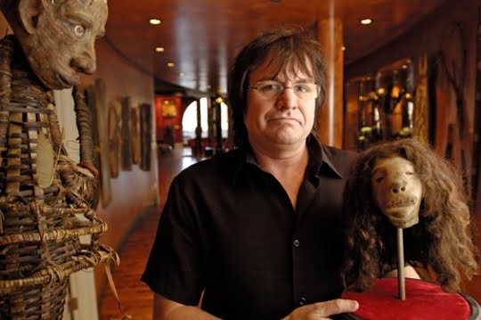
RIP Bill Jamieson


Much has been said about the sad and sudden passing of epic collector and friend to many (including myself!) Billy Jamieson. I am not sure I have anything to add to this often eloquent outpouring of disbelief and grief, except to add note of my own sadness, and to take this moment to mark his passing.

James Taylor put it best, perhaps, on his website "Shocked and Amazed":

Hearing of Bill Jamieson’s death yesterday was about as shocking an occurrence as can be imagined in this business. Still a young man, truly, and a man whose importance to collecting and “spreading the word” had yet to be fully felt, his passing leaves a hole at least 10X larger in the business than the enormous hoard of attractions he leaves behind...

My own experience with Billy was marked by kindness, generosity of spirit, and a sharp and roving intelligence. He loaned us a variety of artifacts from The Niagara Falls Museum--a circa 1827 dime museum whose entire contents he had purchased in 1999--for use in The Coney Island Spectacularium. He also joined us at Coney Island a few weeks back, where we enjoyed the pleasure of his company on the judges stand of The Mermaid Parade followed by a memorable and inspiring lecture in the museum.

I still cannot quite believe he is really dead. He was one of the most full-of-life and inspiring men it has ever been my pleasure to meet.

Rest in peace, Billy. You are--and will continue to be--sorely missed.

Photo sourced from Colorslab.

"Get stuffed: The Disturbing Animals Created when Taxidermy Goes Wrong," The Daily Mail, 2011


The Daily Mail's article "Get stuffed: The disturbing animals created when taxidermy goes wrong"--which profiles the activity of the Facebook group "Badly Stuffed Animals"--has been rapidly making the internet rounds. In an oddly proud moment, I found that the article showcases one of the more disturbing pieces from The Niagara Falls Museum that we have on show as part of The Great Coney Island Spectacularium. Can you guess which one it is? Hint: it is unable to stand on its own two legs...

To see this piece in person, come down to The Great Coney Island Spectacularium! You can read the article and see the full collection by clicking here.

Thanks to Eleanor Crook and Matt Haber for sending this my way.

All images from the article.

THIS SATURDAY! Brains in Jars, Old Libraries, and Underground Crypts in New Haven, Connecticut


We have a few more slots open for our awesome all day field trip this Saturday. See following for details, and email me at morbidanatomy [at] gmail.com if you'd like to come along!

FIELD TRIP: Day of Brains in Jars, Old Libraries, and Underground Crypts in New Haven, Connecticut
A chartered bus field trip to New Haven, Connecticut with guided tours of The Cushing Brain Collection, The Institute Library, and The Center Church Crypt and an unguided visit to the Beinecke Rare Book and Manuscript Library
Date: This Saturday, July 16th
Time: 10:00 AM- 7 PM
Admission: $60
Presented by Morbid Anatomy

*** MUST RSVP to morbidanatomy [at] gmail.com

This Saturday, July 16th join Observatory and Morbid Anatomy for a special field trip to New Haven, Connecticut. Our first stop will be the amazing Cushing Collection, with its over 500 human brains in glass jars and haunting pre- and post-operative photographs amassed by "father of modern neurosurgery" Dr. Harvey Cushing. We will be introduced to this collection-- newly open to the public--via a guided tour by Terry Dagradi, curator of the collection. Our next stop will be the historic and lovely Institute Library (founded 1826), Connecticut's oldest living independent literary institution and one of the last remaining membership libraries in North America, where director Will Baker will give us a tour followed by an opportunity for unguided exploration and lunch. Next, we will be treated to a special after-hours tour of the Center Church Crypt, an underground cemetery featuring 137 grave stones of New Haven's founders and earliest citizens going back to 1687. Our final stop will be an unguided visit to the incredible Beinecke Rare Book and Manuscript Library before hopping on the bus for our return home.

Trip Details: The $60 event cost of this event includes round trip transportation on a special chartered bus from Observatory to New Haven and back again as well as tour costs. Please bring your lunch, which we will have an opportunity to eat at our second stop. The bus will pick up and drop off in front of the 543 Union Street (at Nevins Street) entrance to Observatory. Pick up is 10:00 AM sharp and drop off approximately 7:00 PM depending on traffic.

There is a limit for this trip, so please RSVP to morbidanatomy [at] gmail.com if interested.

Images: Of and from The Cushing Collection as featured in The New York Times.

"What We leave Out: The Art of Science Fiction Writing," 92YTribeca, Tomorrow Night


Just found out about this pretty great looking panel discussion featuring former Observatory lecturer Carl Schoonover and Mr. Wilson's Cabinet of Wonder auther Lawrence Wechsler:

What We leave Out: The Art of Science Fiction Writing
Date: Mon, Jul 18, 2011, 7 pm
Venue: 92YTribeca MAINSTAGE
Location: 92YTribeca, 200 Hudson Street
Event Code: TS12D11185

Scientific experimentation is as much about what we choose not to study as what we do—researchers must always ignore some elements of a problem to focus on others.

Similarly, writing is a process of taking out as much as putting in, of deciding what we can include and what we must leave untouched. Writers who work with the future tend to care most about a particular aspect—about changes in technology, society or the environment.

Panelists Anna North, Carl Schoonover and Lawrence Wechsler discuss the ways writers fashion a future by whittling down an infinity of possibilities until that which most interests them remains. Join them as they shed light on the processes of writing and experimentation and how the eventual results are only part of the story.

The panel takes place tomorrow night! You can purchase tickets here.

Thanks, Carl, for sending this--and the photograph--along.

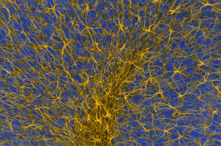
Image; Credit: Thomas Deerinck and Mark Ellisman, 2008.
This image depiects non-neuronal cells in the cerebellum, an area in the back of the brain that play a role in motor coordination. These cells, called glia (yellow), support the activity of neurons, providing nutrients and oxygen. They have been illuminated using a highly, specific antibody-based staining technique, leaving everything else--including the neurons they nurture--invisible in the background (blue).