Bioinformatics: The Early Years

Bioinformatics historians take note: PLOS Computational Biology has published a comprehensive backgrounder on the field's biggest annual meeting the Intelligent Systems for Molecular Biology conference, which celebrated its 20th anniversary this year.

Larry Hunter, who spearheaded the first ISMB in 1993, shares details on how the meeting grew out of a small group of artificial intelligence researchers with an interest in molecular biology. Other early organizers like Richard Lathrop, Russ Altman, Peter Karp, David Searls, and Alfonso Valencia weigh in on how the meeting and the field in general has changed during the past 20 years.

The first meeting was done "on a shoestring," Hunter says. "The program covers one 8.5 x 11 sheet, both sides, which I folded into three and made a nice, neat schedule, complete with the poster session. I kind of remember, now, putting that together on my early Mac. We did things pretty much on the cheap."

At the time, Lathrop says, "nobody had even broached the idea of sequencing the human genome. It was just too gargantuan and mammoth a task, and was considered almost heresy in its early days."

And despite the huge amount of progress during the last 20 years, there's still plenty of room for better bioinformatics tools. "We can sequence somebody's genome, and we've learned virtually nothing with any reasonable amount of certainty," Karp says. "I sure hope that in 10 or 20 years, we can do a lot more with a personal genome predict not just that they're five percent more likely to get some disease, but that they're 90 percent more likely to get one or more diseases. And the treatment that will help that individual."

For more reflections on ISMB's first 20 years, check out a recent interview with Hunter at our sister publication BioInform.

See original here:
Bioinformatics: The Early Years

Waste cooking oil makes bioplastics cheaper

"Bioplastics" that are naturally synthesized by microbes could be made commercially viable by using waste cooking oil as a starting material. This would reduce environmental contamination and also give high-quality plastics suitable for medical implants, according to scientists presenting their work at the Society for General Microbiology's Autumn Conference at the University of Warwick.

The Polyhydroxyalkanoate (PHA) family of polyesters is synthesized by a wide variety of bacteria as an energy source when their carbon supply is plentiful. Poly 3-hydroxybutyrate (PHB) is the most commonly produced polymer in the PHA family. Currently, growing bacteria in large fermenters to produce high quantities of this bioplastic is expensive because glucose is used as a starting material.

Work by a research team at the University of Wolverhampton suggests that using waste cooking oil as a starting material reduces production costs of the plastic. "Our bioplastic-producing bacterium, Ralstonia eutropha H16, grew much better in oil over 48 hours and consequently produced three times more PHB than when it was grown in glucose," explained Victor Irorere who carried out the research. "Electrospinning experiments, performed in collaboration with researchers from the University of Birmingham, showed that nanofibres of the plastic produced from oils were also less crystalline, which means the plastic is more suited to medical applications."

Previous research has shown that PHB is an attractive polymer for use as a microcapsule for effective drug delivery in cancer therapy and also as medical implants, due to its biodegradability and non-toxic properties. Improved quality of PHB combined with low production costs would enable it to be used more widely.

The disposal of used plastics - which are largely non-biodegradable - is a major environmental issue. Plastic waste on UK beaches has been steadily increasing over the past two decades and now accounts for about 60% of marine debris. "The use of biodegradable plastics such as PHB is encouraged to help reduce environmental contamination. Unfortunately the cost of glucose as a starting material has seriously hampered the commercialization of bioplastics," said Dr Iza Radecka who is leading the research. "Using waste cooking oil is a double benefit for the environment as it enables the production of bioplastics but also reduces environmental contamination caused by disposal of waste oil."

The next challenge for the group is to do appropriate scale-up experiments, to enable the manufacture of bioplastics on an industrial level.

More information: Dr Radecka's poster presentation "Making bioplastic from different oils" will take place on Monday 3 September at the Society for General Microbiology's Autumn Conference 2012.

Provided by Society for General Microbiology

Go here to read the rest:
Waste cooking oil makes bioplastics cheaper

Coconut oil could combat tooth decay

Digested coconut oil is able to attack the bacteria that cause tooth decay. It is a natural antibiotic that could be incorporated into commercial dental care products, say scientists presenting their work at the Society for General Microbiology's Autumn Conference at the University of Warwick.

The team from the Athlone Institute of Technology in Ireland tested the antibacterial action of coconut oil in its natural state and coconut oil that had been treated with enzymes, in a process similar to digestion. The oils were tested against strains of Streptococcus bacteria which are common inhabitants of the mouth. They found that enzyme-modified coconut oil strongly inhibited the growth of most strains of Streptococcus bacteria including Streptococcus mutans an acid-producing bacterium that is a major cause of tooth decay.

Many previous studies have shown that partially digested foodstuffs are active against micro-organisms. Earlier work on enzyme-modified milk showed that it was able to reduce the binding of S. mutans to tooth enamel, which prompted the group to investigate the effect of other enzyme-modified foods on bacteria.

Further work will examine how coconut oil interacts with Streptococcus bacteria at the molecular level and which other strains of harmful bacteria and yeasts it is active against. Additional testing by the group at the Athlone Institute of Technology found that enzyme-modified coconut oil was also harmful to the yeast Candida albicans that can cause thrush.

The researchers suggest that enzyme-modified coconut oil has potential as a marketable antimicrobial which could be of particular interest to the oral healthcare industry. Dr Damien Brady who is leading the research said, "Dental caries is a commonly overlooked health problem affecting 60-90% of children and the majority of adults in industrialized countries. Incorporating enzyme-modified coconut oil into dental hygiene products would be an attractive alternative to chemical additives, particularly as it works at relatively low concentrations. Also, with increasing antibiotic resistance, it is important that we turn our attention to new ways to combat microbial infection."

The work also contributes to our understanding of antibacterial activity in the human gut. "Our data suggests that products of human digestion show antimicrobial activity. This could have implications for how bacteria colonize the cells lining the digestive tract and for overall gut health," explained Dr Brady. "Our research has shown that digested milk protein not only reduced the adherence of harmful bacteria to human intestinal cells but also prevented some of them from gaining entrance into the cell. We are currently researching coconut oil and other enzyme-modified foodstuffs to identify how they interfere with the way bacteria cause illness and disease," he said.

More information: Dr Brady's poster presentation "Inhibition of cariogenic streptococci using enzyme modified coconut oil" will take place on Monday 3 September at the Society for General Microbiology's Autumn Conference 2012.

Provided by Society for General Microbiology

Go here to read the rest:
Coconut oil could combat tooth decay

Are You a Pre-Health Student?

Do you want to become more involved around campus and the community while becoming more exposed to the healthcare field? You should consider joining Alpha Epsilon Delta, a pre-health honorary at the U of A. We have philanthropy, social and fundraising events as well as exclusive shadowing opportunities and speakers from the health professions at every meeting. Get exclusive opportunities to win scholarships for test prep courses (up to $2000 for Kaplan and Princeton) or $500 cash scholarships. It is a great way for pre-med, pre-pharmacy, public health majors, pre-nursing, pre-dental, and any other pre-health field to network with professionals and other students who have similar interests and are willing to help you along your career path. Our first meeting is September 4th at 5pm at Bio Science West. Please visit our website at http://www.alphaepsilondelta.com or add our orgsync page https://orgsync.com/2759/chapter for information on the application, or email Aed_vp@yahoo.com with any questions.

Source:
http://physiologynews.blogspot.com/feeds/posts/default?alt=rss

NovoPath Announces Partnership with McKesson to Provide Anatomic Pathology Solution

This makes perfect sense for the LIS space.  McKesson needed an AP solution to add to their portfolio and increases NovoPath's presence in laboratories under McKesson's corporate strategy.

Princeton, NJ (PRWEB) August 30, 2012


NovopathNovoPath, a market-leading provider of anatomic pathology software solutions, today announced a strategic partnership with McKesson, a healthcare services and information technology leader. As part of the new agreement, McKesson plans to propose the NovoPath Anatomic Pathology Solution in conjunction with its advanced laboratory offering and its integrated health information system. The partnership is expected to benefit providers by offering the market an alternative solution that combines NovoPath’s deep expertise in anatomic pathology with McKesson’s best-of-breed laboratory solutions.

MckessonIn addition to further extending NovoPath’s considerable presence in the anatomic pathology market, this agreement also supports McKesson’s Better Health 2020 strategy to enable better business, care and connectivity across the healthcare continuum including laboratories throughout the country.

Delivering enhanced communications, productivity and accuracy to pathology lab professionals nationwide, NovoPath is a comprehensive workflow and reporting lab information software system specifically designed to support the unique needs of the anatomic pathology laboratory. The NovoPath solution has been installed in small, medium and large hospitals, as well as specialty practice anatomic pathology laboratories, and local, regional and national reference labs.

“We are excited about this new relationship with NovoPath, which has a track record of exceptional service and product offerings for the anatomic pathology laboratory market,” said John Yount, vice president, Lab Solutions.

Both companies look forward to providing a comprehensive anatomic pathology solution that complements McKesson’s suite of laboratory solutions.

“This partnership enables NovoPath to provide McKesson customers and the larger market with a tightly integrated anatomic pathology solution,” commented Rick Callahan, NovoPath’s vice president of sales and marketing. “This integration offers laboratories the best of both worlds; a seamless integration of a best-of-breed with an outstanding enterprise-wide solution.”

About NovoPath, Inc. NovoPath, Inc. develops and markets software solutions for the Anatomic Pathology Laboratory market segment that includes local, regional, national, in-house laboratories as well as community and university teaching hospitals and medical centers. Since the release of its flagship product in 1999, NovoPath, Inc. has focused exclusively on Anatomic Pathology. NovoPath's mission is to provide unique and unparalleled solutions and services to all aspects of the Anatomic Pathology sector in a way that improves workflow, reduces the probability of human error, ensures results accuracy for greater patient safety, protects patient confidentiality, and above all, produces more precise and informative diagnostic outcomes. More information is available at http://www.NovoPath.com.

About McKesson McKesson Corporation, currently ranked 14th on the FORTUNE 500, is a healthcare services and information technology company dedicated to helping its customers deliver high-quality healthcare by reducing costs, streamlining processes, and improving the quality and safety of patient care. Over the course of its 178-year history, McKesson has grown by providing pharmaceutical and medical-surgical supply management across the spectrum of care; healthcare information technology for hospitals, physicians, homecare and payers; hospital and retail pharmacy automation; and services for manufacturers and payers designed to improve outcomes for patients.

Read the full story


Source:
http://feeds.feedburner.com/DigitalPathologyBlog

Digital Pathology Misconceptions Debunked by Digital Pathology Consultants

Amande Lowe, president of Digital Pathology Consultants, LLC has a great article for practicing pathologists about common misconceptions regarding the use of digital pathology in the practice of pathology and how and what the technology means in clinical practice under the current framework of government demands, regulations and consumer expectations. 

Debunked: Digital Pathology Misconceptions

Yes, you can be reimbursed, and no, it won't slow you down.

Posted on: August 29, 2012


MisconceptionsThere is hesitation in the healthcare community to adopt digital pathology for clinical use. But why? Digital pathology is an innovation designed to reduce laboratory expenses, boost operational efficiency, enhance productivity, aid in better treatment decisions and improve patient care. It is used today throughout the world for diagnostic consultations, primary and intraoperative diagnosis, medical student education, resident training, diagnostic decision support, peer review, manual and semi quantitative review of immunohistochemistry (IHC), and tumor boards. Digital pathology is our future, however, forging this vision has not been easy and realization of the vision is a constant work in progress. Likely the hesitation to adopt is due to some common misconceptions about digital pathology. Addressing these misconceptions head on is required if the industry is going to advance. 

Misconception #1: The FDA has not approved digital pathology therefore it must not be safe. The FDA does not regulate laboratories and it does not issue blanket approvals for specific applications like digital pathology. The reality is that the FDA approves the digital pathology hardware and software produced by a manufacturer for a specific intended use (e.g., primary diagnosis of H&E slides). Digital pathology manufacturers are required to go through a premarket approval (PMA) process to have their systems approved as a Class III medical device; this process takes time and is very costly for manufacturers. These hurdles have made it difficult for manufacturers to apply for FDA approval, but they are working on it. However, several digital pathology manufacturers have received FDA 510(k) clearances for specific manual and semi-quantitative analysis of specific IHC assays including HER2, ER/PR, Ki-67, and p53. 

Misconception #2: My lab will not pass a CLIA or CAP inspection if we use digital pathology. The College of American Pathologists (CAP) is a Clinical Laboratory Improvement Amendments (CLIA) accredited organization that offers laboratory checklists which support the accreditation process and are designed to be a roadmap for a successful inspection. The Anatomic Pathology, Laboratory General and Cytopathology checklists all have items that apply to digital pathology. In 2011, a CAP Center working group created thirteen guidelines for validating digital pathology for clinical diagnostic use. CAP is expected to issue a final version of these guidelines soon. The Centers for Medicare and Medicaid Services (CMS) oversees CLIA and approves all CAP laboratory checklists. Therefore, if you use the available resources above then digital pathology will not become a risk to the accreditation of your lab. 

Misconception #3: I cannot be reimbursed for digital pathology. Laboratories across the country are being reimbursed when they use digital pathology today. The most common reimbursements are for CPT codes 88360 and 88361, the manual and semi-quantitative analysis of IHC; 88321 and 88323 for consultations; and 88329, 88331, and 88332 for frozen section interoperative diagnosis. 


Misconceptions2Misconception #4
: My lab cannot afford digital pathology, it is too expensive. Digital pathology is an investment but any lab can afford it and make a smart business decision. The implementation of digital pathology in your lab needs attention and must be thoroughly thought out. To be financially successful with digital pathology, create a business plan, establish a return on investment strategy and gain support within your organization. A business plan is the foundation for financial success. It will explain and illustrate why digital pathology is worth doing now and will identify the consequences of not doing it now. An ROI strategy is an attempt to determine the profitability of an investment and a time frame of when it will occur. Sadly, there is no perfect formula. However a good way to attempt an ROI plan is to preform a cost benefit analysis (CBA). With a CBA you analyze the benefits of a given situation or business-related action, sum them up, and then subtract all associated costs with putting that action into place. Do not overlook the value of improvements, including gains in efficiency or cost savings for reduced rework. This is extremely important especially when considering your laboratory workflow. 


Misconception #5
: Digital pathology disrupts my laboratory workflow. How do you break something that is already broken? Many laboratories have a workflow that is manual, fragmented and extremely inefficient. Digital pathology will not be disruptive, instead digital pathology will make these workflow inefficiencies more apparent. These inefficiencies will need to be addressed to be a more productive and progressive laboratory. Digital pathology is often described as the scanning of a glass slide into a whole slide image; yet, it is much more. The pieces of digital pathology include acquisition, integration, data management and interpretation. If all of these pieces interlock together, then laboratory efficiency and the pathologists satisfaction with the technology will improve. Traditionally, when glass slides are prepared they are manually matched with the patient paperwork including patient history, requisition and gross review; then the slides and information are delivered to the pathologist. With digital pathology, the process starts to look a lot different. Now you have whole slide images that can be reconciled to the digital patient paperwork; this information is then electronically delivered to the pathologist. The only way to do this effectively is with a laboratory information system (LIS), electronic medical record (EMR) integration and barcodes. Barcodes will reduce human error, save time on the constant need for verification and rechecks, and improve quality assurance by clear tracking of all specimens throughout the histology process. 

Government demands and consumer expectations are growing for transparency in medicine, improvements in patient safety and identification, electronic medical records, and more personalized treatment plans. Digital pathology supports this healthcare evolution, and enables the digital transformation of pathology. Change is hard; however, if we become complacent with how pathology is practiced today we will underestimate the significance of what the practice of pathology could become tomorrow. 


Related Content

Source: Advance for Administrators of the Laboratory


Source:
http://feeds.feedburner.com/DigitalPathologyBlog

Coagulation App from CDC


PttappscreenThere has to be an app for that!  

 Actually there is one now from informatics folks at the Centers for Diease Control (CDC).

Downloaded this yesterday and everything a (medical) app should be -- Low cost (free), intuitive, easy to use, practical, educational and informative. Not hard to use, navigate or get infrormation from.

iTunes preview

CDC's PTT Advisor offers clinicians a tool to quickly select the appropriate follow-up tests to evaluate patients with a prolonged partial thromboplastin time (PTT) laboratory result and a normal prothrombin time (PT) laboratory result. PTT Advisor will run on your iPhone, iPod touch or iPad. PTT Advisor has been created by CDC in collaboration with experts in diagnostic coagulation with a collective experience of more than 50 years in the field. 

CDC works 24/7 keeping America safe from health, safety and security threats, both foreign and domestic. Whether diseases start at home or abroad, are chronic or acute, curable or preventable, human error or deliberate attack, CDC fights it and supports communities and citizens to prevent it. CDC is the nation's health protection agency — saving lives, protecting people from health threats, and saving money through prevention. 

iTunes preview

FEATURES:

  • Simple and easy-to-use interface
    Pttapp2
  • Quickly navigate through the decision tree
  • Additional information/footnotes provided at key decision points
  • Quickly modify your answers and review all your selections at any point
  • Can be used for both pediatric and adult populations
  • No registration or login required
  • No data is collected or stored

Contact e-mail:  InformaticsLab@cdc.gov

Q: How do I get PTT Advisor? 
A: You can download PTT Advisor for your iPhone, iPod touch, or iPad from Apple's App Store — free of charge. 

FAQs:

Q: How do I navigate through the application?
A: The toolbar at the bottom of the screen has three navigation buttons: 
Navigation buttons 
(1) Back: Go back one step. 
(2) Next: Go forward one step. 
(3) Go to Last: Go to the last step you were presented, but haven't yet responded to. 

Q: Each screen is labeled at the bottom with Step #. How many total steps are in an evaluation?
A: The number of steps per evaluation is determined by your responses. Certain responses will lead to additional steps, so there is no fixed total number of steps. 

Q: How do I change my response to a question?
A: You can either: 

(1) tap the Back button Back button to navigate to the desired question, then change your response -OR- 

(2) tap the Evaluation Review button Evaluation Review button, then tap the desired question listed under Completed Steps.
You will be returned to the selected question, where you can change your response. 

Q: When I navigate back to previous steps, why are some buttons green with a checkmark?
A: The button that you tapped as your response will be green with a white checkmark. 

Q: I changed a response, and a Change Decision Warning alert appears. What does this mean?
A: If you change your response to a previously answered question, all the responses you gave at steps beyond the question will be discarded. You will receive new questions and information from that point forward. For example, if you are at Step 5, but change the response for Step 2, your responses for Steps 3 through 5 will be discarded. You will get new questions/information at Step 3 and onward. 

Q: What does the Footnotes button Footnotes button do?
A: The Footnotes button will be enabled on a step if footnotes are associated with the information presented. This will be indicated by a note at the end of the question/information that reads: [see footnotes]. Touching the Footnotes button launches the Footnotes screen, where any associated footnotes will be listed. 

Q: What is the Evaluation Review screen?
A: The Evaluation Review screen is launched by touching the Evaluation Review button Evaluation Review button on the toolbar. This screen lists the steps you have completed thus far, as well as the current step (or recommendation, if you've reached the Recommendation step). From here, you can tap any step and return to it (to change your answer, for example), or just review the steps and responses thus far in the evaluation. 

Q: If I restart the patient evaluation with the Restart button Restart button, will my previous responses be lost?
A: Yes, when you restart the patient evaluation, any previously entered responses will be discarded. 

Q: I've made it to the Recommendation screen. What do I do now? 
A: Once you have reached the Recommendation screen and read the recommendation, you can: 
(1) start a new patient evaluation by tapping the Done button Done button or the Restart button Restart button
(2) review the evaluation and your responses by navigating backward with the Back button Back button, or launching the 
Evaluation Review screen with the Evaluation Review button Evaluation Review button
(3) suspend or close PTT Advisor. 

Q: Is the response data collected or stored?
A: No, your responses are discarded once you either (1) close the application, or (2) start a new patient evaluation. 

CDC convened seven Institutes from 1984 to 2007 on critical issues in clinical laboratory practice (http://www.cdc.gov/dls/institutes/). National and international experts focused on the role of the clinical laboratory in providing quality testing to improve patient outcomes. The Clinical Laboratory Integration into Healthcare Collaborative (CLIHC)™,  CDC’s Division of Laboratory Science and Standards (DLSS), is addressing some of the recommendations from these institutes by focusing on important “gaps” that must be filled to optimize the ability of practicing clinicians to effectively utilize laboratory services for better patient care. 
The PPT Advisor was developed through CDC’s Office of Surveillance, Epidemiology and Laboratory Services by the DLSS’s CLIHC™, in partnership with the OSELS Informatics R&D Activity.

Customers also bought:

Source:
http://feeds.feedburner.com/DigitalPathologyBlog

Upcoming Courses to Fulfill your CME, SAMS/MOCS for 2012

Looking to fufill your 2012 CME, SAMS/MOCS accreditation?  Join us this fall  and winter for three NEW outstanding Dermatopathology and Surgical Pathology courses in Maui and the Big Island of Hawaii.  It's not too late to register!  
Scientific Symposiums International is accredited by the Accreditation Council for Continuing Medical Education to provide continuing medical education for physicians.
 
Scientific Symposiums International is approved by the ABP and ABD as a provider of SAMs for those who need to meet MOC’s requirements.  
Sign up Today!  Late Fee Waived for October courses!
UPCOMING FALL AND WINTER COURSES:
Scientific Symposiums
International
5star.gif

Scientific Symposiums International has provided Continuing Medical Education courses to more than 4,500 physicians and healthcare providers from around the world since 1994.

 
For more information, visit our website at:http://www.scientificsymposiums.com
MAUNA KEA RESORT maunakeabeach 4   
Big Island of Hawaii

NEW FOUR-DAY COURSE
October 15-18, 2012 


MAUNA KEA RESORT
Big Island of Hawaii


MKBHPool2

NEW FOUR-DAY COURSE

October 22-25, 2012
Course Director: Philip E. LeBoit, M.D., University of California Medical Center, San Francisco, CA


Click Here for all 2012 and 2013 Course Details and Schedules!
HYATT REGENCY MAUI
Maui, Hawaii


Exterior_Aerial

NEW FIVE-DAY COURSE (Sunday-Monday)

February 17-21, 2013
Course Director: John R. Goldblum, M.D., Cleveland Clinic Learner College of Medicine, Cleveland, OH 

Register Now!

facebook_logo twitter_button 2

Source:
http://feeds.feedburner.com/DigitalPathologyBlog

Testing Application that Increases Sensitivity of FNA Testing for Breast Cancer – Developed by High School Student

We may not be top 10 worldwide in academic performance measured by math and science scores but this can't possibly accurate.  DarkDaily has a story today about another high school student developing some great laboratory advances.  The future is bright for the specialty of pathology!

Courtesy of DarkDaily:

Pathologists take heed! Teenagers are taking off-the-self technology and creating useful new clinical laboratory tests for cancer and other diseases

For the second time in recent weeks, a teenager has made national news for developing a medical laboratory test that can more accurately diagnose disease when compared to methodologies currently used by clinical laboratories and pathology groups. Pathologists and clinical chemists have good reason to ask what is different about the science taught in today’s high school compared to recent years.

Read more: DARK Daily Laboratory and Pathology News http://www.darkdaily.com/#ixzz24wgwvS00


82912Teen-ager Brittany Wenger of Sarasota, Florida, developed a software program that was able to more accurately diagnose breast cancer from specimens collected by fine needle aspiration (FNA). Her effort won second place at the Google Science Fair, along with national news coverage of her accomplishment. (Photo by Andrew Federman of Google.)

Source:
http://feeds.feedburner.com/DigitalPathologyBlog

Remote frozen section examination of breast sentinel lymph nodes by telepathology

From the ANZ Journal of Surgery

    Abstract

    Background

    Intraoperative sentinel lymph node (SLN) examination during breast cancer surgery guides the need for immediate axillary clearance. This may be difficult to implement when surgery is performed distant to the centres where pathological examination is undertaken. We aimed to implement and validate a telepathology service for the remote examination of breast SLN by frozen section (FS).

    Methods

    We tested an Internet-based remote microscopy system to report intraoperative FS in real time from two district hospitals without on-site anatomical pathology services.

    Results

    FS was performed remotely on 52 patients. Seventeen out of 52 patients had metastases, of which there were six false-negative diagnoses comprising four of micrometastatic disease and two of isolated tumour cells (ITCs). There were no false-negative diagnoses for macrometastatic disease and no false-positive diagnoses. As a control, we audited our experience with 239 consecutive SLN FS examined by on-site pathologists. Sixty out of 239 patients had metastases, of which there were 24 false-negative diagnoses comprising 12 cases of ITC, 5 of micrometastases and 7 of macrometastases. The accuracy of remote FS was equivalent to that of in-house FS (88.2% versus 89.9%).

    Conclusion

    Remote FS for breast SLN is an accurate procedure,which is not inferior to FS performed on site.

    Cover image for Vol. 82 Issue 7?8

      Article first published online: 24 AUG 2012

      DOI: 10.1111/j.1445-2197.2012.06191.x

      © 2012 The Authors. ANZ Journal of Surgery © 2012 Royal Australasian College of Surgeons


      Source:
      http://feeds.feedburner.com/DigitalPathologyBlog

      Mobile app helps providers track Meaningful Use compliance

      Providers who want to monitor their compliance with the Meaningful Use requirements now can do so using a new mobile solution. San Antonio-based AirStrip Technologies has expanded its mHealth platform to incorporate a Meaningful Use tracker to follow and measure clinical quality and meaningful use in a mobile solution.

      Claiming to be the only vendor-neutral Meaningful Use compliance tool available, the tracker integrates and mobilizes data from electronic health records, medical devices and other sources, enabling clinicians to track their compliance with Meaningful Use in real time, and daily analytics on a customized dashboard, according to AirStrip's announcement. It can follow EHR usage for just one department, or for an entire health system.

      The announcement couldn't be more timely, as both the Centers for Medicare & Medicaid Services and the Office of the National Coordinator for Health IT released their final rules implementing Stage 2 of the Meaningful Use incentive program just last week. AirStrip is in the process of identifying sites to pilot the platform.

      "The Meaningful Use Tracker helps clinicians reach compliance and patient care goals while measuring success against their peers," Care Automation CEO JF Lancelot said in the announcement. "When coupled with the information already provided by AirStrip's mobile medical monitoring solutions and EHR enhancer, analytics provided by the tracker can support earlier warnings about compliance issues, to foster more informed responses to both internal benchmarks and overall patient needs."

      The platform also is configured for future expansion to accommodate new stages of Meaningful Use requirements. This is significant, as ONC already is working on Stage 3 of the Meaningful Use program, which won't be implemented until 2016 at the earliest.

      To learn more:
      - here's AirStrip's announcement

      Related Articles:
      ONC committees advancing to Meaningful Use Stage 3
      CMS unveils final rule for Stage 2 of Meaningful Use
      Meaningful use: A springboard for innovation

      Read more about: Meaningful UsemHealth

      Courtesy of FierceMobileHealthcare

      Source:
      http://feeds.feedburner.com/DigitalPathologyBlog

      Erasmus MC and Philips aim to speed up research for cancer and other diseases with digital analysis of tumors

      Erasmus MC and Philips aim to speed up research for cancer and other diseases with digital analysis of tumors

      • Erasmus MC first hospital in the Netherlands to digitize cell and tissue research using a digital pathology system from Philips
      • Switching to digital allows pathology images and knowledge to be shared amongst cancer research institutes


      Rotterdam, The Netherlands – Erasmus MC is the first hospital in the Netherlands to switch to digital for their experimental laboratory analysis of cell and tissue samples using a digital pathology system from Philips. This switch to digital is an important step in tumor research and ultimately aims to speed up and improve the diagnosis and treatment of cancer and other diseases. Erasmus MC is working with Philips, which has developed a completely new, very fast technology for scanning, image processing and analysis that makes it possible to obtain digital images of suspect tissue at very high resolution. This enables medical researchers to view the images efficiently from any given workplace and to gain new insight into diseases such as cancer.


      If cancer is suspected in a patient, tissue is removed surgically or by means of a biopsy. The tissue is then examined by a pathologist at microscopic level and sometimes also tested at molecular level. This makes it possible to ascertain whether or not the tissue is cancerous and, if so, to what extent the cancer is malignant. This process also plays a very important role in the analysis of large numbers of test samples for experimental cancer research in order to gain a better understanding of the causes and mechanisms of diseases at cellular and molecular level. These new insights may enable new diagnostic approaches and therapeutic treatments.


      The analysis of small tissue samples can create quite a problem in medical investigations. A great deal of time and effort is spent sending, recording and processing the hundreds of microscope slides of tissue samples. Consultation with a colleague at another location can be a lengthy process, as the tissue slide first has to be sent over by courier, with the added risk of damage or loss.


      Digital

      By scanning the tissue slide using the very fast Philips digital pathology system, the examining pathologist can gain direct access to the digital files and the work can be distributed more effectively among the available researchers. Cancer cells in the tissue can be identified and analyzed quickly using advanced image analysis software. It also becomes easy to share information and images with cancer research institutes all over the world.


      "In recent years the demand for cell and tissue examination has risen enormously with more complex cases," says Peter Riegman PhD, Head of the Erasmus MC Tissue Bank. "The changeover in the future to digital pathology will help our team of pathologists, biomedical researchers, technical specialists as well as the management team to ensure we maintain our high standard and further speed up experimental medical research."


      "For over a hundred years now pathologists have used an optical microscope to examine the stained tissue on a microscope slide," says Perry van Rijsingen, General Manager of Philips Digital Pathology. "By integrating digital pathology in the existing information system at the research laboratory, Erasmus MC now has at its disposal a digital platform that offers new opportunities for intensive cooperation in education and research with other disciplines such as radiology."


      The Philips system for digital pathology consists of an ultra-fast scanner and image management system with software for viewing, analyzing and interpreting the images. At Erasmus MC the changeover from analog to digital will be made first in experimental research and pathology education, and this may well be followed by digital diagnostics in the coming years.


      High resolution images available

      http://www.newscenter.philips.com/main/standard/resources/research/ErasmusMC/Tissue-slides-Philips-digital-pathology-at-Erasmus-MC.jpg

      http://www.newscenter.philips.com/main/standard/resources/research/ErasmusMC/Scanner-Philips-digital-pathology-at-Erasmus-MC.jpg

      http://www.newscenter.philips.com/main/standard/resources/research/ErasmusMC/Display-Philips-digital-pathology-at-Erasmus-MC.jpg


      For further information, please contact:

      Erasmus MC

      Tel +31 10 703 3289

      Email: press@erasmusmc.nl

      http://www.erasmusmc.nl/perskamer 


      Philips Digital Pathology

      Hans Driessen

      Mobile + 31 610610417

      E-mail: hans.driessen@philips.com

      http://www.philips.com/digitalpathology:


      About Erasmus MC

      Erasmus MC is the largest and most authoritative scientific University Medical Center in the Netherlands. Almost 13,000 staff members work within the core tasks of patient care, education, and scientific research on the continuous improvement and enforcement of individual patient care and social healthcare. They develop high-level knowledge, pass this on to future professionals, and apply it in everyday patient care. Over the next five years, Erasmus MC wants to grow into one of the best medical institutes in the world. Erasmus MC is part of the Dutch Federation of University Medical Centers (NFU): http://www.nfu.nl 


      About Royal Philips Electronics

      Royal Philips Electronics (NYSE: PHG, AEX: PHI) is a diversified health and well-being company, focused on improving people’s lives through meaningful innovation in the areas of Healthcare, Consumer Lifestyle and Lighting. Headquartered in the Netherlands, Philips posted 2011 sales of EUR 22.6 billion and employs approximately 122,000 employees with sales and services in more than 100 countries. The company is a leader in cardiac care, acute care and home healthcare, energy efficient lighting solutions and new lighting applications, as well as male shaving and grooming, portable entertainment and oral healthcare. News from Philips is located athttp://www.philips.com/newscenter.


      Source:
      http://feeds.feedburner.com/DigitalPathologyBlog

      Join us at Innovator’s Edge, Charlotte, NC – September 20


      Innovator's Edge - sponsored by Ventana Medical Systems, Inc. 
      Thursday, September 20, 2012 10:00 AM - 3:00 PM (Eastern Time)
      Charlotte Convention Center
      704-339-6000
      501 South College Street
      Rooms 204 and 205
      Charlotte, North Carolina 28202
      United States 
      Map and Directions

      Join us at Innovator's Edge

       A mobile symposium to empower pathology professionals 

      Join us at Innovator’s Edge, a dynamic mobile symposium that brings the power of education, connection,and industry-leading solutions to you. From thought leaders in breast cancer diagnostics and digital pathology, to experts in lean workflow, we empower you to bring personalized cancer care to the glass slide. Trust the industry-leading team with more tissue companion diagnostics in the pipeline than any other pathologycompany to keep you on the cutting edge of our rapidly evolving profession. 

      Choose the program that interests you most, to optimize your time with our team of global experts: 

              Registration is free of charge. Space is limited.

       
      Hands-on demonstrations of emerging technologies will be available on-site for those who are
      interested in applying new insights in the laboratory setting: 


      • VENTANA iScan HT* – high throughput digital slide scanner
      • Benchmark ULTRA – continuous access IHC/ISH staining platform
      • VENTANA Benchmark Special Stains – fully automated special stains platform
      • VANTAGE – workflow management solution
      *The VENTANA iScan HT slide scanner is for research use only. Not for use in diagnostic procedures.
      Agenda

      Advances and Controversies: 
      Digital Pathology and Breast Cancer Management 
      4.0 AMA-PRA Category 1 credits accredited by Charlotte AHEC

      10:00 am   Welcome
                       Kevin Smith, MD

                       President, Celligent Diagnostics
                       
                       Introductions and Pre-Test Information
                       Peter Banks, MD

                       Pathologist, Scientific Affairs,
                       Ventana Medical Systems, Inc.

      10:15 am   Digital Pathology: Current Status and Issues
                       Keith J. Kaplan, MD

                       Director of Information Technology

                       Carolinas Pathology Group

                       Breast Cancer: Current Diagnostic Issues and 
                       Biomarkers for Prognosis and Management
                       Chad A.Livasy, MD

                       Pathologist, Breast and Gynecologic Pathology

                       Carolinas Pathology Group

      12:15 pm   Lunch

      1:00 pm     Breast Cancer: Current Clinical Management Issues
                       Steven A. Limentani, MD
                       Associate Medical Director, Levine Cancer Institute
                       Clinical Professor of Medicine, UNC-Chapel Hill

      1:30 pm     Evidence-Based Pathology: A Word of Caution
                       Mark R. Wick, MD

                       Professor of Pathology
                       University of Virginia Health System               

      2:30 pm     Faculty Panel Discussion with Open Q & A
                       Post-test performance      

                 
      3:00 pm     Adjournment   

      3:00pm      Reception 

      In the AP Lab:
      Best Practices for Continuous Performance Improvement

      Kelsi Currier, MPH
      Ventana Medical Systems, Inc.
      10:00 am - 2:00 pm
      Lunch Provided
      Three hour session approved for 3 CEUs
        
      Course Content
      • See the AP Lab through the Eyes of a Lean Expert
      • AP Industry Challenges
      • Change Management
      • Lean Overview
      • Lean Definitions and Interactive Exercises
      • Six Sigma Overview Case Study: Leaning the AP Laboratory
      • Open Q&A Discussion
      • Closing Remarks  

      Contact Information


      Source:
      http://feeds.feedburner.com/DigitalPathologyBlog

      Pathologists temporarily saved from unfair EHR penalties


      GI_0_51stlogoThe Pathology Blawg has a nice piece and comments on the ridiculous notion that pathologists (and radiologists and anesthesiologists), given the nature of their clinical practices (non-direct, non-patient care, non-outcomes focused for payment) could and would be penalized for not meeting EHR meaningful use criteria like "real" doctors; those who are on the front lines, in the trenches, managing smoking cessation, weight control, diabetes, hypertension, hyperlipidemia, COPD, etc...


      On Friday August 24, 2012, the CAP released a STATLINE special alert detailing how the Centers for Medicare and Medicaid Services (CMS) granted a temporary “stay” in penalizing pathologists, anesthesiologists and radiologists from the MU benchmarks.  In addition, HR 4066 now has 38 co-sponsors.

      Read more here.


      Source:
      http://feeds.feedburner.com/DigitalPathologyBlog

      Letter from Aperio on Recent Sale

      Dear Aperio
      Customer
      ,

      On behalf of everyone at Aperio, I want to take
      this opportunity to tell you about a new and exciting development. I am pleased
      to announce that Leica Biosystems is acquiring Aperio. Aperio began as a
      start-up business 12 years ago and today has grown into the leader of
      ePathology solutions. By integrating with Leica Biosystems, we will improve our
      global reach and provide the broadest portfolio of pathology solutions to serve
      this growing and changing market.

      At Aperio, we believe pathology plays a vital
      role in patient care and research, with an ever-increasing importance due to
      advances in diagnostic testing, more targeted drug therapy options, and
      advances in personalized medicine. Leica Biosystems shares that belief.

      Leica Biosystems’ goal is to provide solutions
      that meet all your anatomical pathology needs and help advance cancer
      diagnostics. They are represented in over 100 countries, with manufacturing
      facilities in 7 countries, sales and service organizations in 19 countries, and
      an international network of dealers. Leica Biosystems provides end-to-end
      solutions that help customers improve workflow efficiency and diagnostic
      confidence.

      Leica Biosystems is acquiring Aperio because of
      our leadership in ePathology solutions, our great products, and our dedicated
      team. We believe that together we can advance our product portfolio more
      effectively, expand our global reach, and continue to improve our customer
      service. The Aperio ePathology solutions are complementary to Leica’s own broad
      portfolio of pathology solutions. The integrated business will enhance both
      solutions going forward. For the foreseeable future we expect business to
      remain as usual.

      The transaction is subject to various closing
      conditions, including customary regulatory approvals, and we will continue to
      operate independently until the closing.

      Please do not hesitate to contact me if you have any
      questions
      . We look forward to continuing to serve you in the years
      to come as Leica Biosystems and Aperio embark on this exciting new journey
      together.

      Best regards, ???

      David Schlotterbeck ?

      CEO, Aperio


      Source:
      http://feeds.feedburner.com/DigitalPathologyBlog

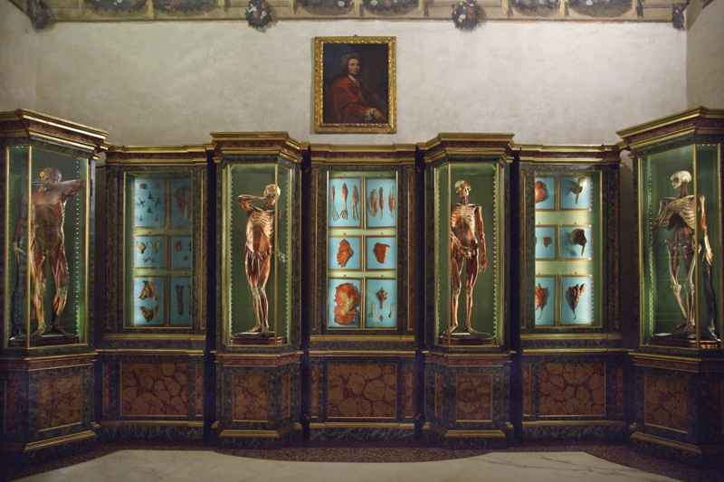
      Morbid Anatomy Exhibition and Event Series, Viktor Wynd Fine Art/Last Tuesday Society, London, September 2012


      As mentioned in a recent post, beginning in just a few days, Morbid Anatomy will be artist-in-residence at Viktor Wynd Fine Art and The Last Tuesday Society in London, England. The residency will span the entire month, and will include an exhibition (photographs from which you see above), as well as a full month's worth of "Morbid Anatomy Presents" programming that will include some seriously amazing lectures, a screening, a "Congress for Curious Peoples" symposium, and a field trip to the rarely open-to-the-public St Bartholomew's Hospital Pathology Museum where I will also give a lecture on the art and history of anatomical museums.

      The exhibition, "Ecstatic Raptures and Immaculate Corpses: Visions of Death Made Beautiful in Italy," will open with a party next Thursday, September 6 and will premiere a new body of work based on my latest obsession: the through-lines connecting the beautiful, immaculately preserved corpse found in both  the churches and enlightenment-era anatomical museums of Italy. The exhibition, which will feature my own photographs and waxworks by the über-talented Eleanor Crook and Sigrid Sarda. You can download a postcard invitation which contains full information by clicking here.

      I have just created pages for each event, which you can find at the Morbid Anatomy Facebook page by clicking here. The list also follows here, for your convenience:
      FULL LIST OF EVENTS

      Monday, 3rd September 2012, 7 PM
      Granta Magazine - Medicine Issue Launch

      Tuesday, 4th September 2012, 7 PM
      Robert Marbury - Rogue Taxidermy in the Digital Age

      Wednesday, 5th September 2012, 7 PM
      Dr Sam Alberti of The Hunterian Museum on the History of Medical Museums

      Thursday, 6 September 2012, 6-8 PM
      Opening Reception for "Ecstatic Raptures and Immaculate Corpses: Visions of Death Made Beautiful in Italy," Sponsored by Hendricks Gin

      Saturday, 8th September 2012, 11 AM - 5:30 PM
      'Congress for Curious People' Seminar - London Edition

      Monday, 10th September 2012, 7 PM
      Ronni Thomas and The Real Tuesday Weld - 'Midnight Archive' screening

      Tuesday, 11th September 2012, 7 PM
      Martin Clayton on Leonardo Da Vinci and Dissection

      Wednesday, 12th September 2012, 7 PM
      Curious Cafés of the Belle Epoque with Vadim Kosmos

      Monday, 17th September 2012, 7 PM
      Gemma Angel on the History of Human Tattoos

      Wednesday, 19th September 2012, 7 PM
      Field Trip to St Bart's Pathology Museum with Lecture by Joanna Ebenstein

      Thursday, 20th September 2012, 7 PM
      Paul Craddock - History of Blood Transfusions

      Tuesday, 25th September 2012, 7 PM
      Dr. James Kennaway - Bad Vibrations

      Wednesday, 26th September 2012, 7 PM
      Dr. Pat Morris - Extreme Taxidermy: Elephants and Humans

      Thursday, 27th September 2012, 7 PM
      Royal Raymond Rife and his Oscillating Beam Ray with Mark Pilkington

      Sunday, 30th September 2012, 7 PM
      Eleanor Crook on Plastic Surgery of the World Wars

      You can find out more about the exhibition here and more about the events here. All of the above images are drawn from the exhibition "Ecstatic Raptures and Immaculate Corpses: Visions of Death Made Beautiful in Italy," opening at Viktor Wynde's Fine Art on September 6th with a reception from 6-8, and will be on view through the end of the month. And a special shout out to Jessica Pepper, who so expertly and beautifully retouched these images.

      Source:
      http://morbidanatomy.blogspot.com/feeds/posts/default?alt=rss

      It Came from the Stores, Exhibition, Grant Museum of Zoology, London, Through August 31, 2012

      “A lovely skeleton, but sadly lacking a skull,” laments one of the tags afforded to the remains of a Capuchin monkey in this show of the unseen at the ever-exotic Grant Museum. “Rarely do ‘incomplete’ specimens make the grade for display.”

      When I am in London, I will most certainly be checking out the wonderful sounding exhibition "It Came from the Stores," on view at the incomparable Grant Museum  until August 31st.

      You can find out more here.

      Image caption: An elephant shrew is among the specimens on show at the Grant Museum of Zoology

      © UCL, Grant Museum of Zoology

      Source:
      http://morbidanatomy.blogspot.com/feeds/posts/default?alt=rss

      Boost nutrition with foods that burn more calories than they contain

      So-called zero-calorie foods, like celery and cucumbers, contain fewer calories than the body uses to break them down. And although nutritionists account for the energy it takes to chew and digest them when they calculate how many calories we need, these eats deserve prime spots on our plates. You can eat them in large quantities without busting your gut, and low-calorie doesn't mean low nutrients.

      "And, obviously, if eating very low-calorie foods keeps you from eating higher calorie foods, that's a win," says Monica Reinagel, licensed nutritionist and creator of the Nutrition Diva podcast. So fill up your fridge with the following foods that are loaded with vitamins and minerals -- not calories.

      Cucumbers

      If you're tired of fending off hunger by guzzling glass after glass of water, snack on cucumber slices, instead. "Eating foods that are high in water can help you feel full at least temporarily by taking up a lot of space in your stomach," notes Reinagel. Cucumbers also pack vitamins K and C, potassium, and silica, which helps build and maintain connective tissue, like muscle, tendons, ligaments and bone.

      Citrus fruit

      Don't wait until cold season to fill up on oranges, tangerines, and grapefruit -- they may help whittle your middle. People with higher vitamin C levels have lower waist-to-hip ratios than those whose bodies contain less of the antioxidant, according to a study published in the American Journal of Clinical Nutrition. What's more, University of Arizona researchers found that those with higher levels of vitamin C oxidized 25% more fat during treadmill sessions than those with lower levels of the vitamin.

      Leafy greens

      Whatever variety you pick, you can't go wrong with piling a plate with salad greens. At 4 calories per cup, watercress is loaded with vitamins A, C and K, and a study in the American Journal of Clinical Nutrition found that eating 3 ounces of the peppery green daily increases levels of the cancer-fighting antioxidants lutein and beta-carotene. Spinach (7 calories per cup) is brimming with vitamin K, calcium, phosphorus, potassium, zinc and selenium and contains a hormone that allows muscle tissue to repair itself faster, according to research from Rutgers University.

      Asparagus

      A half-cup of cooked asparagus will set you back only 20 calories. Plus, you'll get hefty doses of vitamins K and A, and B vitamins such as folic acid. Since B vitamins play a role in breaking down sugars and starches, eating asparagus may help regulate blood sugar and fend off type 2 diabetes.

      Follow this link:
      Boost nutrition with foods that burn more calories than they contain

      Dr. Mao's Wellness Living: Heal Headaches Naturally To Avoid Side Effects

      Courtesy Photo

      One in six Americans experience chronic headaches. Luckily, Chinese medicine has an arsenal of natural remedies to help soothe your aching head.

      According to Chinese medicine, pain is your bodys signal that there is a blockage of energy and blood. Your body is asking you to notice, evaluate, and change the behavior that caused the pain in the first place. For example, a headache may be pointing to an improperly balanced diet. It is best to treat pain using natural methods so you can avoid side effects and also become more aware of your bodys unique needs.

      In rare cases, headaches may be due to severe medical conditions, such as glaucoma or brain hemorrhages. If you experience severe, debilitating headaches that dont respond to over-the-counter medication, visit your physician or the emergency room immediately.

      Here are a few all-natural remedies to alleviate headaches:

      1. Press Valley of Harmony for headache relief

      Recently, a patients eight-year-old child was suffering from a headache. I was able to bring her the benefits of acupuncture without the needles. I simply used my own fingers to administer acupressure on an acupoint in the web between her thumb and index finger called Valley of Harmony. Within five minutes, her headache was gone.

      Find the acupoint Valley of Harmony (Li 4) on yourself. Its in the web between the thumb and index finger on both hands. Apply steady pressure with your thumb until you feel soreness. Hold for 2 minutes. Repeat on the other hand.

      Find the acupoint Great Yang (taiyang), which is in the indentation of the temples. Stimulate the point with the knuckles of your thumbs or the tips of your index fingers. Massage in circular motion for five minutes.

      2. Massage your headache away

      Read the original:
      Dr. Mao's Wellness Living: Heal Headaches Naturally To Avoid Side Effects

      H. Samuel Case, McDaniel professor

      H. Samuel Case, who taught human physiology and exercise science at what is now McDaniel College for four decades, died Aug. 22 of complications from leukemia at Carroll Hospice Center's Dove House in Westminster.

      The longtime Westminster resident was 70.

      "I've been here for two years, but I very quickly came to see how much Sam embodied the essence of McDaniel College. His death is such a painful loss, " said Roger Casey, president of the college.

      "In my speeches to prospective students, I tell them that the faculty is not in your life for just four years, but 40. And I got that from observing Sam," he said. "He lived life to the fullest, and his death reminds us of our own mortality and that we only have a short time to make a difference, as Sam did."

      "Sam's life is a life worth celebrating. He was a fine human being and totally selfless," said Joan Develin Coley, former McDaniel College president and friend of 40 years.

      "When I think of Sam, he led a life that was exemplary and one of service. He never needed or wanted to toot his own horn," she said. "People were just drawn to him. The students loved him and the faculty loved him."

      The son of a farmer and an educator, Howard Samuel Case, who never used his first name, was born and raised in Three Bridges, N.J.

      After graduating in 1959 from Hunterdon Central Regional High School in Flemington, N.J., where he played football and wrestled, he earned a bachelor's degree in 1963 in physical education at what was then Western Maryland College.

      "The starting line of his career emerged by accident while he was still an undergraduate at Western Maryland College," said Joyce Muller, associate vice president of communications and marketing at McDaniel.

      "He suffered a concussion while playing football in his sophomore year and opted to assist with coaching, and by the time he graduated, had racked up three years of coaching experience," said Ms. Muller.

      Go here to see the original:
      H. Samuel Case, McDaniel professor