‘Contemporary Medical Science and Technology as a Challenge to Museums’, 15th Bi-Annual EAMHMS Congress, Copenhagen, September 16-18


In a few weeks--from September 16th through 18th--the 15th biannual conference of the European Association of Museums for the History of Medical Sciences--to be hosted by the Medical Museion--will be in full effect at the Medical Museion of Copenhagen, Denmark.

The topic of this year's congress--"Contemporary medical science and technology as a challenge to museums"--will be debated and discussed by a variety of museum practitioners and enthusiasts, from curators to museum directors to artists to bloggers such as myself. I will be presenting my paper--"The private, curious, and niche collection: what they can teach us about exhibiting new medicine"-- on Thursday the 16th. Other participants will include New York based artist and SVA professor Suzanne Anker, the Wellcome Collection's Ken Arnold, and friend-of-Morbid-Anatomy James Edmonson of the Dittrick Museum.

This is sure to be a thought provoking and fascinating congress. Hope very much to see you there!

Full schedule follows:

THURSDAY, 16 SEPTEMBER

  • Thomas Söderqvist: Why this conference now?
  • Kim Sawchuk: Biotourism and biomediation
  • Kerstin Hulter Åsberg: Uppsala Biomedical Center: A mirror of modern medical history – how can it be displayed?
  • Wendy Atkinson and René Mornex: A major health museum in Lyon
  • Robert Martensen: Integrating the physical and the virtual in exhibitions, archives, and historical research at the National Institutes of Health
  • Ramunas Kondratas: The use of new media in medical history museums
  • Danny Birchall: ‘Medical London’, Flickr, and the photography of everyday medicine
  • Joanna Ebenstein: The private, curious, and niche collection: what they can teach us about exhibiting new medicine

FRIDAY, 17 SEPTEMBER

  • Judy M. Chelnick: The challenges of collecting contemporary medical science and technology at the Smithsonian Institution
  • James Edmonson: Collection plan for endoscopy, documenting the period 1996-2011
  • John Durant: Preserving the material culture of contemporary life science and technology
  • Stella Mason: Medical museums, contemporary medicine and the casual visitor
  • Alex Tyrell: New voices: what can co-curation bring to a contemporary medical gallery?
  • Jan Eric Olsén: The portable clinic: healthcare gadgets for home use
  • Yin Chung Au: Seeing is communicating: possible roles of Med-Art in communicating contemporary scientific process with the general public in digital age
  • Nina Czegledy: At the intersection of art and medicine
  • Lucy Lyons: What am I looking at?
  • Henrik Treimo: Invisible World
  • Victoria Höög: The optic invasion of the body. Epistemic approaches to current biomedical images
  • Ken Arnold and Thomas Söderqvist: A manifesto for making science, technology and medicine museums

SATURDAY, 18 SEPTEMBER

  • Morten Skydsgaard: The exhibition ‘The Incomplete Child’: boundaries of the body and the guest
  • Sniff Andersen Nexø: Showing fetal realities: visibility, display, performance
  • Suzanne Anker: Inside/Out: fetal specimens through a 21st Century lens
  • Yves Thomas and Catherine Cuenca: Multimedia contributions to contemporary medical museology
  • Nurin Veis: How do we tell the story of the cochlear implant?
  • Jim Garretts: Bringing William Astbury into the 21st Century: the Thackray Museum and the Astbury Centre for Structural Molecular Biology in partnership
  • Adam Bencard: Being molecular
  • Roger Cooter and Claudia Stein: Visual things and universal meanings: aids posters, the politics of globalization, and history
  • Karen Ingham: Medicine, materiality and museology: collaborations between art, medicine and the museum space
  • Silvia Casini: Curating the biomedical archive-fever
  • Thomas Schnalke: Dissolving matters. The end of all medical museums’ games?

You can find out more about the congress by clicking here. You can read more about the Medical Museion via a few recent blog posts, the first being the Dittrick Museum's (here) and the other being that of The Sterile Eye (here).

Image: From The Sterile Eye; Caption: Copy of écorché statue by Theobald Stein. Original from 1869.

Amazing Medical Woodblock Prints, Japan, 19th Century


Just came across this really fantastic and unusual collection of 19th Century Japanese medical woodblock prints on the Pink Tentacle website, which sourced them from the extensive University of California San Francisco (UCSF) Japanese Woodblock Print Collection website.

As the UCSF website explains:

The UCSF Japanese Woodblock Print Collection consists of four hundred Japanese woodblock prints on health-related themes. Of those, more than half are colorfully illustrated in the ukiyo-e manner, the remainder being printed single-sheet texts. From the treatment and prevention of diseases like smallpox, measles, and cholera, to the stages of pregnancy and drug advertisements, these prints offer a unique window into traditional Japanese attitudes toward health and illness.

The majority of the prints date to the mid- to late nineteenth century, when Japan was opening to the West after almost two hundred and fifty years of self-imposed isolation. Thus, they provide valuable pictorial evidence for the effect of Western medical science on traditional beliefs and practices.

Five subject areas broadly define the collection. The treatment and prevention of three contagious diseases; smallpox, measles, and cholera; are topics for eighty of the prints. A related category includes prints in which Buddhist or Shinto deities intervene to ensure a cure. Pregnancy and women's health issues form a distinct theme, including several images of the stages of gestation. Because foreigners were thought to carry disease to Japan, the collection also includes several maps of Nagasaki, where the Dutch were confined during the Edo period, as well as prints depicting foreigners and their ships. Drug advertisements from the nineteenth century make up the largest category...

The woodblock prints in this collection offer a fascinating visual account of Japanese medical knowledge in the late Edo and Meiji periods. Collectively, they record a gradual shift, by the late nineteenth century, from the reliance on gods and charms for succor from disease, to the adoption of Western, scientific principles as the basis for medical knowledge. They show the introduction of imported drugs and vaccines and increased use of printed advertisements to promote new medicinal products.

You can view the entire collection on the UCSF website--arranged by the themes Contagious Disease, Drug Advertisements, Foreigners & Disease, Religion & Health and Women’s Health--by clicking here.

I HIGHLY recommend clicking on each image to view larger, richer, and more detailed image.

Image captions top to bottom:

  1. Shinto god from Izumo province for preventing measles -- Taiso Yoshitoshi, 1862
  2. Pregnancy guide -- Hamano Teisuke, 1880
  3. Illustrated guide to parental obligations -- Utagawa Yoshitora, 1880
  4. Pregnant women playing in summer heat (5 heads, 10 bodies) -- Utagawa Kunitoshi, 1881
  5. Chasing measles away -- Utagawa Yoshimori, 1862

From Pink Tentacle via Ellettrogenica.

Seeking Private, Curious, Arcane and Overlooked Scandinavian Museums, Collections, Sights and Curiosities


Greetings folks.

I am in the midst of planning a trip to Scandinavia around the upcoming Congress for the European Association of Museums for the History of Medical Sciences on the theme of ‘Contemporary medical science and technology as a challenge to museums' to be held at Medical Museion in Copenhangen, September 16-18.

More on that conference--at which I will be delivering a paper--soon (though the impatient among you can see the full line up here); for now, I mention it only as an excuse for soliciting suggestions from Morbid Anatomy readers regarding private, curious, arcane, and overlooked museums, collections, sights and curiosities that one shouldn't miss in this part of the world, of which I know frightfully little.

Any suggestions very much appreciated! Suggesters can leave comments on this post or email me at morbidanatomy@gmail.com.

Thanks so much!

Image: The Biologiska Museet, Stockholm--which is very much on my list!--from Picasa user Traci Brandon.

This Tuesday at Observatory: “Behind the Scenes and Under the Skin” or, “The Body at Blythe," with Lisa O’Sullivan, Medicine Curator of Science Museum


Next Tuesday at Observatory please join Morbid Anatomy for “Behind the Scenes and Under the Skin” or, “The Body at Blythe” an illustrated lecture by Lisa O'Sullivan, Senior Curator of Medicine at the Science Museum of London. As many of you no doubt know, the Science Museum houses the incredible collection of 100,000 or so of the artifacs amassed by Henry Wellcome, tiny glimpses of which you see above; the collection will be discussed and pictured at much greater length in Tuesday's lecture.

Hope very much to see you there.

“Behind the Scenes and Under the Skin” or, “The Body at Blythe”
An illustrated talk by Lisa O’Sullivan, Senior Curator of Medicine at the Science Museum, London
Date: Tuesday, September 7th
Time: 8:00 PM
Admission: $5
Presented by Morbid Anatomy

Join Lisa O’Sullivan, Senior Curator of Medicine at the Science Museum, London for a behind-the-scenes look at the Museum stores. As is the case for many large museums, only about 5% of the Science Museum’s objects are ever on display. This is an opportunity to see some of the other 95% - as photographed by Morbid Anatomy’s Joanna Ebenstein on a recent visit.

The Museum’s ‘small objects’, all 203,000 of them, are stored at Blythe house, an early 20th century office building (larger objects live in aircraft hangars in the West of England). Over 100,000 of these artefacts are medical, the majority from the Wellcome collection. Over 30 rooms hold objects from Roman votives to mediaeval saints, x-ray machines to. The collection displays the breadth of Henry Wellcome’s collecting, and vision of medicine.

Lisa will conduct a virtual tour of some of the rooms, highlight some of her – anatomical – favourites amongst the objects, and take questions about life ‘backstage’ at the Museum.

Lisa O’Sullivan is the Senior Curator of Medicine at the Science Museum in London, where she curates the Wellcome collections. She is responsible for the anatomical collections, and all issues relating to human remains in the museum. Lisa’s doctorate looked at the construction of nostalgia as a clinical category in nineteenth-century France. In addition she has degrees in medical anthropology, history and literature. In 2010, she is a postdoctoral fellow at the University of Sydney.

You can find out more about this presentation by clicking here. To find out more, see this recent post, from which the above images were drawn.You can get directions to Observatory--which is next door to the Morbid Anatomy Library (more on that here)--by clicking here. You can find out more about Observatory here, join our mailing list by clicking here, and join us on Facebook by clicking here.

The Edward Gorey House Museum, Yarmouth Port, Massachusetts


My boyfriend and I finally made a much-anticiated pilgrimage to the wonderful Edward Gorey Museum in Yarmouth Port, Massachusetts a few weeks ago. We were far from disappointed by what we found!

The museum (see photos above) is located in the home of the recently deceased author/illustrator/eccentric Edward Gorey. The home has, sadly, largely been cleared of Gorey's legendary clutter--though a few tantalizing fragments from his private collection can be found scattered about--and transformed into a compact house-museum dedicated to the man's life and work.

Part of me wishes they had simply left the place as it was at Gorey's death, and allowed visitors the opportunity to wander around the famously idiosyncratic environment in which the man produced so many of his iconic works. However, I was quickly won over by the museum's small-town- quirky charm, and the pretty great displays, which included original drawings and half-finished inked works, reproductions of his sketchbooks, amazing ephemera and souvenirs from Mystery and his Broadway production of Dracula, one of his raccoon fur coats, many of his Doubleday book covers, a number of his handmade stuffed animals, many coveted rare works such as his fantastic peepshow, and scores of other artifacts. The gift-shop was also seriously incredible--with scores of Gorey-themed souvenirs I had never seen before--and the folks running the museum were lovely to talk to, knowledgeable and passionately devoted to the man and his work.

In all, the museum really managed to capture the atmosphere and spirit of Edward Gorey's peculiar and alluring universe, with all its whimsy, quirkiness, and elegance; one gets the feeling that Edward Gorey himself might almost have approved, if he had been capable of approving of any museum devoted to his own life and work.

If you are a fan of Mr. Gorey, I cannot more highly recommend making a pilgrimage of your own to the Edward Gorey House Museum. You can find out more about it--including hours and directions--by clicking here; you can visit their awesome gift shop by clicking here. If you would like to make a virtual visit, you can view the full set of photos documenting my own trip--from which the sample above is drawn--by clicking here.

Tomorrow Night at Observatory: "Documenting the Invisible: Spiritualism, Lily Dale, and Talking to the Dead," Illustrated Lecture by Shannon Taggart


Tomorrow night at Observatory! Hope very much to see you there.

Documenting the Invisible: Spiritualism, Lily Dale, and Talking to the Dead
An illustrated lecture by photographer Shannon Taggart
Date: Tuesday, August 31
Time: 8:00 PM
Admission: $5
Presented by Morbid Anatomy

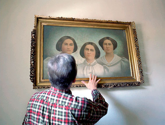
Spiritualism is a loosely organized religion based primarily on a belief in the ability to communicate with spirits of the dead. The movement began in upstate New York in 1848 when two young girls named Margaret and Kate Fox claimed to be in contact with the spirit of a dead peddler buried beneath their home. Photographer Shannon Taggart first became aware of Spiritualism as a teenager when her cousin received a reading in Lily Dale, NY, The World’s Largest Spiritualist Community. A medium there revealed a strange family secret about the death of their grandfather that proved to be true. Taggart became deeply curious about how someone could possibly know such a thing.

Thus began a five year photography project focused on Modern Spiritualism. During her image making she immersed myself in the history and philosophy of Spiritualism, had more readings than she can count, experienced spiritual healings, took part in séances, attended a psychic college and sat in a medium’s cabinet, all with her camera. Despite this exposure she finds herself no closer to any definitive answer of what it all means. She feels as if she has peered into a mystery.

Shannon Taggart is a freelance photographer based in Brooklyn, New York. She received her BFA in Applied Photography from the Rochester Institute of Technology. Her images have appeared in numerous publications including Blind Spot, Tokion, TIME and Newsweek. Her work has been recognized by the Inge Morath Foundation, American Photography, the International Photography Awards, Photo District News and the Alexia Foundation for World Peace, among others. Her photographs have been shown at Photoworks in Brighton, England, The Photographic Resource Center in Boston, Redux Pictures in New York, the Stephen Cohen Gallery in Los Angeles and most recently at FotoFest 2010 in Houston. For more about Shannon Taggart, visit http://www.shannontaggart.com.

You can find out more about this event here. You can get directions to Observatory--which is next door to the Morbid Anatomy Library (more on that here)--by clicking here. You can find out more about Observatory here, join our mailing list by clicking here, and join us on Facebook by clicking here.

Image: Ron with the Fox Sisters, Founders of Spiritualism; Lily Dale, NY © Shannon Taggart

'Skin,' Wellcome Collection, Through September 26, 2010


The last decade has revealed a burgeoning interest and fascination with human skin, particularly among philosophers, writers, artists and designers. Meanwhile, regenerative medicine has seen major advances in the development of artificial skin designed to improve the structure, function and appearance of the body surface that has been damaged by disease, injury or ageing. So there couldn't be a better time to get under the surface of this subject.--Lucy Shanahan, Wellcome Collection Curator and co-curator of 'Skin'

I have been hearing excellent reports from scores of people about the new Wellcome Collection exhibition entitled, simply, 'Skin.' Sadly, I will not be able to see it in person (as it closes on September 2th), but the images above--most drawn from the exhibition website--and the web exhibition text make it clear that the Wellcome has done it again: a thoughtful, broadly considered, and lovely investigation and survey into the science, meaning, art, and implications of the notion of 'skin.'

More about the show, from the press release:

The skin is our largest organ. It gives us a vital protective layer, is crucial for our sense of touch and provides us with a highly sensitive and visible interface between our inner body and the outside world. Spots, scars, moles, wrinkles, tans and tattoos: the look of skin can reveal much about an individual's lifestyle, health, age and personality, as well as their cultural and religious background. The skin is also remarkable for its ability to regenerate and repair itself.

The multidisciplinary exhibition 'Skin' takes a predominantly historical approach, beginning with early anatomical thought in the 16th and 17th centuries, when, for anatomists, the skin was simply something to be removed and discarded in order to study the internal organs. The story continues through the 18th and 19th centuries and approaches its conclusion in the 20th century, by which time the skin was considered to be of much greater significance and studied as an organ in its own right.

The exhibition will incorporate early medical drawings, 19th-century paintings, anatomical models and cultural artefacts juxtaposed with sculpture, photography and film works by artists including Damien Hirst, Helen Chadwick and Wim Delvoye.

The 'Skin' exhibition will be complemented by the 'Skin Lab', which features artistic responses to developments in plastic surgery, scar treatments and synthetic skin technologies, including two newly commissioned works by the artists Rhian Solomon and Gemma Anderson. Visitors are invited to participate in an interactive and sensory experience - experimenting with skin-flap models used in plastic surgery, trying on latex skin-suits or studying biological jewelery.

For more about the exhibition including hours and visiting information, visit the Wellcome Collection website by clicking here. You can visit the image galleries--from which most of the above images were pulled and which contain many more riches--by clicking here.; Credits and captions for images follow. Also, if you are, like me, a fan of the Wellcome and its work, you won't want to miss tonight's lecture at Observatory featuring Wellcome Collection curator Kate Forde; click here for more on that.

Images:

  1. Wax Model, Tiña favosa generalizada (Widespread tinea favosa), c. 1881, by Enrique Zofío Dávila, courtesy of Olavide Museum, Madrid
  2. Xteriors VIII' by Desiree Dolron, 2001-08. Reminiscent of Dutch Old Master painting, this ethereal photograph seamlessly blends the everyday with the historical and the mythical. It creates an atmosphere of melancholy associated with death, which is implied in the gaunt form and ghostly pallor of the child's skin, though the true narrative remains a mystery.
  3. Superficial blood vessels of the head and neck. Coloured mezzotint by J F Gautier d'Agoty, 1748. In some écorché drawings, the skin is only partially removed.
  4. Vertebral column with dissections of nerves and blood vessels, with skin in the background, and (left) the figure of a man representing Ecclesiastes. After Johann Georg Pintz, 1731.
  5. Vagina, perineum and anus, from 'Nouvelles Demonstrations d'Accouchemens'. Jacques-Pierre Maygrier, 1822-25.
  6. Human skin hanging in a frame. Thomas Bartholin, 1651.
  7. Démence Précoce Catatonique Dermographisme. L Trepsat, 1893. From 'Nouvelle Iconographie de la Salpêtrière', 1904. During the second hald of the 19th century, the belief spread that the phenomenon of dermatographism (or 'dermographism', or 'skin writing') was linked to hysteria and other mental or nervous disorders. Here a female patient at the Salpêtrière hospital in Paris has had her diagnosis 'Démence précoce' (dementia praecox) 'written' on
    her back.
  8. Areas of psoriasis on the back of a 30-year-old man, c. 1905.
  9. A notebook allegedly covered in human skin, c. 1770-1850. The label reads: "The cover of this book is made of Tanned skin of the Negro whose Execution caused the War of Independence". This presumably refers to Crispus Attucks, who was the only black victim of the Boston Massacre of 1770, and who was immediately celebrated as an American hero. In 1888 a memorial to him was erected on Boston Common. If authentic, this exhibit might therefore, somewhat couterintuitively, suggest an act of honour and acclaim. Close examination suggests that the cover is probably not made of human skin.
  10. A selection of tattoos on human skin. Anonymous, 1850-1920. Selected from over 300 examples of human skin collected by Henry Wellcome, these specimens are most likely to be French in origin and date from 1850 to1920. The tattoos were bought in Paris in June 1929 by Peter Johnston-Saint, one of Wellcome's purchasing agents. The seller was osteologist and anatomist, La Vallete, who had obtained some of his collection of specimens through his work at Parisian military establishments and prisons. The crude designs in this selection are mainly of nude female figures, which were often worn by prostitutes as markers of their trade, but were also popular among seamen, soldiers and prisoners as reminders of a woman left behind, or as general sexual fantasies.

Tonight at Observatory: "The W.D. Trotter Anatomy Museum – A Brief History" A virtual tour by Chris Smith, Curator of the W.D. Trotter Anatomy Museum


Tonight at Observatory! Hope to see you there.

It’s Scotland Jim, But Not As We Know it: The W.D. Trotter Anatomy Museum - A Brief History
An illustrated lecture and virtual tour by Chris Smith, Curator of the W.D. Trotter Anatomy Museum, University of Otago, Dunedin, New Zealand
Date: Friday, August 27
Time: 8:00 PM
Admission: $5
Presented by Morbid Anatomy

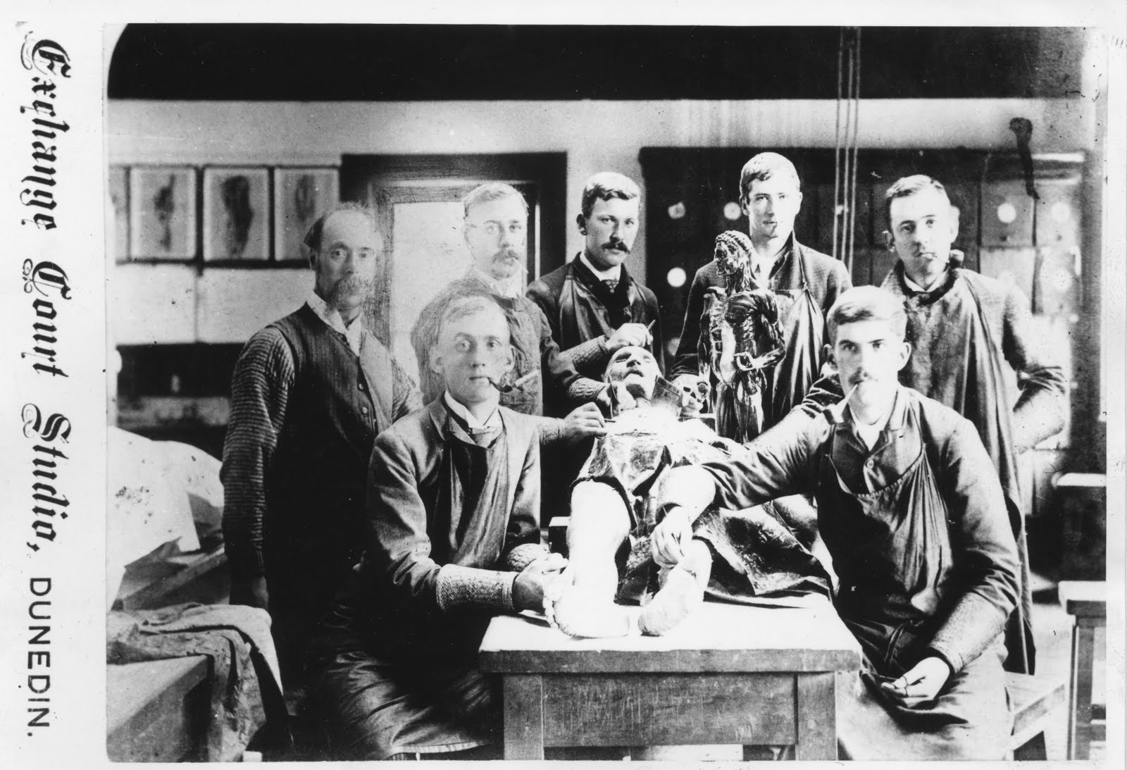
Tonight, Chris Smith, curator of the W.D. Trotter Anatomy Museum of the University of Otago in Dunedin, New Zealand, will give a brief history of the Museum, its collections and the role it plays today. As part of the southern-most Medical School in the world, this isolation can be both a hindrance as well as of benefit; but with its foundation built upon a strong Scottish heritage, the traditions of Anatomical Teaching have been sustained and continue to strengthen in this proud institution. From the early plaster, wax and papier-mâché through to todays technologies of 3D imaging and plastination, you will be given a whirlwind tour of this collection and some of the personalities responsible for its creation and development over the last 135 years.

Chris Smith is a trained Secondary School Teacher with 10 years experience in teaching and education and a passion for the collection, teaching and preservation of history. Chris changed gears in 2005 to take up the role as Anatomy Museum Curator and Anatomy Department Photographer at the University of Otago. In this role Chris has maintained and further developed the use of anatomical specimens, both historic and modern, for teaching and research, as well as increasing public awareness of the collection and the history of the museum and department. In 2007 and 2008 he traveled to Thailand as part of the Bio-archaeology team to excavate and photograph human remains at Ban Non Wat (Origins of Angkor Project), a prehistoric Neolithic to Iron Age site. He regularly attends conferences within New Zealand and neighboring Australia, visiting institutions and collections and in 2008 received a Queen Elizabeth the 2nd (QEII) Technicians’ Study Award, which enabled him to visit institutions and collections in United Kingdom and attend the European Association of Museums of the History of Medical Science Congress held that year in Edinburgh. It was at this event that he and Joanna crossed paths and as such with a visit to meet new family in the US in 2010 and making contact with Joanna, he has been put in this privileged position of being able to share a little about ‘his’ museum.

You can find out more about this presentation here. You can get directions to Observatory--which is next door to the Morbid Anatomy Library (more on that here)--by clicking here. You can find out more about Observatory here, join our mailing list by clicking here, and join us on Facebook by clicking here.

Tomorrow Night at Observatory : "Exquisite Bodies: or the Curious and Grotesque History of the Anatomical Model" with the Wellcome's Kate Forde


Tomorrow night at Observatory, join Kate Forde--curator at the amazing Wellcome Collection in London--to learn
about the rise and fall of the popular anatomical museum in 19th century Europe as detailed in The Wellcome’s recent ‘Exquisite Bodies’ exhibition. Long-time readers might recall this blog's extensive coverage of the exhibit, for which I served as curatorial adviser and designer. Images above all depict artifacts included in the exhibition.

You can find out more about the show here, here and here and see a preview of the lecture here.

Full details below; this is going to be a truly stellar event; hope to see you there!

Exquisite Bodies: or the Curious and Grotesque History of the Anatomical Model
An illustrated lecture by Wellcome Collection Curator Kate Forde
Date: Thursday, August 26
Time: 8:00 PM
Admission: $5
Presented by Morbid Anatomy

Tonight, Kate Forde of London’s Wellcome Collection will deliver an illustrated lecture detailing the rise and fall of the popular anatomical museum in 19th century Europe as detailed in The Wellcome’s recent ‘Exquisite Bodies’ exhibition.

The ‘Exquisite Bodies’ exhibition, which was curated with the assistance of Morbid Anatomy’s Joanna Ebenstein, was inspired by the craze for anatomy museums and their artifacts–particularly wax anatomical models–in 19th century Europe. In London, Paris, Brussels and Barcelona displays of wax models became popular with visitors seeking an unusual afternoon’s entertainment. The public were invited to learn about the body’s internal structure, its reproductive system and its vulnerability to disease–especially the sexually transmitted kind–through displays that combined serious science with more than a touch of prurience and horror.

At a time when scandal surrounded the practice of dissection, the medical establishment gave these collections of human surrogates a cautious welcome; yet only a few decades later they fell into disrepute, some even facing prosecution for obscenity. This talk will trace the trajectory of these museums in a highly illustrated lecture featuring many of the historical artifacts featured in the show.

To find out more about the exhibition ‘Exquisite Bodies,’ click here and here.

Kate Forde is Curator of Temporary Exhibitions at the Wellcome Collection, London. She is interested in the role of museums in the shaping of cultural memory and in the display of fine art within science-based institutions. Her current research is taking her from the great dust-heaps of Victorian London to Staten Island’s landfill Fresh Kills for an exhibition with the working title ‘Dirt’. You can see a preview of tonight's lecture by clicking here.

You can find out more about this presentation here; As mentioned above, you can find out more about the exhibition here, here and here and see a preview of the lecture here. You can get directions to Observatory--which is next door to the Morbid Anatomy Library (more on that here)--by clicking here. You can find out more about Obs
ervatory here, join our mailing list by clicking here, and join us on Facebook by clicking here.

New Exhibition at Observatory: Paradiso Contrapasso, Curated by GF Newland with Lord Whimsy and Susan Crawford, Opening Party this Thursday, Oct. 14!


Our newest Observatory exhibition "Paradiso Contrapasso"--curated by my boyfriend GF Newland along with our good friends Lord Whimsy and Susan Crawford of Plankton Art Co-- will be opening its doors this Thursday with a free reception running from 8 PM till the wine runs out.

Full details follow; very much hope to see you there!

Paradiso Contrapasso: An exhibition curated by Observatory’s G.F. Newland with Lord Whimsy and Susan Crawford of Plankton Art Co.
OPENING RECEPTION: October 14th, 8:00 PM
ON VIEW: October 14th - November 28th, 2010
HOURS: Thursdays & Fridays 3-6; Saturdays & Sundays 12-6

To compliment the recent opening of Paradise, a year long event at Proteus Gowanus, Observatory explores the theme: Paradiso Contrapasso. In Dante’s Inferno, Paradiso Contrapasso distinguishes each sinner by making his or her punishment uniquely appropriate to the committed sin, so that every soul inhabits a Hell all its own.

For example, consider the story of Paolo and Francesca:
An unlikely marriage is proposed between the beautiful Francesca and the rich, but ugly Gianciotto. Paolo, the handsome brother of Gianciotto seduces the young bride and they become lovers. When Gianciotto discovers their indiscretion, he murders them both. In Hell, Paolo and Francesca are fused together in an eternal embrace, wishing only to be separated.

As Dante journeys through Purgatorio and Paradisio, he does not revisit this technique of contrapasso. For our event, however, Observatory encourages artists to consider divine comedic retribution in all of its possible representations, and from sources such as the Bible and religious and esoteric cosmologies, the ethical philosophies of Aristotle and Thomas Aquinas, Symbolist poetry, the works of Roald Dahl, the Wizard of Oz, Cautionary Tales, Folklore and Fairy Stories, the Twilight Zone, Modern Dystopias, etc. The emphasis is on “Divine” and “Comedy”, and on our superstitious fear of getting what we wish for!

ARTISTS INCLUDE:
Martin Bland
Erich Carlle
Dennis Corrigan

Matthew Cox

Lord Whimsy / Allen Crawford
John Dilworth
Arlene Ducao
Joanna Ebenstein
Crista Grauer
Andrew Keating
Ilias Koen
Luis Locarno
Heather Masciandaro
Richard Meyer
GF Newland
Alison Overton
Herbert Pfostl
Rebecca Posner
James Prochnik
Rachel Rabhan
R. Sikoryak
Melissa Stern
Alison Termine
Friese Undine

To find out more, click here. You can get directions to Observatory--which is next door to the Morbid Anatomy Library (more on that here)--by clicking here. You can find out more about Observatory here, join our mailing list by clicking here, and join us on Facebook by clicking here.

Image: "Holiday in Hell," Denis Corrigan, Oil on Canvas

"Le Livre de Sante," Joseph Handler, 1967 (Volume 6)


All images from Joseph Handler's Le Livre de Sante (Monte Carlo: Andre Sauret, 1967)
volume 6: "La bouche et les dents. Le systeme digestif. Les reins" and found on the wonderful blog Journey Around my Skull. You can visit the full post--with additional images--by clicking here.

Images top to bottom:

  1. Dents temporaires et germes des dents permanentes, illus. Pasqualini
  2. Les levres, illus. Aslan
  3. Constitution du rein, illus. W. Hess

Philips News from European Newsletter

Courtesy of theEngineer:

17 August 2010 | By Siobhan Wagner 

Philips has announced plans for commercialising a digital technology that will ease the workload of pathologists detecting cancer in tissue cells.

The company revealed it will be introducing digital pathology systems for sale in the US in October. This follows an announcement by the company in July that it will be partnering with Dako, a Danish specialist in tissue-based cancer diagnostics, to integrate its image analysis technology into Philips’s future digital pathology systems.

Currently pathologists use microscopes to examine tissue sections mounted on glass slides and treated with different stains. The staining enhances the contrast between cellular and molecular components such as nuclei or specific proteins.

Glass slide in prototype slide scanner

Glass slide in prototype slide scanner

Accurate interpretation of the results is critical to the diagnosis and staging of each individual patient’s disease and requires a great deal of skill and experience.

Philips believes digitising the images that pathologists normally view through a microscope may enable the introduction of objective and quantitative image analysis tools.

‘The issue with pathology itself is reproducibility. It’s a very qualitative profession,’ said Guido Du Pree, vice-president of marketing for digital pathology at Philips. ‘By going digital and using image analysis algorithms you can increase the reproducibility.’

Du Pree said Philips’s fast pathology slide scanner can scan standard slides at a speed of one slide every 50 seconds.

‘The speed of scanning is one of the reasons why digital pathology today is only used in research and education environments and not for clinical purposes,’ he added. ‘The systems today are too slow and cannot handle the average workload of a pathology lab.’

Philips’s system also incorporates digital image archive and analysis software for feature recognition and quantification.

The company said it will initially focus on leveraging Dako’s image analysis software for tissue-based breast cancer diagnosis using its compounds for staining specific proteins associated with the disease.

With success in this area, Philips and Dako will explore the possibility of extending the collaboration to include image analysis software for diagnosing prostate and colon cancers.

Du Pree said the technology will ease the workload of pathologists and also improve communication with doctors who care for patients eagerly awaiting the results of a biopsy.

‘The work in a pathologist department involves a lot of referrals and today that is all sent over physically by courier or mail,’ he added. ‘By going digital it will be much faster and much easier.’

Du Pree said the company will initially be marketing its systems in the US because of the sheer number of commercial laboratories demanding the technology.

‘We’re a Dutch company with strong angles in the European market so next year we will look to launch in Europe,’ he added

Digital uptake

Over the last several years the NHS’s National Programme for IT has seen radiology departments throughout health trusts in England trade in traditional film images for digital ones.

The technology, known as Picture Archiving and Communications Systems (PACS), allows doctors to download X-rays and other images onto their computer screen. As of December 2007 PACS were rolled out throughout every health trust in England, and there is increasing uptake of the systems throughout Scotland and Wales.

However, pathology departments, not just in the UK but elsewhere throughout the world, have yet to catch up with the digital revolution.

One of the main reasons for this is the technical hurdles presented by the size of the images that need to be acquired in pathology.

The digital images that would be acquired by microscopes in the pathology department are much larger than CT files and can be difficult to manage and analyse.

Medical imaging specialists throughout the world have been working on the problem with commercial products waiting to be rolled out. 

Du Pree estimated there would be an increasing demand for digital pathology in the coming years.

‘With the ageing population we will have more and more patients and cancer is typical of elderly people,’ he said. ‘Next to that there will be a greater demand on pathology departments because of developments in what can be tested. This means more work for the same amount of pathologists. Therefore, there is a huge need for efficiency.’

Read more: http://www.theengineer.co.uk/news/news-analysis/philips-to-commercialise-digital-pathology-technology/1004414.article#ixzz0wv9J87Ok

"Still Life: The Art of Anatomy," Dunedin Public Art Gallery, Dunedin, New Zealand, Through September 12


I just found out about an excellent looking exhibition now on in Dunedin, New Zealand; the exhibition is called "Still Life: The Art of Anatomy," and it frames a variety of historical and contemporary anatomical teaching tools held in public and private hands--including models and illustrations--as artworks in a fine art setting.

Images of the exhibition above and full details below; if you are based in New Zealand, be sure to check this out!

Still Life: The Art of Anatomy
Saturday, 10 July 2010 - 12 September 2010
Dunedin Public Art Gallery
Dunedin, New Zealand

Noted Dunedin based filmmaker and medical doctor Paul Trotman, has worked closely with the Dunedin Public Art Gallery in researching Dunedin's rich collections towards the realization of Still Life: The Art of Anatomy. This exhibition brings together an array of historical and contemporary items, such as Dr John Halliday Scott's elegant anatomical drawings and old master prints, through to porcelain and wax casts of aspects of the body and the latest interactive computer generated 3D anatomical models. Still Life provides a stunning insight into this complex subject and also reveals the important lineage that science and art shares through the analysis, distillation and depiction of the human form.

You can find out more by clicking here or here.

7-year-old girl dies after Botox injections

A 7-year-old girl died after a Botox injection paralyzed her lungs, her family says, and they are suing pharmaceutical company Allergan for wrongful death.

Botox is the trade name for the botulinim toxin, which is produced by the botulism bacteria. Botulinim toxin blocks nerve signaling, leading to muscle paralysis, and has been called the single most toxic protein known. In small doses, it is approved for use to smooth away facial wrinkles. Although it is not approved in the United States for the treatment of muscle spasms such as those caused by cerebral palsy, U.S. law allows doctors to prescribe drugs for unapproved uses if they wish.

Kristen Spears began Botox treatment for cerebral palsy-related spasms at age six. In November 2007, Spears died from pneumonia and respiratory failure, which her family claims was caused when the botulinim toxin spread to her lungs and weakened her breathing muscles. Read more...

Symptoms of prostate cancer

New Double-Positivity Tool for MultiColor IHC and IF

Join CRi, the newest educational sponsor on the Digital Pathology Blog for a webinar on a new imaging tool.  These technologies are important because unlike light microscopy where single or double immunostains or IF stains performed and reviewed manualy typically look at tumor biology, digital imaging technologies allow us to study cell(ular) biology with much greater sensitivity and specificity than conventional microscopy.  These technologies provide a slide-based surrogate for molecular analysis by being able to look at multiple antibodies or markers simultaneously in individual cells rather than single stains on a single tumor section.

Learn how a new imaging tool developed by CRi can enable you to explore new molecular combinations in a single tissue section, expanding what’s possible in biomarker discovery, oncology translational research, and molecular epidemiology. Define how many cells are double-negative, single-positive for one label, single-positive for another label, or double-positive from your immunohistochemical or immunofluorescence tissue sample. ??You can now start asking more demanding scientific questions, such as how many tumor cells are proliferating, what is the normalized phosphor-EGFR expression, how many cancer stem cells are present, or how many cells have had a phospho-epitope travel across the nuclear membrane. New commercially-available multilabel antibody kits will also be discussed

Title:

New Double-Positivity Tool for MultiColor IHC and IF

Date:

Thursday, August 26, 2010

Time:

1:00 PM - 2:00 PM EDT

After registering you will receive a confirmation email containing information about joining the Webinar.

System Requirements? - PC-based attendees? - Required: Windows® 7, Vista, XP, 2003 Server or 2000

Macintosh®-based attendees? - Required: Mac OS® X 10.4.11 (Tiger®) or newer

Space is limited.?Reserve your Webinar seat now at:

https://www1.gotomeeting.com/register/557136369


A healthy heart slows brain aging

A team of researchers from Boston University has determined that the healthier your heart is, the slower your brain ages. On the flip side, those with less-than-optimal heart health experience more rapid brain aging than those whose hearts have a more healthy blood flow.

The team evaluated 1,500 people for the study and found that, as the brain ages, it actually begins to shrink. When the heart is pumping blood at a healthy rate, the brain is able to keep "fit". But in people whose blood flow is restricted by poor cardiac function, their brains age roughly two years quicker on average.

Interestingly, it is not just old people with heart disease whose brains age quicker; otherwise healthy people in their 30s who have less-than-par blood flow to the heart experience more rapidly aging brains than those with healthy flow. Read more...

Improve your memory

Digital pathology in clinical consultation practice

Interesting article in Journal of Pathology Informatics on digital pathology in clinical consultation practice by Subodh Lele at the University of Nebraska.

Abstract:

Digital pathology or whole slide imaging technology in clinical consultation practice to me translates into convenient, effective and efficient communication. A not so old method of communication that we still use today is the wired telephone. As we all know, it has limitations in its usage due to it being "wired" and therefore not convenient to use, say for example, while walking down the street. Search for a better device eventually led to the development of the cellular phone. This provides for the required convenience factor without compromising the other features of the standard telephone. Cellular phones have evolved quite dramatically over the last few years such that the most recent versions of the phone with their numerous applications have become almost indispensable for daily use for many. However, the full potential of this device has still not been realized, especially in the field of telemedicine and telepathology. With the development of the new tablet computer/phone, one may not only read the newspaper but could also view whole scanned slides. Soon, it may be possible to read whole slide scans and also sign them out as one would do in his/her office, essentially from anywhere at anytime, using such portable devices!

Evan Michelson of Obscura Antiques and Oddities, "The Culture of Curiosity," Lecture, Coney Island Museum, Sunday August 15, 4:30


Next Sunday at 4:30 PM as part of the Coney Island Museum's "Ask the Experts" Series, Evan Michelson--co-proprietor of Obscura Antiques and Oddities and Morbid Anatomy Library Scholar in Residence--will be giving a reprise of her popular "Culture of Curiosity" lecture, which some of you might have seen at Observatory last November.

If you missed it the first time, or were made curious enough [sic] about the topic to want to know more, do yourself the favor of heading down to Coney to hear Evan wax poetic [sic] in a new and expanded discussion of "the continuing appeal of curated chaos in the home."

Full details follow; very much hope to see you there!

"The Culture of Curiosity"
An illustrated lecture by Evan Michelson of Obscura Antiques and Oddities
Date: Sunday, August 15
Time: 4:30 PM
Admission: $5
Location: The Coney Island Museum

From humble parlor to Princely treasury, the Culture of Curiosity has endured for hundreds of years. In equal parts uncanny obsession, camp statement, melancholy musing, frivolous commentary and timeless metaphor, ultimately it's all about mystery.

Come and join Evan Michelson (Morbid Anatomy Scholar in Residence) in an exploration of the continuing appeal of curated chaos in the home.

Evan Michelson is an inveterate collector and museum aficionado. She has spent a lifetime obsessing over specimens, lurking in crypts, touring necropoli and gathering information on all things fading from the collective memory.

For about two decades Evan was in and out of various aggressively confrontational/decadent bands. She is currently co-owner of Obscura Antiques and Oddities, and Scholar-in-Residence at the Morbid Anatomy Library. She lives in Victorian excess with her husband, a few pets, and many esoteric and uncanny objects.

You can find out more about the event by clicking here. Hope to see you there!

The above photo is a Wax Department Store Mannequin from the Early 20th Century from Evan Michelson's incredible home collection, as seen in my recent exhibition The Secret Museum. You can find more images of her collection here.