Aperio’s SecondSlide® Digital Slide Sharing Service Exceeds 3,000 Users

Rapid Growth Fueled by Improved Turnaround Time, Efficiency and Cost Savings 

VISTA, Calif.--(BUSINESS WIRE)--Aperio, a global leader in digital pathology for the healthcare and life sciences industries, today announced that its SecondSlide® digital slide sharing service has exceeded 3,000 users.

Launched in 2009, the number of SecondSlide users has increased steadily, with adoption accelerating virally throughout the pathology community and growth soaring 60% since first quarter 2010. Users can easily join the network and access or share digital slides with colleagues in a matter of minutes.

Jared N. Schwartz, MD, PhD, Aperio’s chief medical officer, stated, “SecondSlide is very powerful, especially for cases that need immediate review. It is also an excellent tool for allowing small community hospitals to access sub-specialty expertise located at academic medical centers.”

SecondSlide is a web-based service for pathologists that enables the secure, encrypted exchange of whole-slide images, photomicrographs, documents and commentary. These data reside at a secure global data center “in the cloud,” where they can be readily accessed by authorized parties. The slide sharing service is compatible with a variety of commercially available image capture devices, including whole-slide scanners and digital cameras attached to microscopes.

“In just over a year, SecondSlide has been validated by the global marketplace as a cost-effective and IT-friendly digital pathology solution,” stated Dirk G. Soenksen, CEO of Aperio. “SecondSlide dramatically reduces the time, effort and expense of pathology consultations and facilitates improved quality assurance.”

SecondSlide enables pathologists with subspecialty expertise to expand their consulting practice, and makes access to their knowledge and experience significantly easier for community hospitals and pathology labs located remotely. SecondSlide is also being used extensively for consultations, medical education and publication support. The service provides access to digital slide conferencing and a simple and secure “message board” for each case, allowing the pathology community to engage in dialog and exchange information.

The SecondSlide digital slide sharing service is presently available to pathologists free of charge; for more information please visit http://www.secondslide.com.


CBLPath Launches Best Practice™ Partnership Program

Back in May I posted a press release from CBLPath announcing their integrated diagnostics services.  This weeks at Pathology Informatics 2010 they have announced their Best Practice Partnership Program. If you are unable to attend the meeting this week in Boston, more details will be showcased next week in Chicago at CAP 2010.  

CBLPath's approach to this is interesting.  By utilizing their services and expertise from sales to billing, this model will enable community pathologists to partner with CBLPath to grow their practice in a unique model historically for anatomic pathology laboratories, both for community-based groups and traditional large AP laboratories such as CBLPath.

Another factor at force here is the recognized convergence of diagnostic surgical pathology, digital pathology and molecular pathology.  I think there is a realization now that these services enable one another through convergence and each is enhanced by the others.  My feelings from Boston in one word is: convergence.  Digital pathology, surgical pathology, image analysis, workflow, image delivery and fast rendering are all starting to finally converge towards increased adoption for diagnostic surgical pathology. More on this to follow.

Full press release below from CBLPath:

Enabling revenue growth, enhancing patient care for community-based pathologists 

Community-based pathologists have been watching specialty anatomic pathology laboratories erode their market share.  These national labs have a significant advantage due to access to technology and electronic connectivity, and service features which are driving pathology out of the local community and into their centralized laboratories.  Today, combined with their historically rich local relationships, community-based pathologists have an advantage of their own – CBLPath’s Best Practice™ Partnership Program.

The Best Practice Partnership Program is comprised of a full range of business and technology services enabling community-based pathologists to more successfully compete against sub-specialized, larger, centralized testing laboratories.  The program consists of five components: Sales and Marketing; Information Technology through LabIS™, CBLPath’s proprietary pathology management system; Medical Operations; Lab Operations; and Managed Care and Billing.

“Our business has experienced tremendous growth since its inception,” said Tom Curtis, CBLPath’s Vice President of Marketing.  “Our growth is attributed to the same five components we are offering our clients through Best Practice.  By becoming a Best Practice Partner, community-based pathologists can enhance the level of patient care they provide with the resources of a national reference laboratory behind them.”

CBLPath has embraced the industry convergence of anatomic, molecular and digital pathology.  The organization is providing its Partners with 400 employees, more than 30 of which are board-certified sub-specialty pathologists, access to accurate, definitive molecular testing and landmark digital pathology systems.   Through Best Practice, community-based pathologists are able to provide better medicine to their patients and a wider breadth of service to their own clients.

“While other centralized labs are competing with the local pathologist for business, CBLPath recognizes a natural synergy between our lab and the local pathologist,” said David Bryant, CBLPath’s President and Chief Commercial Officer.  “We created the Best Practice Partnership Program to empower local pathologists to thrive in this competitive market, keeping medicine local.”

CBLPath will showcase the Best Practice Partnership Program at the CAP ’10 meeting next week in Chicago. For more information about Best Practice, visit http://www.CBLPath.com/bestpractice.


About CBLPath

CBLPath is a national specialty lab offering a full convergence of anatomic, molecular and digital pathology services. The company provides a one-stop solution for comprehensive sub-specialized diagnostics, and timely, accurate, patient-centered disease management guidance. Through its Best Practice ™ Partnership Program, CBLPath partners with pathologists to help them grow their practices, while giving them the ability to stay independent and “keep medicine local.” The company also provides sub-specialty physicians access to comprehensive, high-quality testing in their local market. Founded in 1988, CBLPath established a reputation for providing timely, highly accurate diagnoses along with extraordinary customer service and a true patient-centered commitment. For more about the company, please visit http://www.CBLPath.com.


Virtual technology still needs a local pathologist touch – Flagship Biosciences opens digital pathology services office in Boston area


Who would have expected that the use of a truly virtual service would lead to customers wanting a pathologist nearby?

Companies such as Flagship Biosciences provide digital pathology services company where they receive glass slides, scan them, run image analysis with pathologist QA, and then have discussions with their biotech clients about what the results mean. This entire service is usually done without any travel or
face-to-face meetings. Flagship Biosciences operates a centralized data storage operation in a highly secure area in Northern Arizona (many of the largest data storage providers like GoDaddy.com are based in low-cost Arizona or Nevada). It shouldn't matter that most of their clients are on the other end of the nation, either in Cambridge, Massachusetts or the New Jersey/Philadelphia pharma corridor. It shouldn't matter...but it does.

For some time now they have found that despite the virtual nature of digital pathology, biotech researchers still want the pathologist "down-the-hall". Looking at a slide together and understanding what a particular efficacy or toxicology effect means. Much of this is trust in the pathologist. This is not a surprise, the technology is digital PATHOLOGY, not DIGITAL pathology -- like bicycling, it is the cyclist that matters more than the bike.
To establish trust still means building relationships, and this is still easiest to happen with local interactions. Studies normally start with a few slides, discussing the best approaches for quantitation -- optimizing stains, software techniques, histology preparation, etc. Only once both the researcher and pathologist are convinced the measurement approach is going to work do they move to larger studies. This means actually a lot of travel and personal interactions, so the customer knows what they are doing, and Flagship Biosciences knows that the biology to be measured is in line with their customer's therapeutic development goals. 
To leverage the technology's virtual advantages, but still maintain personal interaction, they are trying something new. If it works, it is a window into the pathologist's office of the future. Their new office in the Boston suburbs has three advantages -- a board-certified pathologist, a technician and a scanner. The pathologist supervises all aspects of the work. The technician performs much of the laborious image analysis steps (especially with pattern recognition and large studies, there is a lot of manual work involved in QA'ing what the computer finds). The scanner is there locally to get the results back to the customer as soon as possible.
Perhaps your city my be the next site for a scanning biotech center if the Boston Flagship Biosciences experiment is successful.
Press release below: 

New office offers local access for Cambridge biotech companies to pathologist, technician, scanning and image analysis

Flagstaff, AZ – September 13, 2010 – Flagship Biosciences LLC, a provider of digital pathology services, has opened a local digital pathology office in the Boston suburbs to better support the growing demand for local quantitative histopathology services. The office is staffed by a board-certified pathologist, a scanning and image analysis technician, and digital pathology scanners.

“While digital pathology has the remarkable ability to remove geographic barriers, Flagship’s Cambridge area customers have expressed interest in local access to quantitative histopathology services,” said Dr. Frank Voelker, DVM, DACVP, who leads the new office. The ability for pharmaceutical researchers to sit down in person and discuss study designs and image analysis approaches in tissue with a local pathologist is very helpful. While our customers can view all of our tissue results remotely on each slide via a web-based portal, there is still no replacement for individual interaction in person.  Many of these studies contain hundreds of glass slides, and being able to teach and share information with our Cambridge area customers can be very useful.”

Dr. Frank Voelker leads the Boston Flagship office, and has had a distinguished career in pathology in the Boston area, including 12 years at Novartis Institutes for Biomedical Research, where he was founder and head of the Tissue Biomarker Laboratory and later Section Head of the Marker Localization and Assays Group.

“We believe this type of laboratory office for quantitative pathology work is the wave of the future, said Dr. David Young, DVM, DABT, DACVP, President of Flagship Biosciences. “Rather than pathologists setting up an office with a microscope and boxes of glass slides, the Flagship office includes a board-certified pathologist, with a digital workspace of image analysis tools and a web-based portal. A technician who helps with the scanning and image analysis is part of the pathologist’s office of the future”.

The new Flagship Office opened in September in Quincy, Massachussetts. It is part of a general trend of movement of biotech companies into the Boston suburbs.

About Digital Pathology

Digital pathology is the use of whole slide scanning rather than glass slides for analysis of tissue. The technology facilitates remote and distributed pathology environments, and is considered a transforming technology within the discipline of anatomic pathology. The use of whole slide images not only removes geographic barriers, but also allows for computer assisted quantitative analysis. This is an advantage that can be tremendously helpful in making efficacy and toxicity measurements more reproducible and accurate in pharmaceutical drug development, especially in discovery science and oncology companion diagnostics programs.

About Flagship Biosciences

Flagship Biosciences is a pathologist-owned company whose mission is to improve tissue assessment in pharmaceutical and medical device development. Flagship’s services include quantitative pathology assessment on-demand, low-cost digital pathology slide scanning, secure hosting, pathologist-supervised IHC and histology placement, and custom image analysis and companion diagnostics development. All services are reviewed and supervised by board-certified pathologists.


Leica Microsystems Extends its Product Portfolio in Virtual Microscopy

(Nanowerk News) With its unprecedented scanning speed and top-quality on-screen imaging, the Leica SCN400 Slide Scanner has already set new standards in virtual imaging. Through the acquisition of Genetix Ltd. at the beginning of this year, Leica Microsystems is now able to considerably expand its solutions offering. With the software solutions Ariol and SlidePath, Leica Microsystems now adds the ability to store, manage, analyze, and report on digital images created with the Leica SCN400 Slide Scanner or the Leica DM6000 B Research Microscope.

High throughput solution for in vitro diagnostics: Ariol on the Leica SCN400 
Ariol on Leica SCN400 combines leading scanning technology with advanced analysis experience
Ariol on Leica SCN400 combines leading scanning technology with advanced analysis experience.

Combining Ariol with the Leica SCN400 Slide Scanner provides a complete solution for laboratories dealing with a high volume of slides. The product is a high throughput solution for biomarker assessment. Ariol on Leica SCN400 combines leading scanning technology with advanced analysis experience, giving the customer the good feeling to get the best product in all respects. From sample to result, Leica Microsystems is striving to provide the total histology solution and be the pathologist's integrated partner in every step of the process. 

Web-enabled image handling and e-learning: SlidePath on the Leica SCN400 

SlidePath software on the Leica SCN400 Slide Scanner opens the world of image and data handling in a flexible and powerful web-enabled solution. With the Digital Image Hub (DIH) module from SlidePath, Leica Microsystems provides the complete end-to-end solution for virtual microscopy, especially optimized to provide rapid whole slide imaging coupled with an online management solution that can handle multiple formats including DICOM and other standard imaging formats. DIH is also designed for use in research under the guideline of regulatory bodies, such as preclinical research or toxicological studies, which profit from multiple features especially designed for this customer group. Furthermore, DIH can be combined with diverse specialized software modules from SlidePath, such as a special module for TMA management – called OpTMA – or the Digital SlideBox, which provides tools for e-learning.


Pathology workstation for in vitro diagnostics: Ariol on the Leica DM6000 B 
A perfect pathology workstation for the assessment of clinical brightfield and fluorescent biomarkers

The in vitro diagnostics software Ariol on the Leica DM6000 B Research Microscope creates a perfect pathology workstation for the assessment of clinical brightfield and fluorescent biomarkers. The system integrates seamlessly into the pathologist's workflow with the means to digitize whole slides or regions of interest. With this solution customers enter the world of Virtual Microscopy while staying with the highest possible flexibility in terms of hardware configurations. 

The modules Hersight, ERsight, PRsight and PathVysion are available in the US for in vitro diagnostic use. 

Research workstation for fluorescent capture and analysis: Ariol on the Leica DM6000 B Centered on the Leica DM6000 B, the Ariol platform provides the complete solution for capturing, re¬viewing, and analyzing tissue slides labeled with fluorescent markers. TMA as well as full tissue sec¬tions can be captured and analyzed with multiple markers and multiple planes. The powerful analysis tool combined with the versatile hardware creates an ideal workstation for researchers in advanced approaches to fluorescent capturing.

LabCorp to buy Genzyme

This has been rumored for some time - LabCorp to buy Genzyme

Laboratory Corp. of America Holdings (LH) has agreed to buy the genetic testing business Genzyme Corp. (GENZ) for $925 million as the medical-testing giant looks to expand into areas such as reproductive medicine and oncology.

Meanwhile, the sale comes as Genzyme has been struggling for more than a year with manufacturing problems at its most important plant, crimping supply of its two biggest drugs. As such, Genzyme in May said planned to shed three noncore operations--including genetic testing--by year's end "strategic alternatives" for three units while repurchasing $2 billion of stock.

Genzyme said Monday that proceeds from the genetic-testing sale would go toward that end. The company's stock-market value is about $18 billion. Divestiture of the other two units--diagnostics and pharmaceutical intermediates--remains on track, said Genzyme.

"This acquisition will substantially expand our capabilities in reproductive, genetic, hematology-oncology and clinical trials central laboratory testing," said David P. King, chairman and chief executive of LabCorp.

Genzyme Genetics performs more than 1.5 million tests a year. It had about $371 million in revenue in 2009.

LabCorp in July said its second-quarter profit rose 13% following prior-year charges as revenue improved more than expected.

Genzyme closed at $70.79 Friday while LabCorp finished at $76.65. Neither traded premarket.


How to get Perfect Abs

Some of the biggest things holding you back in your weight loss efforts and your dream of perfect abs are not named 'Protein','Fat' or 'Carbs'.

It's probably not even 'Calories'.

(...well actually it is Calories, but it's just a tiny bit more complex then that.)

No, today I'm talking about two non-calorie things.

Self-Confidence and Perfection.

So let's clear this up right away.

It is a misconception that you have to be 'special' to be successful at weight loss.

This misconception stems from the fact that we like to think that the people giving us diet advice are somehow 'perfect'.

Living perfect lives, with the perfect bodies with the perfect jobs etc.

This creates a situation where it becomes difficult to have confidence in ourselves because we simply do not measure up to these seemingly perfect people.

However, Nobody is perfect.

So here's the truth...

That diet guru with the awesome abs that everyone thinks eats 'perfectly'...He's a chocolate FEIND.

The 600 pound bench presser who constantly gives advice on training..He's had three shoulder surgeries and bone spurs removed from his elbows...as a direct result of his benching.

The Hollywood celebrity touting his super dedicated workout program he used to get in shape for "Kiss of Death 32"...He forgot to tell you he's on various anabolic steroids.

The Paleo Princess who thinks that HFCS is made directly form the essence of the devil...She secretly loves the occasional Coca Cola when at the movies.

The shredded diet guru...who is actually about 60 pounds overweight (the pictures you see on-line are from 2001)

The dozen or so magazine covers you see every month...They are a result of photoshop mastery.

The point is:

No one is perfect, so don't freak out when you discover that you're not perfect either.

The idea of having shredded abs 24/7 without even flexing is just as foreign to the professional fitness model as it is to you.

We need to realize that the illusion of perfection is really, really hurting us and our abilities to reach our goals..or even define them.

So here are some givens:

You are going to have off days of dieting.

You weight loss is going to plateau, or even go up occasionally.

You are going to love the way you look some days, and be less then impressed on other days...and you don't probably look any different on either day.

This doesn't make you a failure, it makes you human...keep at it.

You are going to break a fast early, eat more than you want to, miss a workout, eat a 'bad' food.

The idea of eating perfectly for 4 months worth of hardcore deiting is just plain silly, even for the most dedicated fitness proffesional.

So if you slip, don't worry about it, just keep moving towards your goals.

Remember: No one is perfect.

This makes it a little easier to ramp up the self-confidence.

It's an even playing feild.

Realizing this is a giant step into becoming awesome. Because once you realize that no one is perfect, then you have ZERO reason not to be self-confident.

We're all flawed in our own special way, which of course means we can all be awesome if we choose to be.

BP

As a note...this post was highly influenced by this blog post -->  http://bit.ly/b9DzcI

Social Diet Hack

I’d like to let you in on one of my all time favorite ‘diet hacks’

No, it’s not fasting (that’s not really a ‘Diet hack’ – it’s more of a lifestyle)

Actually, it’s scotch.

Yep, good ol’ Scottish Whiskey (Irish is pretty darn good too, but I digress)

So you’re probably wondering how a ounce of the good stuff (neat of course) can be a diet hack?

Simple..

It’ll save you calories…. lots of them.

An ounce of scotch has about 70 Calories. That’s less than a glass of wine (120 ish) and a bottle of beer (140 ish)…but that’s not the ‘Hack’.

The “Hack” is in the social graces of Scotch.

Let’s say you go out for drinks with your friends…guys order beer, the girls order wine and you order a scotch.

Right away, you’ll probably get some looks… mostly because people have this weird idea that an ounce of Scotch has a TON more alcohol in it then a bottle of beer or a glass of wine (which is false, they are all equal).

And this is where the ‘hack’ comes in.

You can ‘nurse’ a glass of scotch for an hour and no one will say anything.

(I’ve never been in the situation where I’ve been nursing a scotch and someone has asked me if I was ‘on a diet’)

In a social setting, for some reason Scotch trumps people’s crazy need to push you to keep up with them when they drink.

So your friends can be 3 beers into the night, or a half bottle of wine..and you can still be nursing your first ounce of alcohol, and no one will say anything.

In a hypothetical situation where you’ve had two ounces of scotch, while your buddy has had 6 beers, you’ve saved yourself well over 500 calories…and a whole bunch of bloating and probably a really crappy morning..

Bottom line – Obviously you could just have a water, but with this diet hack you get to enjoy a Scotch, nice and slow…the way you should enjoy it (Without ice of course)…you drink about 70 Calories while your friends drink hundreds of calories in their beer, wine and assorted mix drinks (Oh and you still get the anti-inflammatory actions of alcohol too, just without any of the extra calories).

In settings like weddings, or awkward family reunions, this could save you hundreds of unneeded calories, while still allowing you to be ’social’.

Again, The obvious ultimate hack is water, but if you’re going to drink, you may as well drink the good stuff (just remember to drink it slow).

Drink Stop Drink

BP

PS- No this is not an excuse to allow alcohol when you are fasting!

Monoclonal antibody production by hybridoma technique explained

I shall explain the hybridoma technique used in production of monoclonal antibodies in a simplified way.

An antigen (unless it is a small peptide) is a complex molecule with several antigenic determinants (or epitopes). When the immune system encounters such an antigen, it is usually processed to result in several fragments. Humoral (antibody-mediated) response may occur against some of these fragments. There are multiple clones of B cell, each against a specific epitope; resulting in production of antibodies against several epitopes. Such a response is said to be polyclonal. This is what that happens when our body encounters a microbial antigen following infection or immunization.

There are situations when it becomes necessary to have antibodies against a single antigenic determinant produced by a single clone of B cell. Such a response is said to be monoclonal. In order to produce monoclonal antibody, it is necessary to possess a purified antigen.

Hybridoma technique was developed by Georges Kolher and Cesar Milstein in 1975, for which they were awarded the Nobel Prize. The chief participants of this technique are the B cells and the myeloma cells. The B cells are obtained from the mouse which has been immunized with the antigen of choice. Myeloma cells are malignant B cells that are immortal and multiply continuously. Myeloma cells that have lost the ability to produce antibodies are chosen for this technique. In addition, these cells lack the ability to produce hypoxanthine-guanine phosphoribosyl transferase (HGPRT-) and thymidine kinase (TK-) enzymes through an induced mutation. Normal B cells have functional enzymes (HGPRT+ and TK+) and are able to produce antibodies. However, their life span is not beyond two weeks. Hybridoma technique involves physical fusion of both these cells so that the resulting hybrid (called hybridoma) has the features of both these cell types.

The first step towards the production of monoclonal antibodies is the immunization of the animal by antigen of choice. Mouse is the commonly used animal, but rat or hamster too can be used. Following repeated immunization (booster doses) the blood of the animal is tested for antibodies against the immunized antigen. Once it is determined that the animal has produced sufficient antibodies, it is killed and its spleen removed. The spleen is rich in B cells and would contain B cells specific to the immunized antigen among B cells of other specificities. The B-cells are separated from other cells and cultured. They are then mixed with cultured myeloma cells and allowed to fuse. Fusions of the cells are aided by polyethylene glycol (PEG). Not all cells fuse; present in the reaction mixture are unfused B cells, unfused myeloma cells and fused hybridoma cells. The next step involves separation of hybridoma cells from the unfused cells using a special selective medium.

In order to understand the functioning of selective medium, one must be aware of the following facts. Multiplying cells need to produce their DNA. Most cells produce their purines nucleotides and thymidylate (both precursors of DNA) utilizing tetrahydrofolates by a De-Novo pathway. This can be blocked using anti-folate drugs such as Aminopterin. The cells can then adopt Salvage pathway to synthesize DNA if hypoxanthine and thymidine are exogenously supplied. Purine nucleotides are produced from hypoxanthine using hypoxanthine-guanine phosphoribosyl transferase enzyme and thymidylate is produced from thymidine using thymidine kinase. The selective medium (HAT medium) used to select the hybridoma contains aminopterin, hypoxanthine and thymidine. Amiopterin inhibits the De-Novo pathway and presence of hypoxanthine and thymidine facilitates salvage pathway.

Normal unfused B cells can’t produce DNA by De-Novo pathway because of aminopterin but are able to undertake salvage pathway. This is because they contain functional enzymes (HGPRT+ and TK+). However, since they are mortal, they die after few multiplications. Unfused myeloma cells too can’t use De-Novo pathway because of aminopterin. They are unable to utilize the salvage pathway either because of deficient enzymes (HGPRT- and TK-). These cells die despite being immortal. However, fused hybridoma cells receive (HGPRT+ and TK+) trait from normal B cells and immortality from myeloma cells. These cells can utilize salvage pathway for DNA synthesis and yet be immortal. After two weeks, only the hybridoma cells survive in the selective medium.

The surviving hybridoma cells would have formed against different epitopes. The next step is to select the hybridoma produced against the desired antigen. The cultures are diluted to such an extent that only a single cell gets transferred to the wells of microtitre plate. The cells are allowed to multiply. These cells produce antibodies that can be readily detected in their supernatant fluids. Supernatant fluids from all the wells are tested for antibodies against the antigen of choice and the well that contains desired antibodies is selected and the rest may be discarded. Finally, a hybridoma cell producing antibodies against the epitope of choice is available.

These hybridoma cells may be lyophilized, cultured in vitro or injected intra-peritoneally into a mouse and monoclonal antibodies raised whenever required.

Humoral immunity

Humoral immunity: How does the antibody production occur?

Depending on the nature of the antigen and the physiology of the individual, the immune response of the body to a foreign antigen may include only antibody production or a cellular response (T cell) or even both. It is still not very clear how the body makes this choice. Some antigens induce only an antibody response while others induce both humoral and cell mediated immunity. Sometimes, there are no apparent responses at all, yet in some cases these responses become exaggerated and harmful.

I shall concentrate only on humoral (antibody-mediated) response. Body responds to different kinds of antigens differently even with antibody production. Antibody production may be quick in some cases and delayed in other cases. Immune system may retain memory cells towards some antigen and none in case of some other antigens. Although the primary antibody response is IgM, some people produce IgE or IgG class of antibodies. It all depends on the body’s constitution and the immune response genes.

The cells that actually produce are the B cells and their derivatives, the plasma cells. In fact, most of the antibody production is by plasma cells and B cells produce only little. Plasma cells are derived from B-cell only after appropriate stimulation and activation of B cells. B cells are predominantly located in lymphoid organs such as bone marrow, spleen and lymph nodes. They may also be found in circulation, but their numbers are less compared to T cells.

Antigens can be classified into two types (T-independent antigens and T-dependent antigens) based on their ability to induce B-cell activation.

Some antigens have multiple repeating identical units; such antigens can bind to several receptors on the B-cell surface and effectively cross link them. B lymphocytes are not perfectly spherical cells as is often depicted; in fact their surfaces have long projections and have 106 numbers of receptors on their surface. These surface receptors are either monomeric IgM or IgG immunoglobulins. Their role is to bind with the antigenic epitopes. When an antigen such as flagellin, which has multiple repeating identical units, binds to many of the surface receptors, the receptors are said to be cross-linked. This is the first signal in B-cell activation. Apart from antigens cross linking the surface receptors, B cells have another mechanism of cross-linking surface receptors that involves immunoglobulin receptor and complement receptor. If an antigen that has C3d bound to it binds to surface immunoglobulin, another receptor (CR2) can bind with the deposited C3d on the antigen. This process too can cross link the surface receptors and activate the B cell. Upon activation by these signals, B cells undergo proliferation and start producing antibodies. Since there is no involvement of T cells in antibody production, these antigens are said to be T-independent. Antibody response to non-protein antigens, such as polysaccharides and lipids do not need participation of antigen-specific helper T cells. Since T cells have role here, the antibody production is typically quicker. An unfortunate effect of this response is that the immune system does not retain any memory of antibody production. Antibodies to T-independent antigens are mainly of low-affinity and responses are simple and mainly consist of IgG and IgM.

Humoral immune response to protein antigens is more complex; these antigens are said to be T-dependent as it requires participation of T cells for B-cell activation. Since the CD4 T lymphocytes stimulate B cells, they are called helper T cells. Antigen-specific CD4+ T cells recognize a protein antigen only when it is presented by antigen presenting cell (B cell, macrophage, dendritic cell etc) along with MHC class II molecules. B cells are also capable of antigen presentation to T cells. When an antigen binds to a specific B-cell through the surface immunoglobulin receptor, it is endocytosed by a receptor mediated endocytosis process. The processed antigen is then presented on its surface coupled with MHC II molecules for recognition by specific T cell. The initial encounter between the antigen-specific T cell and B-cell occur at the interface of the primary follicles and T cell area. Following this initial binding and the binding of other co-receptors (such as B7 of B-cell and CD28 of T cell, CD40 of B-cell and CD40L of T cell), the T cell also gets activated. Cytokines such as IL-2, IL4 and IL-5, which are secreted by activated helper T cell acts on B-cell to induce B-cell proliferation. Many of these B cell clones transform into effector cells called plasma cells and start producing antibodies. Within the lymphoid tissue, antibody secreting cells are found mainly in extrafollicular sites, such as red pulp of spleen and medulla of lymph node. These cells also migrate to bone marrow at 2-3 weeks after antigen exposure, and bone marrow becomes the principal site of antibody production. Antibody secreting cells do not circulate actively. Some of the Antibody producing cells that migrate to the bone marrow and live for several years, where they continue to produce antibodies even when antigen has been eliminated
The secreted antibodies have same specificity to the surface Ig receptor that captured the antigen. The antibodies that are secreted initially are predominantly of the heavy chain µ (IgM) isotype. In response to CD40 engagement and cytokines, some of the progeny of activated B cells undergo the process of heavy chain isotype switch. This leads to production of antibodies with heavy chains of different classes such as ? (IgG), ? (IgA) and ? (IgE).
Some of the antigen-activated B cells do not develop into antibody secretors. Instead, they acquire the ability to survive for long periods without antigenic stimulation. These memory cells are capable of mounting rapid antibody responses to subsequent introduction of antigen.

Conference Report: ‘Contemporary Medical Science and Technology as a Challenge to Museums’, 15th Bi-Annual EAMHMS Congress, Copenhagen

For medical museums, whose collections are typically composed of evocative historical objects, developments in contemporary biomedicine offer a twofold challenge to collecting and exhibiting. The first challenge is the nature of contemporary biomedical equipment: large, expensive, and without immediately obvious function (think fMRI scanner). Where a display of surgeons’ tools can be both instructive and chilling, a collection of grey-box scanners and robotic surgical suites is likely to offer both historians and visitors less. The second challenge is more fundamental: medical investigation and treatment now operates beyond the limits of the visible, at the level of genes and proteins, a scale which it is hard to relate to our own bodies and lived experience. Even the beautifully-limned image of an SEMmed protein can’t offer the visceral thrill of corporeal recognition that a pickled heart in a jar does...

For the curious among you: The Wellcome Collection's Danny Birchall has written a very nice conference report--as excerpted above--about last month's ‘Contemporary Medical Science and Technology as a Challenge to Museums’ EAMHMS Congress in Copenhagen.

Click here to read full report on Danny's blog "Museum Cultures."

Image: Installation view of Medical Museion, the host institution in Copenhagen.

Lecture: "Anatomical Venuses, The Slashed Beauty, and Fetuses Dancing a Jig," University College London, Thursday October 7, 6:00 PM


For all you Londoner's out there: on Thursday, October 7th at 6:00 PM, I will be giving a free lecture at University College London about anatomical museums and their curious denizens, heavily illustrated with many photographs I have been collecting over the years, such as those seen above.

The lecture is free and open-to-the-public. Full details follow; hope to see you there!

Anatomical Venuses, The Slashed Beauty, and Fetuses Dancing a Jig:
A Journey into the Curious World of the Medical Museum

Date: Thursday, October 7th
Time:
6 PM
Location: UCL, Department of History of Art
20 Gordon Square, WC1E 6BT London, Room 3-4 (first floor)

Tonight's lecture will introduce you to the the Medical Museum and its curious denizens, from the Anatomical Venus (see above) to the Slashed Beauty, the allegorical fetal skeleton tableau to the taxidermied bearded lady, the flayed horseman of the apocalypse to the three fetuses dancing a jig. The lecture will contextualize these artifacts by situating them within their historical context via a discussion of the history of medical modeling, a survey of the great artists of the genre, and an examination of the other death-related diversions which made up the cultural landscape at the time that these objects were originally created, collected, and exhibited.

You can download an invitation to the event by clicking here.

All Images From The Secret Museum Exhibition;" Top to bottom:

  1. Anatomical Venuses," Wax Models with human hair in rosewood and Venetian glass cases,The Josephinum, Workshop of Clemente Susini of Florence circa 1780s, Vienna, Austria
  2. Wax Model of Eye Surgery, Musée Orfila, Paris. Courtesy Université Paris Descartes
  3. Fetal Skeleton Tableau, 17th Century, University Backroom, Paris

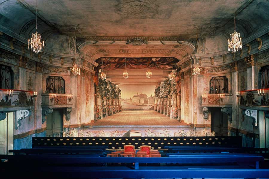
Drottningholm Court Theatre, 1764-1766, Stockholm

Today I visited one of the most incredible spaces I have ever had the honor to momentarily inhabit: the Drottningholm Court Theatre, a former royal summer theatre at the Drottningholm Royal Park, a UNESCO World Heritage site in Stockholm, Sweden.

The Drottningholm Court Theatre was described memorably by our tour guide as "an intact baroque theater unique for never having been restored." This intactness includes not just paint, chandeliers, stage, and viewing boxes but also extends--astoundingly!--to the stage machinery and special effects, which are not only original but also still used in productions! The sets and "side flats" are not original, but are utterly convincing and painstaking reproductions in canvas and wood from originals found under a meter of dust when the theatre was rediscovered in 1921. The theatre also has--and continues to use!--machinery for lowering a person from the ceiling, as in the case of a goddess descending on a cloud, as well and a trap door to be used in such cases as "drowning heroines or sudden appearances."

During the summer, the Drottningholm Court Theatre stages 18th Century operas and ballets using these replica sets and the original stage machinery to create a completely immersive 18th Century theatre experience; unfortunately for me, the productions had already ended for the season before I arrived, so I had to content myself with volunteering to operate the "gale machine" (a wooden wheel whirled around inside a tight canvas strip producing with its friction a sound remarkably like howling winds) while my volunteer-partner worked the thunder machine--a box of rocks tossed this way and that by the pull of a rope.

The video above helps give a sense of the charm and wonder of these wonderful antique sets and machineries in motion where my words fail; both the video and a visit to this really fantastic--in ever sense of the word--theatre are highly recommended! I am already fantasizing about a return trip just to see The Magic Flute in this environment.

To find out more about the Drottningholm Court Theatre, click here.

Thanks so much to friend, friend-0f-the-blog, and author of the wonderful book Death, Modernity, and the Body: Sweden 1870-1940 Eva Åhrén for telling me about this incredible place, and for all her other wonderful Sweden tips as well.

Morbid Anatomy Library and Observatory, Open Studios, This Saturday, October 2nd, 12-6


This Saturday, October 2, please join the Morbid Anatomy Library (as seen above) and sister space Observatory as we open our spaces to the public as part of the 14th annual Gowanus Artists Studio Tour, or "A.G.A.S.T." There will be snacks, beverages, art, artifacts, and, of course, books.

Following are the full details; Very much hope to see you there!

14th annual Gowanus Artists Studio Tour (A.G.A.S.T.)
Saturday October 2nd
12-6 PM
543 Union Street at Nevins, Brooklyn
Free and Open to the Public

Directions: Enter the Morbid Anatomy Library and Observatory via Proteus Gowanus Gallery

R or M train to Union Street in Brooklyn: Walk two long blocks on Union (towards the Gowanus Canal) to Nevins Street. 543 Union Street is the large red brick building on right. Go right on Nevins and left down alley through large black gates. Gallery is the second door on the left.

F or G train to Carroll Street: Walk one block to Union. Turn right, walk two long blocks on Union towards the Gowanus Canal, cross the bridge, take left on Nevins, go down the alley to the second door on the left.

For more about the Morbid Anatomy Library, click here. You can find out more information about A.G.A.S.T., and get a full list of participants, by clicking here. You can find out more about Observatory and the exhibition now on view by clicking here.

Beautiful Irish Medical Photographs, The Burns Archive, 1870s


The photographs above, dating from the 1870s, picture patients who were operated upon by 19th Century surgeon Edward Stamer O’Grady; these photos, all drawn from the incredible Burns Archive, were featured--paired with their original case histories!--in the most recent issue of Scope Medicine in Focus.

Full story can be found here; you can see a PDF of the article--with additional images--by clicking here.

All images ©2010 The Burns Archive; From top to bottom:

  1. Patient of Edward Stamer O'Grady
  2. A 50-Year-Old Laborer "MM," Admitted Feb 17, 187
  3. Once the 27 Ounce Tumor Was Removed, the Patient "Went Home Quite Well."

“Memento Mori: The Birth and Resurrection of Postmortem Photography,” Merchant's House Museum with the Burns Archive, New York, Through November 29th


The New York Times blog just ran a brief story on a new exhibit I have been dying (sic) to see: “Memento Mori: The Birth and Resurrection of Postmortem Photography,” an exhibition exploring memorial photography then and now, curated in a collaboration between Eva Ulz and the incomparable Burns Archive of New York.

The exhibition--on view at the Merchant's House Museum in lower Manhattan until November 29th--features a collection of antique memorial photography drawn from the incredible Burns Archive curated alongside similarly-themed photographs by contemporary artists such as Joel-Peter Witkin, Sally Mann, Hal Hirshorn, Marian St. Laurent and Sarah Lohman. This intriguing looking exhibition takes as its theme the role of post-mortem photographs at different cultural moments.

As the article explains:

“People dealt with death differently in the 19th century,” says Eva Ulz, the curator of “Memento Mori: The Birth and Resurrection of Postmortem Photography” at the Merchant’s House Museum. “People looked forward to a reunion in heaven. Creating portraits was considered a precursor to that heavenly reunion. They shouldn’t be thought of as creepy.”

As much as it is about religious belief, the show — which was organized in conjunction with the Burns Archive and includes some 145 postmortem images and ephemera taken between the 1840s and the early 1900s — takes as its main subject the role of photography in everyday life, then and now. As the 20th century began to unfold and photography became much more common, Ulz says, postmortem images were sapped of their ritualistic importance.

To help put that shift in context, Ulz asked five contemporary shooters, including Sally Mann, Joel-Peter Witkin, Hal Shirshorn and Sarah Lohman, to contribute their own take on the postmortem photograph. The photographer Marian St. Laurent, who created an actual coffin called “Our Darling: A Memorial of Photography,” sees the exhibit as “a remembrance of photo negatives in the digital age. As we push the limits of advanced seeing in technology, we’ve never been more blind to the power of images.”

Ulz hopes “Momento Mori” will create in viewers a deeper understanding of their relationship with photography today. “I hope they get an idea of where they and their images fit in the cycle of life,” she says. “If you had to choose, which one picture would you want to represent you for all eternity?”

The show--which celebrates the publication of the Burns Archives’ latest book Sleeping Beauty III Memorial Photography: The Children--is on view at the Merchant's House Museum until November 29; you can find out more about the exhibition by clicking here. You can find out more about the new book Sleeping Beauty III Memorial Photography: The Children--which joins its predecessors Sleeping Beauty I and II--by clicking here. To find out more about The Burns Archive, click here; to check out its new and wonderful blog, click here. You can read this NY Times Blog post in its entirety by clicking here.

Thanks to Jim Edmonson of the Dittrick Museum for drawing my attention to this article!

All images from Sleeping Beauty III Memorial Photography the Children/Stanley B. Burns, MD, as found on the article website.

"Paradiso Contrapasso," Upcoming Exhibition at Observatory, Call for Works, Deadline October 5


Hi All! Sorry for the silence; I am on the road, and the internet has been scarce!

I break the silence with an exciting announcement; the next Observatory exhibition is in the works, and the curators--Gerry Newland of Observatory along with friend of Morbid Anatomy Lord Whimsy and his wife Susan--have put out a call for works! You can find out more about the show--wonderfully entitled "Paradiso Contrapasso,"--and all the specifications for submissions below; hope to see your work in the show, or, if not, than to see you at the opening party on Thursday October 14th!

Full call for works follows:

A Call To Artists: Observatory presents: Paradiso Contrapasso

Submissions due: October 5, 2010
Pieces Due: October 11, 2010
Exhibition dates: October 14 - November 28th, 2010

To compliment the recent opening of Paradise, a year long event at Proteus Gowanus, Observatory explores the theme: Paradiso Contrapasso. In Dante's Inferno, Paradiso Contrapasso distinguishes each sinner by making his or
her punishment uniquely appropriate to the committed sin, so that every soul inhabits a Hell all its own.

For example, consider the story of Paolo and Francesca:
An unlikely marriage is proposed between the beautiful Francesca and the rich, but ugly Gianciotto. Paolo, the handsome brother of Gianciotto seduces the young bride and they become lovers. When Gianciotto discovers their indiscretion, he murders them both. In Hell, Paolo and Francesca are fused together in an eternal embrace, wishing only to be separated.

As Dante journeys through Purgatorio and Paradisio, he does not revisit this technique of contrapasso. For our event, however, Observatory encourages artists to consider divine comedic retribution in all of its possible representations, and from sources such as the Bible and religious and esoteric cosmologies, the ethical
philosophies of Aristotle and Thomas Aquinas, Symbolist poetry, the works of Roald Dahl, the Wizard of Oz, Cautionary Tales, Folklore and Fairy Stories, the Twilight Zone, Modern Dystopias, etc. The emphasis should be on "Divine" and "Comedy", and on our superstitious fear of getting what we wish for!

G.F. Newland, co-founder of Observatory Gallery, and Allen and Susan Crawford of Plankton Art Co. will co-curate.

Eligibility: All artists working in any media. (There is limited space for sculptural and free-standing works.)

Submissions: (Please include all information. Late or incomplete submissions will not be considered.)

  1. Up to 5 images. Digital file submissions will be accepted via email. Digital files must be in JPEG or PDF format, resolution set to 72 dpi. Please number images to correspond to Image List.
  2. Image list. Numbered to correspond with your image submissions. Include image #, your name, title, date of work, medium, size and price. You may also include a brief description for each image, however this isn’t required.
  3. A one page résumé. Please include a three line bio, your contact information and an email address.
  4. An artist’s statement. No longer than 300 words.

Fees: NO FEE TO ENTER

Deadline: Submissions must be received by October 5; All pieces must be received no later than October 11th, 2010.

Gallery commission for sold art: 30%

Return of Submission Materials: Include a SASE if you want your materials to be returned. Make sure there is sufficient postage. Materials without postage will not be returned.

Drop Off: Drop off of accepted artwork will be October 11th and 12th noon to 2pm. Mailed artwork must arrive by October 11th and include return shipping label/postage/etc.

Pick Up: Artists are responsible for picking up artwork on TBA. Return of mailed artwork with return postage will begin on November 28th.

Email submissions to: gfnewland@gmail.com.
By post: Observatory 543 Union St #1E, Brooklyn, NY 11215

See below for full details; for more information or to submit work, email Gerry Newland at gfnewland@gmail.com.

Image: The Souls of Paola and Francesca from Gustave Doré's Illustrations to Dante's Divine Comedy

"The Wunderkammer," Installation by Georg Laue, Me Collectors Room, Berlin


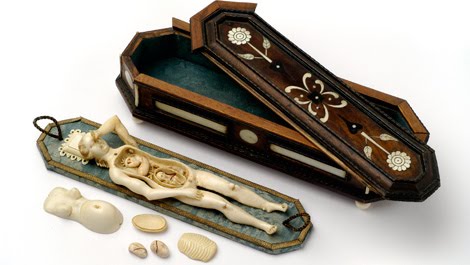
I just came across a rather interesting looking new exhibition at a gallery called the Me Collectors Room in Berlin. Entitled "The Wunderkammer," this new permanent installation is the work of antique dealer/cabinetist Georg Laue, proprietor of the famed Kunstkammer Georg Laue in Munich, Germany, and seems--as you can see in the images above--to include a pretty astounding collection of fine memento mori, ivory Anatomical Venuses, and turned ivory wonders.

From the website:

THE WUNDERKAMMER

ASTONISHMENT
The WUNDERKAMMER rekindles the tradition of the Kunst- and Wunderkammer of the Renaissance and Baroque periods. It imparts an insight into the world view and the standard of knowledge of past centuries and does just what a Wunderkammer was able to do between 200 and 500 years ago: transport the visitor into a realm of sheer astonishment — whether by means of the legendary unicorn, exposed latterly by the cognoscenti as the tusk of a narwhal, an amber mirror flooded with light, or cabinets that only reveal their mysteries to the observant viewer.

DISCOVERY
The quality of the exhibits, numbering in excess of 150, is unique and makes the WUNDERKAMMER one of the most significant private collections of its kind. The juxtaposition of works from different cultures generates its very own effect. The permanent collection places an emphasis on Vanitas (“Consider the fact that you will die”). In the Baroque period, death was already staged with a mixture of devotion, interest, and humour. The scope for interpretation of this topic is manifested by an anatomical model dating from the second half of the 17th century. The organs and the foetus of the laid out body of a pregnant woman can be removed and prompt one to indulge in a playful handling of this miniature.

UNDERSTANDING
The objects in the WUNDERKAMMER exert an incredible fascination and will captivate the curious with a vision of a small, encyclopaedic, unique universe, which ultimately contributing to a deeper understanding of the correlations between art, nature, and science.

This exhibition definitely looks worth a visit! And, for the more curious among you, theme-specific tours of the collection are also available in which, as the website explains, "existential themes such as Eros, death, and transience, as well as the genesis of the collection, form the central focus."

You can find out more about Georg Laue and his Munich shop clicking here. You can find out about more about the exhibit by clicking here.

Found via Wunderkammer. All images from the Me Collectors Room Berlin website.

Seeking Private Collections in Scandinavia, Great Britain, and Germany for "Private Cabinets" Photography Project


As alluded to in a few recent posts [1, 2], a week from tomorrow I will be embarking on a trip to Scandinavia and Great Britain with perhaps (time depending) a touch of Germany along the way.

Regular readers might recall that I currently at work on a long-term project exploring extraordinary private collectors and collections, working title: Private Cabinets (more here); while on my travels, I have scheduled to visit and photograph a few more private collections for inclusion in this series and am on the lookout for yet more.

If any Morbid Anatomy readers out there know of any private wunderkammern or extraordinary private collections featuring medical museum type artifacts, waxworks, human remains, scientific models, old natural history, carnival/circus/sideshow or marvels or curiosities of any sort in these parts of the world, or have, perhaps, such a collection of their own they would like to share, please email me at morbidanatomy@gmail.com. Museum and attraction suggestions in the same topic areas also greatly appreciated!

To find out more about the ongoing "Private Cabinet" series, click here. All images above are from the epic private collection of Tim Knox and Todd Longstaffe-Gowan Collection of London, England as featured in my recent Secret Museum exhibition. Click on images to see much larger, finer images, and click here to see the complete Secret Museum collection.