Flagship Biosciences combines fluorescence scanning with brightfield

The novel staining approach combines the multiplexing capabilities of fluorescence with the tissue context of brightfield whole slide scanning

FLAGSTAFF, Ariz. — March 17, 2011 — Flagship Biosciences announced today a novel whole slide imaging approach called Bridge Staining™, that allows a brightfield background to be aligned and overlaid on a fluorescent image on the same tissue section, allowing visualization of tissue morphologic features on the fluorescent image. The patent-pending technique is versatile, and can be used either with three to four color fluorochrome stained sections or autofluorescence or polarized scanning.

“While generally we prefer to work with brightfield pathology, but fluorescence presents useful applications in multiplexing of proteins in single tissue sections,” said Dr. Frank Voelker of Flagship Biosciences. “We utilize whole slide fluorescence scanning and analysis when two to four antibodies are required to be evaluated in a single section. However, fluorescence makes the determination of the local tissue context difficult for pathologists. This new approach allows immunofluorescence to be conducted as usual, but with the additional step of adding chromogen staining without interfering with fluorescence emissions. Protein expression can be quantified in tissue with fluorescence, with the advantage of using the chromagen staining to assist in pattern recognition of the regions of interest.”

The multimodal scanning is another in a long list of whole slide scanning and image analysis services offered by Flagship Biosciences, utilizing both whole slide brightfield and fluorescence scanning. The Bridge Staining™ approach is being refined in conjunction with a number of tissue-based companion diagnostics programs with Flagship’s pharmaceutical partners. Flagship Biosciences is a pathologist-owned digital pathology services organization. 

Embedded biopolymer with bridge staining 

President Bill Clinton to Keynote 2011 ASCP Annual Meeting

CHICAGO – March 14, 2011 – President Bill Clinton, the Founder of the William J. Clinton Foundation and the 42nd President of the United States and a champion for global health, will be the keynote speaker at the 2011 American Society for Clinical Pathology (ASCP) Annual Meeting on Wednesday, Oct. 19, in Las Vegas.
His address, “Embracing Our Common Humanity,” will set the stage for a truly international conference, which is serving as host to the World Association of Pathology and Laboratory Medicine (WASPaLM) XXVI World Congress. “A Global View of Pathology and Laboratory Medicine” is the theme of ASCP’s signature event, Oct. 19-22 at the Venetian-Palazzo Resort Hotel Casinos.

“President Clinton is the world’s leading advocate for global health initiatives, and ASCP has a corps of dedicated volunteers working to improve laboratory services in resource-limited countries,” said ASCP President John E. Tomaszewski, MD, FASCP.

After his two terms as U.S. President from 1993 to 2001, President Clinton established the William J. Clinton Foundation to strengthen the capacity of people around the world to meet the challenges of global interdependence.  The Clinton Health Access Initiative (CHAI), formerly the Clinton HIV/AIDS Initiative, works to strengthen integrated health systems in the developing world and expand access to care and treatment for HIV/AIDS, malaria and tuberculosis.

ASCP’s Institute for Global Outreach works to improve global health by identifying and implementing innovative methods and partnerships that improve laboratory practice.

ASCP Executive Vice President E. Blair Holladay, PhD, SCT(ASCP)CM, said ASCP and WASPaLM are partnering this year to build stronger bridges between national and international societies for all members of the laboratory team. WASPaLM represents 45 societies of pathologists and laboratory professionals in 34 countries.
Registration for the 2011 ASCP Annual Meeting is now open.

http://www.ascp.org/2011AnnualMeeting/register.html

United States Patent Office Affirms Hamamatsu Photonics’ Pioneering Patent in Digital Pathology

HAMAMATSU, Japan, March 15, 2011 /PRNewswire/ -- Hamamatsu Photonics K.K. (HPK) is pleased to announce that is has been awarded Reissue Patent No. 42,220 ("RE42,220"), confirming the patentability of its original Patent 6,847,729, a pioneering patent directed to slide scanning with remote viewing.

This patent is for the essential mechanism whereby a microscope slide is scanned and then digitized for remote viewing. The patent covers the basic operation of HPK's line of NanoZoomer® digital pathology instruments (including the NanoZoomer® 2.0HT, and the NanoZoomer® 2.0RS). The NanoZoomer® instruments have been widely accepted in the digital pathology market and the strong patent position of HPK should encourage software manufacturers to develop software that supports the NanoZoomer® platform.

Asked to comment on this seminal digital slide scanning patent, Mr. Hitoshi Iida, Managing Director of HPK, stated, "By reissuing this patent, we believe the USPTO has sustained the validity of this most central digital pathology patent. We now feel confident to move forward with enforcement of our patent rights."

The affirmation of the patentability of the patent claims demonstrates the innovation encompassed in scanning products HPK has developed. While HPK is willing to discuss the licensing of this patent on reasonable terms to others, it is studying the slide scanning products on the market with a view to demanding that all infringements of its RE42,220 patent cease.

All inquiries should be directed to David Leinwand, Esq., at Photonics Management Corporation, 360 Foothill Road, Box 6910,Bridgewater, NJ 08807-0910.

About Hamamatsu Photonics KK

Hamamatsu Photonics K.K. ("HPK") (Japan) is a leading manufacturer of devices for the generation and measurement of infrared, visible, and ultraviolet light. HPK is dedicated to the advancement of photonics through extensive research. HPK is also dedicated to the advancement of Digital Pathology. Digital Pathology is an area expected to improve the accuracy and efficiency of cancer diagnosis and life science research. Applications of Digital Pathology include pathology education, telepathology, computer aided diagnosis, cancer research, drug development, and basic research. Whole slide imaging is a core technology supporting Digital Pathology by converting glass slides to digital slides. The patent aids fast and high resolution conversion and efficient viewing and navigation of slides from remote locations.

SOURCE Hamamatsu Corporation

RELATED LINKS
http://www.hamamatsu.com


Primary interpretations, second opinions/consultations with AccelPath

Transforming Pathology

AccelPath has developed a proprietary workflow system to quickly provide high quality, sub-specialized pathology interpretations to referring physicians and laboratories.

Simple steps to state-of-the-art pathology operations:

  • AccelPath installs advanced slide scanners at the customer facility.
  • Slides are scanned locally.
  • Our proprietary workflow creates a digital case and transmits a requisition and images to the most appropriate pathologist based on specialization, credentials and workload.
  • The pathologist performs the interpretation of the digital images and an electronic report is transmitted to the customer, typically within 48 hours.

Further…

  • Images and reports are stored on Accelpath's servers offering a redundant solution to local storage capabilities.
  • AccelPath's proprietary workflow and network is completely HIPAA compliant.
  • Implementation is easy with the help of our experienced team.
  • Customers start to realize the advantages of AccelPath services immediately. 


Our Services, Your Success

AccelPath has invested in top talent and frontline technology to create innovative services designed to

enhance pathology operations. 

Diagnostic Services

  • Primary interpretations
  • Sub-specialty consultations
  • Expert opinions or second opinions

Technical Services

AccelPath's customers receive:

  • State-of-the-art digital scanners and full maintenance
  • Training for lab technicians on easy-to-use digital scanning equipment and Accelpath's workflow software
  • Multiple pathology report delivery options: secure email, fax and online portal
  • 24/7 access to all reports, requisitions and images via HIPPA compliant security

Value-Added Services

We provide value added services at affordable rates, including:

  • Operations consultation including customized financial data analysis and 
    feasibility studies
  • Billing and procedure coding
  • Expert assistance with laboratory design and installation
  • Standard Operating Procedure templates

Fast, sub-specialized reporting is the result of our ability to digitally route pathology images to the best pathologists irrespective of their location. This means customers receive reports and the information they need to promptly meet the needs of their patients. 

Through our unique workflow, we help ensure optimal efficiency, which in turn creates departmental savings.

Finally, we can help practices add to the bottom line if there is interest in building an on site pathology laboratory.

Contact AccelPath today for more information.


Just blame it on your genes

I am becoming increasingly suspicious of articles and media reports that claim something to the effect that ""X" is no longer most common cause of "Y"; Genetic cause found for disease".

Such as the case with this story below at bottom of this post.

In medical school you are taught few absolutes.  There are always "exception to the rule", common diseases may have rare presentations, rare diseases may have common presentations, for that disease but common to a host of other diseases, possibly making recollection and detection of the rare disease harder, because you are also taught that common diseases present commonly, that's why they are called common. 

I do however recall a few absolutes, including that prostitutes get cervical cancer, nuns get endometrial cancer, smoking causes lung cancer and pancreatitis is caused by gallstones and alchohol.  Of course, not all smokers get lung cancer and some cases of lung cancer occur in non-smokers and not all nuns get endometrial cancer (thought to be due to high levels of unopposed estrogen levels due to lack of pregnancy-induced elevated progesterone levels that intermittently oppose the estrogen).

Anyways, you get the idea.  Most medical students are taught a mnemonic for the causes of acute pancreatitis, like the one below (courtesy of Wikipedia).  "I get smashed" is a family-friendly compared to the mnemonic for the bones of the wrist. 

So, now why, for this first time since Hippocrates to Virchow to Will and Charlie Mayo to Dr. Gregory House is alcohol not one of the most common reasons for pancreatitis?  Did our genes change to the point they cause disease we use to credit to other causes?  Have the genes that cause alcoholism mutated to the point they no longer cause alcoholism and therefore potentially some cases of pancreatitis?  Wait, isn't it genes that cause us to drink in the first place? 

Certainly there are certain genotypes and phenotypes that predispose individuals to the development of gallstones which could leave home and travel down the bile duct and get lodged and cause the pancreas to digest itself.  If we are talking specifically about chronic pancreatitis, can eliminate gallstones since gallbladder surgery is one of the most common surgeries performed and without gallstones hard to get recurrent acute or chronic pancreatitis. 

Obviously autoimmune diseases have a genetic basis and thus one could attribute genetic causes to their primary disease and its complications.  Other metabolic conditions like those listed below including high lipids and triglycerides also have a genetic basis. 

Harder to come up with scorpion bites as having a genetic cause unless you are pre-programmed at birth and destined to get a PhD in scorpion biology and it presents an occupational hazard or you live in an area indigenous to such creatures without fear of being stung. 

The truth of the matter is that a number of diseases are caused by human actions, inactions or behaviors that results in modifications of cells and tissues beyond normal and cause pain and suffering.

On the other hand, genetic factors on some level probably do control those actions, inactions or behaviors that prevent or cause disease, so I guess you can attribute every disease to bad genes.

That means I do not need to remember any more mneumonics when all the causes are actually one big single cause.  Think I will have a drink. 

A common mnemonic for the causes of pancreatitis spells "I get smashed", an allusion to heavy drinking (one of the many causes):

This mnemonic is also roughly arranged according to the frequency of its causes. Thus: Gallstone pancreatitis is more common than pancreatitis caused by alcohol, trauma, or steroids.

Alcohol May No Longer Be Primary Cause Of Chronic Pancreatitis
Reuters (3/10, Grens) reports that genetic factors may be replacing alcohol as the major cause of chronic pancreatitis, according to a study in the journal Clinical Gastroenterology and Hepatology. Researchers analyzed the medical records of patients who were treated for chronic inflammation of the pancreas at 20 medical centers. They determined that approximately three out of every 10 cases of pancreatitis were due to genetic diseases, immune system disorders, or other factors, such as physical damage. In another three out of 10 cases, the study team could not determine the cause for chronic pancreatitis, but they noted that patients in the group had higher than average smoking rates.

"Treasures of Heaven: Saints, Relics and Devotion in Medieval Europe" Exhibition, Walters Art Museum, Baltimore, Through May 15th


Wow. WOW.

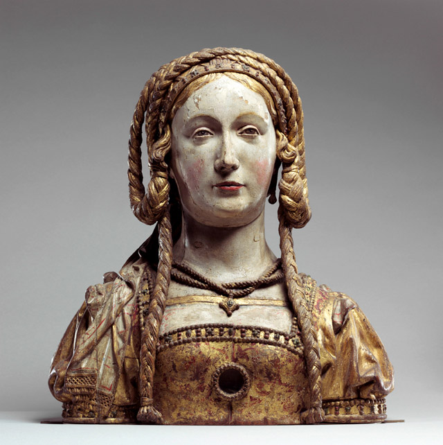
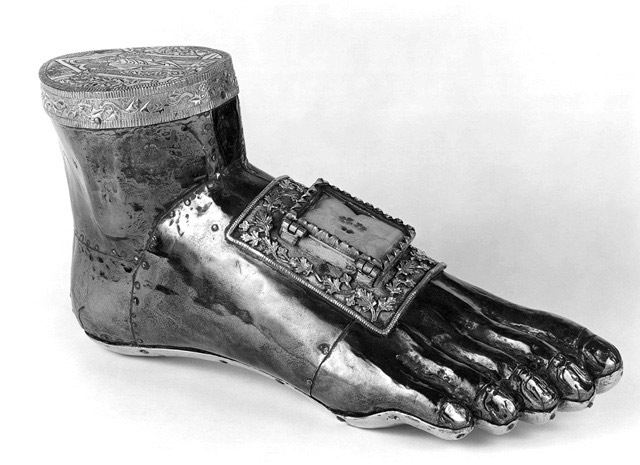
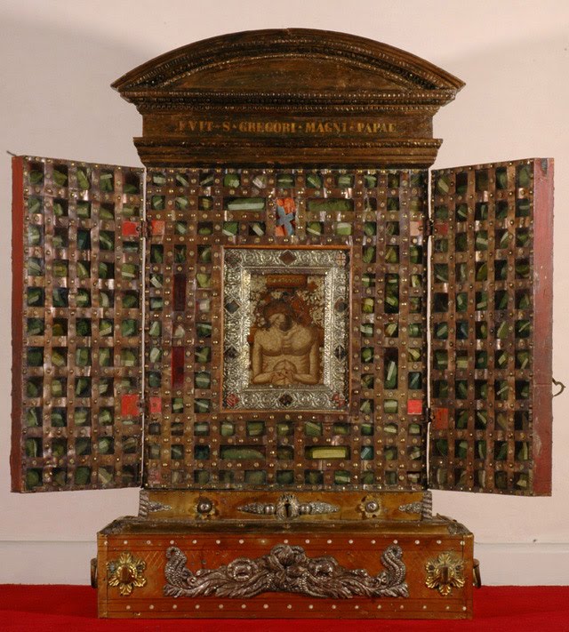
This just in: On view until May 15th of this year at the Walters Art Museum in Baltimore, a new exhibition of relics and reliquaries entitled "Treasures of Heaven: Saints, Relics and Devotion in Medieval Europe."

To get a sense of the kinds of treasures that await, check out the Treasures of Heaven "Digital Monograph" (from which these images were drawn) by clicking here.

Press release for the exhibition follows:

Treasures of Heaven: Saints, Relics and Devotion in Medieval Europe
Feb. 13, 2011 - May 15th, 2011
Walters Art Museum, Baltimore, Maryland

First major U.S. exhibition of Christian relics and reliquaries co-organized by the Walters, the Cleveland Museum of Art and the British Museum

Baltimore—The Walters Art Museum will host an exhibition offering visitors a glimpse into the Middle Ages, a time when art mediated between heaven and earth and wondrous objects of gold, silver and precious gems filled churches and monastic treasuries. Relics, the physical remains of holy people and objects associated with these individuals, play a central role in a number of religions and cultures and were especially important to the development of Christianity as it emerged in the Late Roman world as a powerful new religion. On view at the Walters Feb. 13–May 15, 2011, Treasures of Heaven: Saints, Relics and Devotion in Medieval Europe is the first exhibition in the United States to focus on the history of relics and reliquaries—the special containers to display the holy remains of Christian saints and martyrs. The exhibition is organized by the Walters Art Museum in partnership with the Cleveland Museum of Art and the British Museum.

Reliquaries proclaimed the special status of their sacred contents to worshipers and pilgrims, and for this reason, were often objects of artistic innovation, expressions of civic and religious identity, and focal points of ritual action. This exhibition will feature 133 metalworks, sculptures, paintings and illuminated manuscripts from Late Antiquity through the Reformation and beyond. It will explore the emergence and transformation of several key types of reliquary, moving from an age in which saintly remains were enshrined within closed containers to an era in which relics were increasingly presented directly to worshipers.

Many of the reliquaries in the exhibition have never before been seen outside of their home countries. Objects are drawn from celebrated public and private collections in the U.S. and Europe, and also from important church treasuries. In addition to the three organizing museums, world-renowned institutions, including the Louvre, the Metropolitan Museum of Art, the Art Institute of Chicago and the National Gallery of Art, are lending works to the exhibition. Nine works are traveling from the Vatican collections, including three reliquaries that were once housed in the Sancta Sanctorum, or Holy of Holies, the private relic chapel of the Pope.

Visitors will witness the transformation of reliquaries from simple containers for the earthly remains of Christian holy men and women to lavishly decorated objects of personal and communal devotion.

"As early as the second century AD, the relics of Christian saints—including their bones, ashes and other bodily remains—were thought to be more valuable than the most precious gemstones. They were believed to be a conduit for the power of the saints and to provide a direct link between the living faithful and God," said Martina Bagnoli, Robert and Nancy Hall associate curator of medieval art and exhibition co-curator. "These remains were treated with reverence and often enshrined in containers that used luxurious and precious materials to proclaim the relics' importance."

The medieval devotion to relics gave birth to new forms of architecture and prompted significant developments in the visual arts. The reliquaries showcased in Treasures of Heaven provide evidence of religious objects traveling across tremendous distances and of people making pilgrimages across the Mediterranean to walk in the footsteps of important figures from sacred history. Powerful in inspiring religious devotion among believers, reliquaries became cutting-edge works of art that combined innovative techniques with beautiful design.

"Those who come to the exhibition thinking that the Middle Ages are only a period of darkness will be surprised," said Martina Bagnoli.

Highlights of Treasures of Heaven include:

  • Reliquary Bust of St. Baudime, c. 1180-1200,Parish Church of Saint-Nectaire, Puy-le-Dôme
  • This nearly life-sized bust is one of the earliest surviving objects of its kind and travels outside of France for the first time.
  • Portable Altar of Countess Gertrude, c. 1045, Cleveland Museum of Art
  • This work is from the Guelph Treasure, one of the most important church treasuries to have survived from medieval Germany.
  • Head Reliquary of St. Eustace, c. 1200, British Museum
  • This head-shaped reliquary contained fragments of the skull of the Roman military leader Saint Eustace...

You can find out more about the exhibition here, and more about the topic of relics and reliquaries on the Treasures of Heaven "Digital Monograph" by clicking here. You can purchase the exhibition catalog by clicking here.

All images from the Treasures of Heaven "Digital Monograph;" you can find out more about them the images, and peruse the website, by clicking here. It was unclear how many of these are in the physical exhibition.

"Play Dead," Todd Robbins and Teller, The Players Theatre, New York


The other night, I took my boyfriend out for a night of good, scary, compelling fun at Todd Robbins' and Teller's (of Penn and Teller) new production "Play Dead," on view now at The Players Theatre in New York City.

The production is difficult to describe in normal theater terms; it is kind of like a haunted house meets a 1950s ghost show meets a piss take on a Victorian séance meets a high-end Vegas magic show all staged on a David Lynch film set. The main presence in the production is side-show performer Todd Robbins; he has a wonderfully compelling presence, equal parts passionate story teller, confidence man, and emphatic and empathetic debunker of spiritualist trickery. The piece is a kind of fun yet thoughtful meditation on the mysteries of death, the history of historical monsters in their various forms, and the ways--compellingly demonstrated in the show--that death and loss still make us easy prey for "spiritualist" hucksters.

I don't want to say too much more, as so much of the fun of the production come from the element of surprise, but I will say this: I was seriously amazed; I was amused; and we are still talking about it. I very much recommend checking it out.

"Play Dead" is being staged at The Players Theatre, 115 MacDougal Street, New York. You can find out more--and get tickets--by clicking here.

Full disclosure: I received free tickets from the Play Dead Production Company in exchange for reviewing this show. Lucky for me I really really enjoyed it! And so did my impartial boyfriend.

Image: From the Studio 360 website.

"Proteus" Screening with Film Maker David Lebrun, Observatory, April 1st


This April Fools Day, why not join Morbid Anatomy and Observatory for a screening of one of our absolute favorite films, Proteus, featuring an introduction by--and Q and A with--the film's maker, David Lebrun, in a rare East Coast appearance?

The film Proteus details the biography and struggles of biologist and artist Ernst Haeckel (1834-1919) who, as the copy for the film describes, "found himself torn between seeming irreconcilables: science and art, materialism and religion, rationality and passion, outer and inner worlds." Lebrun tells Haeckel's tale with inventive and almost chillingly beautiful animation constructed almost entirely from 19th Century archival images, with the most stirring and awe-inspiring sequences created from quick successions of scores of Haeckel's astonishing depictions of protista, as seen above in some of his drawings, and in the video clip at about 5:10 minutes in.

We are thrilled to be hosting two screenings of the film, one at 7 PM and one at 9 PM, in conjunction with Proteus Gowanus Interdisciplinary Gallery and Reading Room. Film maker David Lebrun will be on hand at each to introduce the film and to answer any questions you might have.

Please pass this on to any interested parties, and hope very very much to see you there!

Date: Friday, April 1
Time: 7:00 PM and 9:00 PM (2 Screenings)
Admission: $5
Presented by Morbid Anatomy in partnership with Proteus Gowanus

The ocean is a wilderness reaching 'round the globe, wilder than
a Bengal jungle, and fuller of monsters, washing the very wharves
of our cities and the gardens of our sea-side residences.

-- Henry David Thoreau, 1864

For the nineteenth century, the world beneath the sea played much the same role that "outer space" played for the twentieth. The ocean depths were at once the ultimate scientific frontier and what Coleridge called "the reservoir of the soul": the place of the unconscious, of imagination and the fantastic. Proteus uses the undersea world as the locus for a meditation on the troubled intersection of scientific and artistic vision. The one-hour film is based almost entirely on the images of nineteenth century painters, graphic artists, photographers and scientific illustrators, photographed from rare materials in European and American collections and brought to life through innovative animation.

The central figure of the film is biologist and artist Ernst Haeckel (1834-1919). As a young man, Haeckel found himself torn between seeming irreconcilables: science and art, materialism and religion, rationality and passion, outer and inner worlds. Through his discoveries beneath the sea, Haeckel would eventually reconcile these dualities, bringing science and art together in a unitary, almost mystical vision. His work would profoundly influence not only biology but also movements, thinkers and authors as disparate as Art Nouveau and Surrealism, Sigmund Freud and D.H. Lawrence, Vladimir Lenin and Thomas Edison.

422px-haeckel_stephoidea_edit1The key to Haeckel's vision was a tiny undersea organism called the radiolarian. Haeckel discovered, described, classified and painted four thousand species of these one-celled creatures. They are among the earliest forms of life. In their intricate geometric skeletons, Haeckel saw all the future possibilities of organic and created form. Proteus explores their metamorphoses and celebrates their stunning beauty and seemingly infinite variety in animation sequences based on Haeckel's graphic work.

Around Haeckel's story, Proteus weaves a tapestry of poetry and myth, biology and oceanography, scientific history and spiritual biography. The legend of Faust and the alchemical journey of Coleridge's Ancient Mariner are part of the story, together with the laying of the transatlantic telegraphic cable and the epic oceanographic voyage of HMS Challenger. All these threads lead us back to Haeckel and the radiolaria. Ultimately the film is a parable of both the difficulty and the possibility of unitary vision.

DAVID LEBRUN has served as producer, director, writer, cinematographer, animator and/or editor of more than sixty films, among them films on the Mazatec Indians of Oaxaca, a 1960s traveling commune, Tibetan mythology and a year in the life of a Maya village. He edited the Academy-award winning documentary Broken Rainbow, on the Hopi and Navajo of the American Southwest. Proteus premiered at Sundance and has won numerous international awards. The two-hour documentary feature Breaking the Maya Code (2008) tells the story of the 200-year quest to decipher the hieroglyphic script of the ancient Maya of central America; a drastically shortened version was broadcast on the PBS series NOVA and has been seen on television around the world. His experimental and animated works include the animated films Tanka (1976) and Metamorphosis (2010), works for multiple and variable-speed projectors such as Wind Over Water (1983), and a 2007 multimedia performance piece, Maya Variations, created in collaboration with composer Yuval Ron. Lebrun has taught film production and editing at the California Institute of the Arts and has curated numerous art exhibitions. He was president of First Light Video Publishing from 1987-1996, and since then president of Night Fire Films. He was a founding Board Member of the Center for Visual Music (CVM) and is on the Advisory Board of the Chabot Space & Science Center’s Maya Skies project. For a complete biography and filmography, please visit http://www.nightfirefilms.org.

You can find out more about the film by clicking here, and more about this event on the Observatory website by clicking here; you can access these events on Facebook here (7 PM) and here (9
PM). You can get directions to Observatory--which is next door to the Morbid Anatomy Library (more on that here)--by clicking here. You can find out more about Observatory here, join our mailing list by clicking here, and join us on Facebook by clicking here.

Images: From Ernst Haeckel's Die Radiolarien, Berlin, 1862. And special thanks to Ben Cerveny for turning me onto this wonderful film so many years ago.

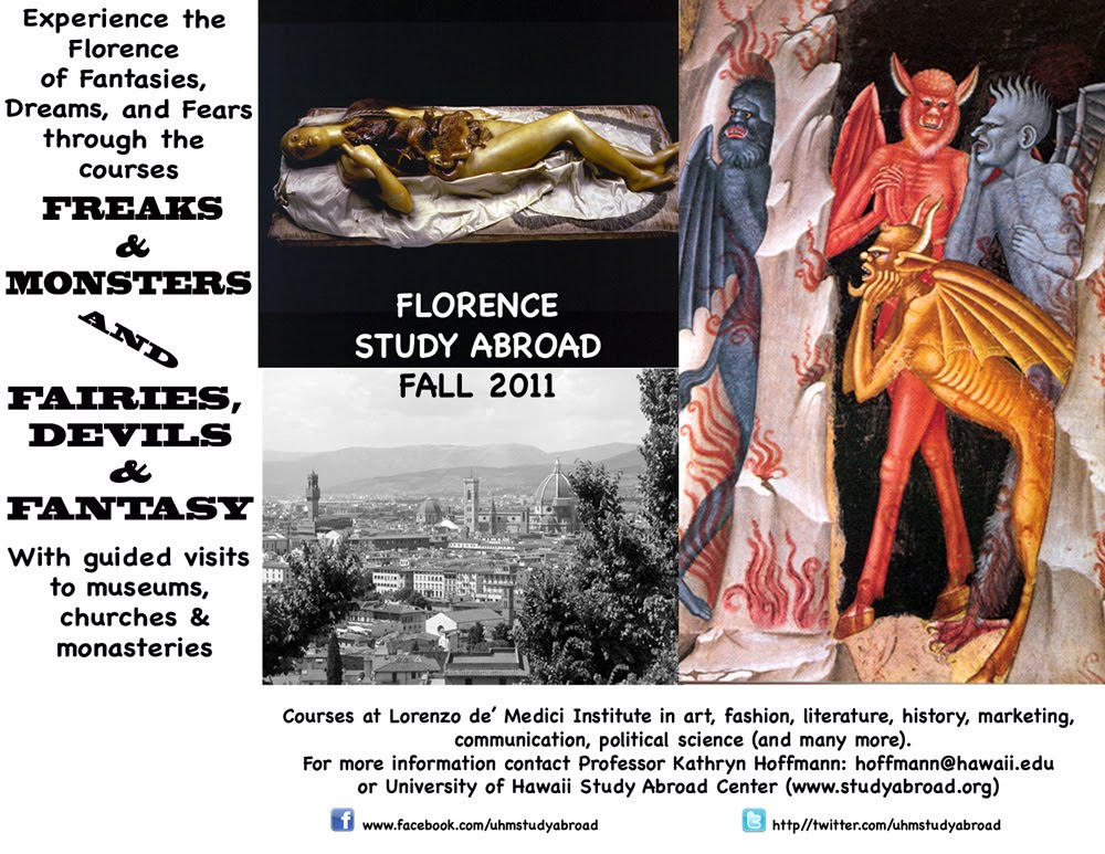
"Freaks and Monsters and Fairies, Devils, and Fantasy Tour of Florence," Fall, 2011, Dr. Kathryn Hoffman


Friend of Morbid Anatomy Kathryn Hoffmann of the University of Hawaii, Manoa has just announced that she will be leading a "Freaks, Monsters, and Fairies, Devils, and Fantasy" tour of Florence, Italy this upcoming fall semester. The tour will take in, in Hoffman's own words, "wax anatomical models of course, as well as the devils of Florence, reliquaries, the history of court and fairground stars with corporeal anomalies, and the original dark version of Pinocchio, where he came to a sad end in Book 5. I'm going to teach and take students out of the classroom and into the museums and churches."

Applications are due on April 1; for more information, email professor Hoffman at hoffmann [at] hawaii.edu.

So wish I could make it!

Please click on the image to see a much larger version.

Host change alters toxic cocktail

Leaf beetles fascinate us because of their amazing variety of shapes and rich coloring. Their larvae, however, are dangerous plant pests. Larvae of the leaf beetle Chrysomela lapponica attack two different tree species: willow and birch. To fend off predator attacks, the beetle larvae produce toxic butyric acid esters or salicylaldehyde, whose precursors they ingest with their leafy food. Researchers of the Max Planck Institute for Chemical Ecology in Jena, Gera number of, now observed that a fundamental change in the genome has emerged in beetles that have specialized on birch: The activity of the salicylaldehyde producing enzyme salicyl alcohol oxidase (SAO) is missing in these populations, whereas it is present in willow feeders. For birch beetles the loss of this enzyme and hereby the loss of salicylaldehyde is advantageous: the enzyme is not needed anymore because its substrate salicyl alcohol is only present in willow leaves, but not in birch. Birch beetles can therefore save resources instead of costly producing the enzyme. First and foremost, however, the loss of salicylaldehyde also means that birch feeding populations do not betray themselves to their own enemies anymore, who can trace them because of the odorous substance. (PNAS Early Edition, DOI 10.1073/pnas.1013846108)........

Gamma-Dynacare Medical Laboratories Implements Leica Microsystems Total Digital Pathology Solution

Leica, with its SCN400 scanner from the latest of the traditional microscope manufacturers continues to move systems and have early success.  Add to that the fact that digital pathology continues to expand in Canada among large healthcare systems and laboratories. With its scanner and software partner, SlidePath, Leica does offer a complete solution for slide scanning, image managment and image analysis with high quality.

Quorum Technologies Inc., a distributor and developer of high-end microscopy products and imaging systems
announced yesterday that Gamma-Dynacare Medical Laboratories is implementing Leica Microsystems Inc. Total Digital Pathology solution. The Leica SCN400 scanner with Digital Image Hub and Conferencing software couples high throughput digital slide scanning with enterprise level software, to provide a complete digital pathology system for Gamma-Dynacare.

The technology is being integrated at the Bomanville laboratory where cancer-screening programs
are being developed. Traditionally, pathologists have used microscopes to analyze specimens on glass slides. The
acquisition of the Leica SCN400 solution provides Gamma Dynacare new tools and flexible workflow options. The
system creates fully digitized images that can be viewed online in a secure and efficient manner from virtually any location. Gamma-Dynacare will also utilize Leica Microsystems' data management and image analysis software to further expand its commercial pathology services across Canada.

"The implementation of Leica Microsystems' SCN400 digital pathology system is exciting for Gamma-Dynacare as it supports our commitment to offer leading technology that can positively impact the delivery of diagnoses and treatment decisions for patients," said Jeff Sumner, Vice President, Clinical Development and Quality Assurance, Gamma-Dynacare Medical Laboratories. "We have been monitoring the technology and our evaluation of Leica Microsystems' total digital pathology platform was positive. We are pleased to be able to provide our clients and patients with the benefits of this new technology."

"The Leica solution implementation at Gamma-Dynacare is an important one as it marks another milestone in Canada's adoption of the digital pathology paradigm," says John Arbuckle, President and CEO of Quorum Technologies.  "Digital pathology is rapidly evolving and the technology has advanced tremendously over the past few years. Our company has implemented and supported approximately 50 digital pathology systems in medical schools, hospitals and research laboratories across Canada. It's great to see one of the country's largest and highly esteemed medical laboratories further validate the technology and prepare to offer a variety of digital pathology services."

About Gamma-Dynacare
Gamma-Dynacare is one of Canada's largest and most respected providers of laboratory services and solutions, with more than 50 years of experience in the industry. Headquartered in Brampton, Ontario, its operations include six laboratory facilities, located in Brampton, London, Ottawa, Bowmanville, Montreal and Winnipeg. In addition, Gamma-Dynacare operates more than 175 Patient Services Centres in Ontario, Quebec, Manitoba, Saskatchewan and Alberta. Gamma-Dynacare' s 2,000 skilled and dedicated employees perform millions of tests each year, playing a key role in the prevention and diagnosis of disease and the treatment of patients. By living its mission of "Caring for People", the company positively impacts the health and wellness of people across Canada, and is a valuable contributor to the betterment of the healthcare system.

About Leica Microsystems
Leica Microsystems is a world leader in microscopes and scientific instruments. Founded as a family business in the nineteenth century, the company's history was marked by unparalleled innovation on its way to becoming a global enterprise. Its historically close cooperation with the scientific community is the key to Leica Microsystems' tradition of innovation, which draws on users' ideas and creates solutions tailored to their requirements. At the global level, Leica Microsystems is organized in four divisions, all of which are among the leaders in their respective fields: the Life Science Division, Industry Division, Biosystems Division and Medical Division. Leica Biosystems, an own operating company, offers histopathology laboratories the most extensive product range with appropriate products for each work step in histology and for a high level of productivity in the working processes of the entire laboratory.
Leica Microsystems is represented in over 100 countries with 12 manufacturing facilities in 7 countries, sales and service organizations in 19 countries and an international network of dealers. The company is headquartered in Wetzlar, Germany.

About Quorum Technologies
Quorum Technologies is a private Canadian enterprise located in Guelph, Ontario. The company develops and
integrates scientific instruments and is the largest independent provider of Microscope based imaging systems in Canada. Quorum is a leader in advanced Live Cell Imaging platforms and holds over half a dozen patents with several others in process. Through its technology transfer relationships with Canadian Universities, Quorum is helping to bring new innovative technologies to market for use in preclinical research, drug development and personalized medicine.
Quorum Technologies is a private Canadian enterprise located in Guelph, Ontario. The company develops and
integrates scientific instruments and is the largest independent provider of Microscope based imaging systems in Canada.

Australia to outlaw thousands of plants, including national flower

In what it claims is an effort to stamp out plants and flowers that can be used as drugs, Australian authorities have proposed insane legislation that will outlaw hundreds, if not thousands, of common plant species, including the golden wattle, its national flower. If the law passes, nurseries, commercial growers, farmers, cactus collectors, and even backyard gardeners will become criminals overnight -- even though many of the plants being targeted are not even used as drugs.

Currently, only five plants are banned from cultivation in Australia because of their alleged inherent drug components. But the new legislation will ban from cultivation any plant that contains dimethyltryptamine (DMT), a naturally-occurring hallucinogen, which according to the Australian Attorney General includes many common and native plants, from various cacti and fodder grasses, to simple backyard ornamental plants. Read more...

AyurGold for Healthy Blood

Digital Pathology: Real Examples, not Just Promises for the Future – Webinar from Aperio

About Aperio
Aperio is the leading provider of digital pathology solutions
in hospitals,
reference labs, and pharmaceutical and research institutions across the world.

Today, our affordable
and complete product portfolio improves patient care by enhancing quality assurance, delivering more efficient workflows, facilitating access to new and more targeted therapies, and improving pathologists’ skills via lifelong education.


Digital Pathology: Real Examples, not Just Promises for the Future

Thursday, March 17, 2011
7:00am PST, 10:00am EST 
(2:00pm London GMT / 8:00am Mexico City Standard)

   or
Monday, March 21, 2011

4:00pm PST, 7:00pm EST 
(8:00am Japan Standard /10:00am Australia Eastern, March 22)

 Join us for an informative panel session to discuss enhanced consultation, quality assurance
and patient outcomes, presented by:

Miguel Burnier, M.D., Ph.D.
McGill University    

Dr. Burnier will discuss the use of digital pathology in ocular pathology with emphasis on the use of digital pathology in consultation cases from abroad and the experience of BIOPSY (Burnier International Ocular Pathology Society) in exchanging difficult cases using digital pathology.

Andrew Evans, M.D., Ph.D.
University Health Network Toronto
   
Dr. Evans will cover the UHN experience on using digital pathology for frozen sections, particularly how to implement the technology, the performance of their system, and lessons they have learned along the way.

Jared N. Schwartz, M.D., Ph.D.
Aperio
  
Dr. Schwartz will talk about the need and ability to use digital pathology for expert infectious disease consultation on a local to worldwide basis and how pathologists, microbiologists, and public health officials can use this technology for education and reference and in reaching rural and/or underserved communities.

To register for the 7am PST on March 17 session, click here.
https://aperio.webex.com/aperio/onstage/g.php?t=a&d=480583239

To register for the 4pm PST on March 21 session, click here.
https://aperio.webex.com/aperio/onstage/g.php?t=a&d=483701118

You may also register by visiting the Aperio Events page.

You will receive a confirmation email upon registration with a web link that will lead you to the online event. Simply visit the link at the assigned time. The session is secure and easy to access. If you have any questions or would like additional information, please contact the Aperio Events Team at events@aperio.com or 760.539.1192. 


Entering Private Practice: What Every Pathology Resident Should Know about Productivity, Management, and Career Development

How to master the secrets of success for finding your first job

dr karim sirgijustin clark

YOUR PRESENTERS:

Karim E. Sirgi, MD, President, UniPath PC

Justin M. Clark (Moderator), Director of Operations, THE DARK REPORT


LIVE Webinar – Register Now

Spring is approaching and as a pathology fellow or fourth-year resident, you’re getting serious about your job search. The stakes are high for young pathologists because both you and the hiring practices want to make the “perfect match.” Do your homework now and learn how to recognize a successful private pathology practice from a struggling one-and this PREP webinar can help!

Pathology Residents Education Program (PREP) provides business education and job-search savvy to third- and fourth-year residents. This session is the latest in a series of events designed especially for pathology residents and fellows.

Take advantage of this opportunity to go inside one of the nation’s fastest-growing multi-specialty, multi-site private pathology groups and learn the secrets of its clinical and financial success. This is essential knowledge you can use to jump-start your job search and position yourself as a smart hire for the nation’s most prominent pathology practices.

Your guide on this “virtual tour” is Karim Sirgi, M.D., President of UniPath, P.C. We’ve asked him to take you behind the scenes of his dynamic anatomic pathology laboratory. It’s a unique opportunity to have one of pathology’s keen business and management minds introduce you to the rhythms and clinical routines of a top-flight practice.

You’ll gain valuable insights into how this professional corporation of 25 pathologists and subspecialist pathologists is organized and managed. Find out what it takes to deliver a full menu of pathology services to local community hospitals and office-based physicians in the region, even as UniPath is building a national referral business in selected pathology subspecialties.

Dr. Sirgi will take you through the essentials of daily and weekly staffing. You’ll master the basics of how to efficiently schedule 25 pathologists at multiple sites, each characterized by distinct competencies and competitive priorities. You’ll learn about specific management techniques that the group uses to protect the time, income, and lifestyle parameters for all the pathologists in the group.

These fundamental management and clinical practice strategies have a practical benefit for you. Armed with this knowledge, you’ll recognize good employment opportunities as you interview. More important, you’ll be able to spot struggling or dysfunctional pathology groups more easily, saving you years of heartache and disappointment working for the wrong pathology group practice.

But there’s more! During this special PREP webinar, Dr. Sirgi will also give you information about which subspecialties are in highest demand and will help you better understand what a private pathology group expects from a new pathologist just leaving residency or fellowship.

The content for this PREP webinar was designed with input from several pathology residence program directors to give you an insider’s understanding of the business, management, and clinical practice issues that make up the daily routine of an anatomic pathologist. Best of all, it prepares you to put your best foot forward in the job interviews that lie ahead.

Ask your Chief Resident or Residency Program Director to register your residency program for this information-packed 75-minute webinar, “Entering Private Practice: What Every Pathology Resident Should Know about Productivity, Management, and Career Development,” on Thursday, March 10, 2011. It’s critical knowledge for any pathology resident or fellow just launching a search for the perfect pathology job.

And don’t forget the question-and-answer session at the end of this conference. It’s your opportunity to get answers to specific questions about the challenges and opportunities of working for a multi-specialty, multi-site private pathology practice. Register today to ensure your participation in this very special event.


THE DARK REPORT WEBINAR AT A GLANCE

DATE: Thursday, March 10, 2011

TIME
: 3 p.m. EST; 2 p.m. CST; 1 p.m. MST; 12 Noon PST

PLACE: Your computer or your residency program’s conference room

COST: $249 per dial-in site (unlimited attendance per site) *Add 30 days of post-conference online access to the recording of this event for $75

TO REGISTER: Click here or call Suzanne Galloway toll-free at 1-800-560-6363


For one low price—just $249—you and your entire team can take part in this fast-paced, insightful webinar. Best of all, you’ll be able to connect personally with either of the panelists when we open up the phone lines for live Q&A


Here’s just some of what you’ll learn during this in-depth 75-minute webinar:

  • The advantages and disadvantages of working in a multi-specialty, multi-site private pathology group.
  • How business and management drivers in a private pathology group practice shape your work routine and compensation.
  • Standard models of pathologist productivity and compensation in private pathology groups.
  • Easy ways to determine the culture of a group and whether it’s a good fit for you.
  • How new technologies, including digital pathology, histology automation, and molecular/genetic tests, can propel your career forward.
  • How to recognize the signs of a thriving, successful private pathology group.
  • How to avoid private pathology groups that are struggling or unhappy places to work.
  • Three ways to identify the long-term stability of a group.
  • How community-based pathologists work and why it’s different from your academic pathology experience.
  • How to talk intelligently in job interviews about the benefits of working in this kind of setting.

…and much more!

Individuals who benefit from this webinar:

  • Pathology chief residents, residents and fellows
  • Academic pathology chairs
  • Residency program directors
  • Pathology department administrators
  • Undergraduate and graduate medical educators
  • Pathology and laboratory professionals

How to Register:

1. Online
2. Call toll free: 800-560-6363

Your webinar registration includes:

  • A site license to attend the conference (invite as many people as you can fit around your speakerphone at no extra charge)
  • A downloadable PowerPoint presentations from our speaker
  • A full transcript emailed to you soon after the conference
  • The opportunity to connect directly with the speaker during the audience Q&A session

Register Now! Or for more information, call us toll-free at 800-560-6363.

Distinguished Expert:

Karim E. Sirgi, M.D. is board certified in anatomic and clinical pathology with subspecialty board certification in cytopathology. He is President of Unipath PC as well as a member of the joint strategy board of Unipath PC-American Pathology Partners LLC. Dr. Sirgi is also Medical Director of laboratories at The Urology Center of Colorado and of the Colorado Center for Laboratory Medical Science, past-president of the medical staff at Presbyterian/Saint Luke’s Medical Center, and past-chair of the HealthOne Presidents Council. He is a diplomate of the Saint Joseph University School of Medicine in Lebanon. Dr. Sirgi completed his residency in anatomic and clinical pathology at Booth Memorial Medical Center, a fellowship in surgical pathology at Barnes Hospital, Washington University Medical Center and an additional subspecialty fellowship in general and interventional cytopathology at MD Anderson Cancer Center. Dr. Sirgi was on the faculty at both institutions after completion of each fellowship. He was chairman of the pathology department at Presbyterian/St. Luke’s Medical Center from 1997-2003 and president of the Colorado Society of Clinical Pathologists in 2003/2004. He is also a former member of the College of American Pathologists Cytopathology Resource Committee and the Gynecology Oncology Group pathology review committee
"Coney Island Spectacularium and 'Oddities' Screening," Coney Island Museum, April 9th, Obscura Day 2011


As part of that noble effort which is Obscura Day, why not come down to Coney Island, take in Morbid Anatomy's brand new exhibition "The Great Coney Island Spectacularium" (which opens the day before), check out Super Freak Weekend, and celebrate the premiere of season two of "Oddities" with free Hendricks Gin cocktails, episode viewing, and general revelry assorted cast members?

Why not indeed!

Full details follow; really hope to see you there!

Title: Coney Island Spectacularium and "Oddites" Screening
Date: Saturday, April 9

Time: 8:00 PM - 10:00 PM
Cost: $15.00

Where: The Coney Island Museum, Surf Avenue, Brooklyn
Click here to purchase tickets

Party in the Coney Island Museum to celebrate the opening of the Coney Island Spectacularium, super freak weekend, and meet the stars of the Discovery Channel show Oddities at the premiere of season two.

At the end of the 19th Century, Coney Island was the pinnacle of an astonishing era of live attractions – the Great Coney Island Spectacularium aims to recreate that momentous age, bringing you sites, sounds, and immersive experiences that can’t be seen anywhere else on earth. The attendees of this event will be the first to experience this taste of Coney Island at the height of its spectacle with the opening of the Coney Island Spectacularium.

But that is only one part of this multi-faceted event! Also taking place is a sideshow performance by some of the countries best sideshow performers gathered in Coney for the annual super freak weekend.

Topping it all off is the premiere of season two of the Discovery Channel show Oddities, with its stars Mike and Evan in attendance! Free Hendrick's Gin cocktails will be served upstairs, and there will be a cash bar (wine and beer) available downstairs.

Details/Special Instructions:
Doors open at 8:00, screening of the season premiere at 8:30.
Tickets are $15.

It’s also day two of the Congress of Curious Peoples, Coney Island USA’s 10-day series of lectures and performances about curiosity and curiosities, broadly conceived, so be sure to check out the schedule for the rest of the week..
Obscura Day is an international celebration of unusual places, happening all over the world on April 9, 2011.

Visit ObscuraDay.com to see all of our 2011 events.

To by tickets, click here. For more about the Great Coney Island Spectacularium, click here. For more about "Oddities," click here.

Caliper Life Sciences And HistoRx To Provide Integrated Solutions For Superior Protein Quantification Of Biomarkers In Tissue

HOPKINTON, Mass. and BRANFORD, Conn., March 10, 2011 /PRNewswire/ — Caliper Life Sciences, Inc. (CALP: 6.285 -0.165 -2.56%) and HistoRx, Inc., today announced a collaboration to co-develop a new generation of tissue-based imaging and analysis solutions, based on immuno-fluorescence, that will deliver accurate, quantitative, and reproducible measurements of proteins in tissue samples.  The collaboration will enable researchers to leverage the combined imaging and analysis power of Caliper’s Vectra™, an automated multiplexed tissue imaging platform, and AQUA® technology, HistoRx’s proprietary image analysis system, to provide an integrated platform to advance the identification and validation of new drug targets and improve assessment of drug response.  

“The Vectra system from Caliper represents a significant move forward for researchers interested in digital pathology with multispectral, brightfield, and fluorescent capabilities,” said Bill Sullivan, Senior Vice President of Operations at HistoRx.  ”By matching AQUA technology’s intensity-based quantitative measurement with Caliper’s market leading multispectral imaging capabilities, users will be afforded a level of quality and performance that is unparalleled.  Caliper is therefore a natural partner for HistoRx in bringing reproducible and truly quantitative immunohistochemistry to the anatomic pathology laboratory.”  

“The combination of Caliper and HistoRx technologies enables an unprecedented level of objective and accurate diagnosis that is currently lacking, both on the research level as well as the clinical level,” said Kevin Hrusovsky, President and Chief Executive Officer of Caliper Life Sciences.  ”We see an opportunity to bring a comprehensive solution to the research market to help expand scientists’ options for capturing key disease signatures and to aid in the development of new drugs and new strategies to battle cancer.”

Under the agreement, Caliper will have exclusive worldwide distribution rights for the integrated product in the life sciences research market as the companies continue to explore additional collaborative opportunities in clinical research and diagnostics.  The AQUA technology is complimentary to Caliper’s inForm™ image analysis software, a flexible investigative tool using trainable pattern recognition algorithms, providing multi-analyte, per-cell quantification in FFPE tissues.  AQUA provides a simple-to-use, pre-configured and automated quantification of single biomarkers, using validated AQUA multi-label kits, producing platform-independent AQUA ‘scores’.  Together, they provide a comprehensive suite of tools to correlate protein expressions with disease progression, clinical outcome or response to therapy.

Caliper expects to launch the AQUA enabled Vectra platform in August of 2011.

About Caliper Life Sciences

Caliper Life Sciences is a premier provider of cutting-edge technologies enabling researchers in the life sciences industry to create life-saving and enhancing medicines and diagnostic tests more quickly and efficiently. Caliper is aggressively innovating new technology to bridge the gap between in vitro assays and in vivo results, enabling the translation of those results into cures for human disease. Caliper’s portfolio of offerings includes state-of-the-art microfluidics, lab automation and liquid handling, optical imaging technologies, and discovery and development outsourcing solutions. For more information please visit http://www.caliperLS.com.

About HistoRx, Inc.

HistoRx, Inc. is a leading developer of tissue-based diagnostic solutions to advance individualized patient care. The company’s products and services are based on proprietary analysis of tissue biomarkers using AQUA technology. AQUA technology is the only platform capable of precisely measuring protein biomarker concentration with sub-cellular resolution in tissue sections, enabling fully objective, standardized, reproducible, and automated analysis to guide drug development and therapeutic decision-making. HistoRx is commercializing a pipeline of proprietary diagnostic products targeting improved treatment decision-making and patient outcomes in cancer care. For more information, please visit http://www.historx.com.

The statements in this press release regarding future events, including the statement that Caliper expects to launch the AQUA enabled Vectra platform in August of 2011, are “forward-looking statements” within the meaning of Section 21E of the Securities Exchange Act of 1934, as amended. These statements are subject to risks and uncertainties that could cause actual results to differ materially from those contemplated by the forward-looking statements as a result of a number of factors, including that Caliper may encounter unexpected problems in integrating its Vectra system with HistoRx’s AQUA technology.  Further information on risks faced by Caliper are detailed under the caption “Risks Related To Our Business” in Caliper’s Annual Report on Form 10-K for the year ended December 31, 2009. Our filings are available on a web site maintained by the Securities and Exchange Commission at http://www.sec.gov. Caliper does not undertake any obligation to update forward-looking or other statements in this release or the conference call.

NOTE:  Caliper is registered trademark and Vectra and inForm are trademarks of Caliper Life Sciences, Inc.  AQUA is a registered trademark of HistoRx, Inc.  

SOURCE Caliper Life Sciences, Inc.

http://www.dailymarkets.com/stock/2011/03/10/caliper-life-sciences-and-historx-to-provide-integrated-solutions-for-superior-protein-quantification-of-biomarkers-in-tissue/


Pompeii in Times Square: “Pompeii the Exhibit: Life and Death in the Shadow of Vesuvius,” Discovery Times Square, New York City


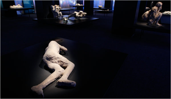
"Experience Pompeii before and after the epic eruption 2,000 years ago. Imagine the moment their world vanished and discover the miraculous artifacts unearthed since. Witness the life and death of those frozen in time by ash - including the largest collection of body casts ever presented.

  • Over 250 artifacts – includes some never-before-seen objects and the largest collection of body casts ever on display including a dramatic skeleton collection
  • A brand-new, immersive movie experience depicting a timelapsed representation starting from the moment of Vesuvius’ massive explosion"

--From the “Pompeii the Exhibit" Website

More than a museum, Discovery Times Square is New York’s destination for discovery through unique and immersive exhibits
--Website for Discovery Times Square

"There is a lot of traffic these days in well-preserved bodies, human and otherwise. They are sliced and pickled for artistic effect or uncannily dissected and plasticized, with every blood vessel visible. They have toured the world, wrapped and mummified in the manner of ancient Egypt, or have been displayed, more modestly preserved by the dry desert sands of the Silk Road. And there are many, many more mummies yet to come.

Why this onslaught of the almost-living dead in museums? Are we latter-day Ezekiels seeking prophetic messages from ancient skeletal remnants? Has the technology used to prepare the dead for world travel suddenly advanced? Or has the need for income by the overseers of mummies suddenly increased?"

--From "When the Dead Arise and Head to Times Square," Edward Rothstein, the New York Times

“Pompeii the Exhibit: Life and Death in the Shadow of Vesuvius”--a new exhibition at Discovery Times Square--activates the same tension between spectacle and education, prurience and propriety, which was exploited to such great financial reward by Gunther von Hangens in Body Worlds and which characterized many 19th Century popular amusements such as tourist visits to the Paris Morgue, popular anatomical museums, and the scores of death- and destruction-themed spectaculars to be found at Coney Island in the late 19th and early 20th Centuries.

In fact, "Pompeii the Exhibit" of 2011 has much in common with a particular Coney Island attraction of 1889--the spectacular “The Last Days of Pompeii”--if not in the particulars than in the shared drive to offer the paying public a fully immersive recreation of the destruction of Pompeii, and in their use of over-the-top hyperbolic detail in describing the wonders of their respective exhibitions.

"The Last Days of Pompeii" of 1889--an immersive spectacle that combined historical vignette, theatre performance, and a pyrotechnic display in recreating the destruction of Pompeii by the fires of Vesuvius--boasted in its press about the number and variety of its cast (over 400 people! "a ballet troupe of 36 dancers trained by Batiste Cherotte... a male chorus..., soldiers, acrobats, jugglers, tumblers, [and] wire-walkers"!)

2011's "Pompeii the Exhibit," on the other hand, focuses on the numbers and authenticity of its artifacts (over 250! Some never seen before! The largest number of body casts ever on display!), bringing to mind the press for such Coney Island Spectaculars such as "The Boer War" (Real British and Boer veterans!) and the "Streets of Delhi" (300 authentic Indian natives in costume! Elephants! Camels! Horses!). To further blur the line between "legitimate museum" and popular attraction, "Pompeii the Exhibit" is hosted at a popular exhibition hall sponsored by a television channel--Discovery Times Square--rather than an "ordained" museum such as AMNH; Also, Pompeii the Exhibit" provides visitors not just artifacts and other traditional ways of experiencing history but also what its website describes as a "brand-new, immersive movie experience" reenacting "the moment of Vesuvius’ massive explosion."

So what to make of it all? I see this new exhibition as excitingly in the tradition of 19th Century popular educational amusements--dime museums, popular anatomical museums, and Coney Island recreations--spaces where spectacle and education, prurience and propriety, coexisted for mass consumption. Fun, didactic, spectacular, and a resounding and thoughtful endorsement in today's Times to boot. I, for one, can't wait to go see it.

You can read a fascinating review of "Pompeii the Exhibit"--as quoted above--by Edward Rothstein in today's New York Times by clicking here. You can find out more about Coney Island's “The Last Days of Pompeii" by clicking here. You can find out more about "Pompeii the Exhibit" by clicking here.

Thanks so much to GF Newland for alerting me to this!

Image: Ruth Fremson/The New York Times

Plaster casts made from hollowed-out molds of rock, where bodies had been captured a moment before they ceased to be.

Pathologist Could Be Held Liable for Remote Review of Biopsy

This case on the surface appears to deal with the issue of appropriate state licensure for remote reads of pathology material.  Pending the outcome of this case, it emphasis the scrutiny and need for ensuring that practioners have met appropriate state licensure requirements and local credentialing requirements.  The problem is that every state has their own requirements concerning telemedicine requirements (and interstate commerce).  

One needs to insure the laws of the both the state where the case is received from as well as the state where a reading is performed are understood and followed.  Issues such as these and potential CLIA implications remain a barrier for digital pathology and in particular an "anytime, anywhere" model.

----------------------------------------------------------------------------------------------------------------------------

A Washington pathologist and her group practice may be subject to liability for the unlicensed practice of medicine under Idaho law stemming from the remote review in Washington of a biopsy from a patient in Idaho, a federal court ruled Dec. 30, 2010 (Smith v. Laboratory Corp. of America, W.D. Wash., No. C09-1662, 12/30/10).

The U.S. District Court for the Western District of Washington said Idaho’s Medical Practices Act (MPA) potentially applies to the Washington pathologist, Dr. Jane J. Yin, if she is found to have rendered a medical diagnosis for an Idaho resident without holding a license to practice medicine in that state.

Source: http://www.ioma.com/issues/GCR/2011_3/1625571-1.html


Omnyx demonstrating Integrated Digital Pathology solution at USCAP

If you are reading this from San Antonio this week attending the USCAP meeting, be sure to check out Omnyx's IDP solution in booth 330. 

Omnyx has focused on workflow since its inception and this demo should highlight their work to date and the efficiencies to be had with digital pathology.

Omnyx, a joint venture of GE Healthcare and UPMC is showing their IDP solution designed by pathologists, for pathologists and built from the ground up with help from a global team of collaborators with the overall goal of digitizing the pathologist's process with intuitive and efficient solutions.  

Omnyx will demonstrate its complete product line, which includes the VL4 and VL120 whole slide scanners, Pathologist Workstation, Histology Workstation, Web Viewer, Algorithms, Workflow Server, and Digital Archive for image storage. These products will be sold initially for research use only.  By digitizing the slides and corresponding workflow, the Omnyx technology is intended to do what a traditional microscope cannot -- unite an entire pathology department and improve collaboration, communication and efficiency.  

Of note, Omnyx will be demonstrating for the first time the VL120 whole slide scanner, which is capable of holding 120 slides that are automatically loaded and scanned.  When launched, this product will be used in environments where continuous scanning is critical to sustain throughput.  The VL120 is a complement to the already unveiled Omnyx VL4 scanner, currently for research use,and uses the identical scanning engine, which includes the patented Independent Dual Sensor imaging technology which can scan slides automatically at 20x or 40x magnification with speed and quality.

The Omnyx IDP solution integrates what are often stand alone products into a comprehensive platform.  It combines high speed, high quality whole slide scanning with scalable information technology products to create a solution that addresses the reliability and process requirements of pathologists and enables single and multi-site organizations to benefit from increased efficiencies and greater access to peers in the field.  It provides pathologists and histologists the tools necessary to work in closer collaboration and efficiently digitize their processes.