Interview with James Archer of Anatomy Blue

James Archer Anatomy Blue clot
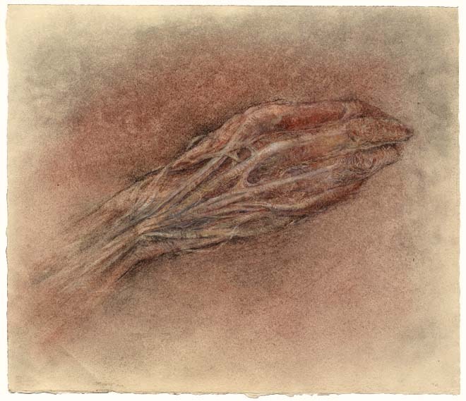
Clot – illustration for print, personal work, ©2012 James Archer

With an MA in Biomedical Communication from UT Southwestern and tremendous experience in the medical illustration/animation scene, James Archer is particularly sought after for his artistic style and approach to work. His 3D medical illustrations captivate the viewer with their diffuse lighting, organic hues, intimate depth of field, and overall softness that make anatomy and science look almost poetic.

I had the opportunity to work with James on a medical animation project recently and for someone with such a high standard of detail and execution, he’s an extremely laid back guy. He’s also impressively nonchalant about his incredible talent. I caught up with James after our project to uncover more about his background, technique, and cool personality.


SA: Tell us a bit about your background and what pulled you into medical illustration.

JA: I grew up in North Georgia in a town called Lilburn. As a child I loved to draw, an activity that would occupy much of my time. As I grew older I developed the ability to create highly detailed, photo-realistic drawings. I would sometimes setup a still life on the kitchen table or my bedroom floor and sit there for hours recording every detail with pencil and paper. Looking back it seemed more like an obsessive-compulsive behavior than a truly artistic endeavor. I now find that there is a strong association between art and obsession.

James Archer Anatomy Blue arteriovenous malformation

SA: Medical illustrators tend to come from an educational background that balances science with formal art training. You received a degree in biology and art history. Why did you choose art history and not a more specific studio art degree? How has a background in art history informed your work?

JA: My decision to major in Art History was one of convenience. After graduating from high school, I was accepted at Emory University on a four-year scholarship. While Emory does not offer a BFA, receiving the scholarship was a blessing. At the time I had no money and could not possibly attend another school without student aid. Attending Emory and majoring in Art History and Biology was the logical choice.

I find that the study of Art History provides context, a sense of place and time. It provides a set of values for which to judge works. And most importantly, it provides a sense of community, a feeling of belonging.


SA: Did you specialize in 3D illustration/animation at UT Southwestern? What motivated you to go in the direction of animation as opposed to traditional medical illustration?

JA: I certainly placed a greater emphasis on 3D illustration assignments compared to traditional illustration assignments. I believe that my interest in 3D illustration and animation was nothing more than an attempt to satisfy a long held obsession to visually recreate the world around me in near photographic form. The creation of synthetic imagery is relatively straightforward with today’s software. The real challenge lies in creating imagery that is emotive.


SA: You worked for quite a few top medical education companies and organizations early in your career such as WebMD, Nucleus Medical Art, and National Library of Medicine. What experiences did you gain from these organizations and when did you decide to start your own business?

JA: Working for Nucleus and WebMD represented a turning point for me. Even though I was given some amazing opportunities at each of those companies, I discovered that I had absolutely no interest in creating purely didactic materials. Instead, I wanted to find ways of reaching people on an emotional level. If your objective is to produce positive health outcomes in a given population then your campaign must include an appeal to emotion.

Working for the National Library of Medicine proved to be the genesis for Anatomy Blue. My primary responsibilities included research and development in 3D animation and production standards for broadcast and video. While working for NLM, I was completely immersed in 3D software and rendering technology. It was during that time that I began to develop lighting and texturing techniques that would later come to define the Anatomy Blue style.


James Archer Anatomy Blue virus
Virus – illustration for print collaterals for pharmaceutical company – ©James Archer

James Archer Anatomy Blue aspergillus
Aspergillus – illustration for print collaterals and booth display for pharmaceutical company – ©James Archer

SA: Flipping through the Medical Illustration Sourcebook, your style always stands out to me for its soft colors, beautiful lighting, and intimate depth of field. You bring such an artistic touch to 3D medical illustration that is unrivaled in my opinion. Can you tell us a bit about how your style evolved into what it is today?

JA: My style is more akin to punctuated equilibrium than gradualism. Aspergillus and Virus were created nearly a decade apart but they look as if they are from the same time period.

There are a number of themes that I’m gradually refining over time, themes like obsession, realism, context and emotion. The image, Virus contains only two elements, a background and a virus, yet there is an implied story, a subtext that is supported by a heightened sense of realism, context in the absence of information. While this may all seem conjectural, I think that the purposeful consideration of these themes have helped me to create work that is unique and engaging.


SA: The name Anatomy Blue comes off as a bit mysterious, soft and melancholic, somewhat reflective of your style. Is there a story behind Anatomy Blue?

JA: I love your analysis, by the way! On its surface, Anatomy Blue is an analog of medical illustration. I agree; the name does embody a certain mystique. Coming from a medical illustrator who is obsessed with [the band] No Depression, a name like Anatomy Blue makes perfect sense.


SA: What is a typical day for James Archer like? What are the challenges of balancing family life and a self-run business?

JA: My greatest challenge is squeezing every second out of every day. I sometimes find myself obsessing over ways to optimize even the most menial of tasks. I’m sure it drives my wife crazy.

First and foremost, I am dedicated to my wife and two children. I would sacrifice my business obligations if I thought they were compromising my family life or values. That being said, balancing family life and work is not easy. The key is to set boundaries. If that means not picking- up a client call while your child is excitedly showing you their latest artistic creation, then you just let the phone ring.


James Archer Anatomy Blue heart
Heart – ©James Archer

SA: How is the market for medical animation doing in the US right now? As a solo medical animator, do you find it difficult to compete with the bigger shops in the US and around the world?

JA: I can’t say that I’ve seriously targeted the animation market. I’m certain that some clients will feel a sense of security knowing that their job is being handled by a small team as opposed to a solo medical animator. However, it is a segment of the market that I’m eyeing more and more each day.


SA: Apart from the ubiquitous use of skulls, what is your take on the growing trend of anatomy and art in pop culture? Does a particular artist that effectively uses anatomy in their work inspire you?

JA: The use of anatomy in art and popular cult raises more questions than answers for me. I suspect that anatomical art may help to demystify fears and ease anxieties related to human anatomy. For others, anatomical art may be a source of anxiety. Either way, I’m fascinated.

I have always found inspiration in works by James Jean. Flip, Homeopathic, and Chemistry are just a few of my favorites. His command of the human form is amazing. He designed a set of three skate decks that depict the expulsion of Adam and Eve from Eden. The tree of knowledge of good and evil is depicted as a fully dissected circulatory system. Every time I look at it I want to take up skateboarding.


View more of James’ incredible work at anatomyblue.com!


[originally published in the AMI spring newsletter]


Source:
http://feeds.feedburner.com/streetanatomy/OQuC

"X-ray Visions: Drawings and Prints from an Artist Residency at the NYU School of Medicine," Works by Laura Ferguson, Through August 13

On through August 13th at MSB Gallery at the NYU Health Sciences Libraries:

X-ray Visions: Drawings and Prints from an Artist Residency at the NYU School of Medicine
MSB Gallery, June 11 - August 13, 2012

What if we could look into the body’s inner space with a kind of enhanced x-ray vision, without the need for dissection or medical imaging – perhaps lit by candlelight, with an eye to the beauty within? As an artist at NYU’s School of Medicine I’ve worked to create this kind of personal vision, making drawings in the Anatomy Lab and from 3D radiology images of my own body.

Art looks beneath the surface of life, and for me the place to look has always been the body. A curving spine – asymmetry at my core – means that for me, walking, moving and even breathing require conscious effort, an engagement with the workings of my bones and muscles, nerves, and senses. Drawing myself lets me work from the inside out, to visually convey the feeling of inhabiting this particular, individual body, and the many ways that personal identity and even consciousness are rooted in physical experience.

I see my work as following in the Renaissance tradition of Leonardo da Vinci and his great anatomy drawings, informed by the new ways of seeing made possible by contemporary medical imaging technologies. As a patient with a lifetime of x-rays, I was always fascinated by these mysterious, shadowy pictures – but felt disconnected from a part of me that seemed to belong more to my doctors than to me. As an artist I’ve been able to reclaim this hidden territory for myself, and hope my work can bring viewers to feel a deeper sense of connection with their own unique inner spaces.

Learn more about the floating colors process.

You can find out more by clicking here.

Source:
http://morbidanatomy.blogspot.com/feeds/posts/default?alt=rss

A Few More Slots Left: "Drawing from the Bestiary: Animal Anatomy of Real and Imagined Creatures," Class with Artist Saul Chernick, Observatory, June 25-July 16

We still have just a few more slots available for the upcoming class "Drawing from the Bestiary: Animal Anatomy of Real and Imagined Creatures" with one of our favorite artists, Saul Chernick. This class will teach students--via illustrated lectures and in-class projects including paper puppets and the creation of bestiary pages--"to use observational and imaginative drawing skills in tandem to capture the essential qualities of their subject" and "learn to draw animals (real, mythic, and otherwise) with greater skill and sensitivity."

You can see some of Chernick's wonderful artworks above; you can see more of them by clicking here. The class will take place on 4 consectutive Mondays, from June 25 to July 16th. The full class description follows. Also, please note that the class size is limited to 15, so if you are interested, please RSVP as soon as possible to morbidanatomy [at] gmail.com.

Drawing from the Bestiary: Animal Anatomy of Real and Imagined Creatures with Saul Chernick
A 4-part class with Artist Saul Chernick, M.F.A., Rutgers University
Dates: Mondays June 25, July 2, July 9 and July 16th (4 consecutive Mondays)
Time: 6:30-9:00 PM
Class Fee: $120
***Class size limited to 15; Must RSVP to morbidanatomy [at] gmail.com
This class is part of The Morbid Anatomy Art Academy

Contemporary artist and arts educator Saul Chernickis renowned for gorgeous artworks featuring convincingly corporeal depictions of imaginary or mythical creatures rendered in the style of Medieval and early Renaissance woodcuts from Northern Europe. Observatory is very pleased to announce a new workshop developed by Saul Chernick specially for the Morbid Anatomy Art Academy. In this class, Chernick will teach students--via illustrated lectures and in-class projects including paper puppets and bestiary pages--"to use observational and imaginative drawing skills in tandem to capture the essential qualities of their subject" and "learn to draw animals (real, mythic, and otherwise) with greater skill and sensitivity."

Full class description follows; you can see more of Chernick's fantastic work by clicking here. Class size limited to 15; Please RSVP to morbidanatomy [at] gmail.com.

Course Description
Open to artists of all levels, the goal of this workshop is help participants learn to draw animals (real, mythic, and otherwise) with greater skill and sensitivity. Through exercises in drawing and paper puppetry, participants will gain a deeper understanding of the skeletal/muscular structures of most mammals, reptiles, and birds. Participants will also learn to use observational and imaginative drawing skills in tandem to capture the essential qualities of their subject and create works of convincing visual fiction!
What to expect

  • Participants will cull images from the web to create a dossier on the animal(s) that interest them
  • Participants will fashion movable paper puppets to understand how their chosen animal moves
  • Participants will draw studies of the skeletal and muscular structures of animals
  • Participants will use the medium of their choice to create a Bestiary page entry that depicts an animal situated in an environment

Materials
What to bring to the first class:

  • Choose 1-3 animals and gather pictures on the web. Be sure to get images of their skeletons in profile. Please print these as they may be hard to use on a phone screen.
  • 3-5 sheets of Bristol Board Paper 9" x 12" or larger
  • Pencils & erasers
  • Scissors
  • Xacto or utility knife
  • Glue

What to bring for subsequent classes:

  • White or tinted drawing paper 16" x 20" or 18" x 24"
  • Tracing paper (same size as drawing paper)
  • Mechanical and/or regular pencils (2h, hb, 2b, 4b)

Optional:

  • Markers, watercolors, gouache, ink, brushes, chalk/oil pastels, colored pencils, Caran D’Ache, collage papers, etc (we’ll discuss further in detail!)

Saul Chernick, M.F.A., Rutgers University, is a visual artist and educator. Chernick has exhibited internationally in galleries and museums including the Aldrich Museum of Contemporary Art, the Bronx Museum of Art, the Jewish Museum of Art, as well as Max Protetch and Meulensteen Galleries in New York City. He has taught art for the public school system, the 92nd Street Y, Cooper Union, Parson's School of Design, and the Museum of Modern Art. He is currently the Professional Development Coordinator for the Joan Mitchell Foundation where he coaches New York artists in teaching art to young people throughout city. His work can be seen at http://www.saulchernick.com.

All images are by Saul Chernick and include, top to bottom:

  1. Field Urchin, 2011, from a series of studies in which he attempted to impose the proportions of cherubs onto horses.
  2. Desktop 2013, 2010, Ink, Watercolor, & Opaque White on Paper
  3. Heavenly Touch , 2009, Ink, Watercolor, & Opaque White on Paper
  4. Guilty Pleasures, 2010, Ink, Watercolor, & Opaque White on Paper
  5. Ars Gratia Artis, 2010, Ink on Paper

You can found out more here. As mentioned above, class size is limited to 15, so if you are interested, please RSVP as soon as you are able to morbidanatomy [at] gmail.com.

Source:
http://morbidanatomy.blogspot.com/feeds/posts/default?alt=rss

The Art of Illusions: Pre-cinematic Entertainment in Mexico, Velaslavasay Panorama, Los Angeles, June 16th


If only I lived in Los Angeles....

This Saturday, at the fantastic Velaslavasay Panorama:

The Art of Illusions
Pre-cinematic Entertainment in Mexico
An Illustrated Lecture by José Antonio Rodríguez
Saturday, June 16th
8 o ’clock pm
Tickets $12 {$10 VPES Members}
Advance tickets are highly recommended and are available at
https://www.brownpapertickets.com/event/242705
_____________________

The Velaslavasay Panorama welcomes José Antonio Rodríguez, who will be here to present an illustrated lecture about pre-cinematic entertainment in Mexico.

Having conducted research on the subject for many years, José Antonio Rodríguez will talk about the multitudinous forms of optical magic, including the magic lantern, diorama, cosmorama, panorama and scientific spectacles which were once popular throughout Mexico and beyond. Professor Rodríguez will discuss the entertainments and forms of visual culture in the eighteenth century as they were experienced in Mexico, which inspired and made possible the proliferation of moving images. He will also address the archaeology of visual media which encouraged the later popularity of the Kinetoscope and the Cinematograph in nineteenth century Mexico. In essence, "The Art of Illusions" will present ideas about how our encounters with visual spectacles guide us in constructing our own vision of the world.

José Antonio Rodríguez is an Art History professor of at the Universidad Nacional Autónoma de México (UNAM) and the author of the landmark publication The Art of Illusions: Pre-cinematic Entertainment in Mexico published through the National Institute of Anthropology and History in Mexico. He is also the author of Edward Weston: The Look of Rupture; Bernice Kolko: Photographer; Ruth D. Lechuga: A Mexican Memory; and Agustin Jimenez: Memories of the Avant-Garde. He is also the editor of the magazine Alquimia, amongst other works. This will be Professor Rodríguez's first appearance in Los Angeles.
This presentation has been funded in part by The Department of Cultural affairs of the City of Los Angeles, The Andy Warhol Foundation for the Visual Arts and The Velaslavasay Panorama Enthusiast Society.

You can find out more by clicking here.

Source:
http://morbidanatomy.blogspot.com/feeds/posts/default?alt=rss

Malaysia Goes Biotechnology Way

Malaysia is realizing the importance of biotechnology and for that purpose it has allocated a sum of around RM210 million in its budget for developing its biotechnology sector. Malaysia has plans to acquire technology through the Biotech Acquisition Programme with a grant of RM60 million. Under the Biotechnology Commercialization Fund, commercialization of R&D findings would be undertaken by international corporations and companies. Along with that around RM59 million have been allocated under the R&D for production of pharmaceutical and nutraceutical products, genomics and molecular biology and promotion of agro-biotechnology activities. The government is also planning to set up Bio Innovation Centre in order to offer site facilities and equipment for new companies in order to undertake bio-manufacturing activities and commercialization. Some of the other incentives which the Malaysian government would be offering are: First: bionexus companies be given income tax exemption for 10 years, beginning from the first year the company is profitable. Second: after the expiry of the 10-year exemption period, a bionexus company will be taxed at a concessionary rate of 20 per cent for another 10 years. Third: tax deduction equivalent to the amount of investment made in seed capital and early stage financing be given to companies or individuals investing in bionexus companies. Fourth: stamp duty and real property gains tax exemptions be given to a bionexus company undertaking merger with or acquisition of a biotechnology company, within a period of five years. Fifth: buildings for research activities related to biotechnology be given Accelerated Industrial Building Allowance, whereby the cost of constructing or acquiring the building is written off over a period of 10 years.

Source:
http://www.biotechblog.org/rss.xml

Ethanol and anaerobic conditions reversibly inhibit commercial cellulase activity in thermophilic simultaneous saccharification and fermentation (tSSF)

Background:
A previously developed mathematical model of low solids thermophilic simultaneous saccharification and fermentation (tSSF) with Avicel was unable to predict performance at high solids using a commercial cellulase preparation (Spezyme CP) and the high ethanol yield Thermoanaerobacterium saccharolyticum strain ALK2. The observed hydrolysis proceeded more slowly than predicted at solids concentrations greater than 50 g/L Avicel. Factors responsible for this inaccuracy were investigated in this study.
Results:
Ethanol dramatically reduced cellulase activity in tSSF. At an Avicel concentration of 20 g/L, the addition of ethanol decreased conversion at 96 hours, from 75% in the absence of added ethanol down to 32% with the addition of 34 g/L initial ethanol. This decrease is much greater than expected based on hydrolysis inhibition results in the absence of a fermenting organism. The enhanced effects of ethanol were attributed to the reduced, anaerobic conditions of tSSF, which were shown to inhibit cellulase activity relative to hydrolysis under aerobic conditions. Cellulose hydrolysis in anaerobic conditions was roughly 30% slower than in the presence of air. However, this anaerobic inhibition was reversed by exposing the cellulase enzymes to air.
Conclusion:
This work demonstrates a previously unrecognized incompatibility of enzymes secreted by an aerobic fungus with the fermentation conditions of an anaerobic bacterium and suggests that enzymes better suited to industrially relevant fermentation conditions would be valuable. The effects observed may be due to inactivation or starvation of oxygen dependent GH61 activity, and manipulation or replacement of this activity may provide an opportunity to improve biomass to fuel process efficiency.Source:
http://www.biotechnologyforbiofuels.com/rss/

An approach for jatropha improvement using pleiotropic QTLs regulating plant growth and seed yield

Background:
Higher seed yield is one of the objectives of jatropha breeding. However, genetic analysis ofthe yield traits has not been done in jatropha. Quantitative trait loci (QTL) mapping wasconducted to identify genetic factors controlling growth and seed yield in jatropha, apromising biofuel crop.
Results:
A linkage map was constructed consisting of 105 SSR (simple sequence repeat) markersconverged into 11 linkage groups. With this map, we identified a total of 28 QTLs for 11growth and seed traits using a population of 296 backcrossing jatropha trees. Two QTLsqTSW-5 and qTSW-7 controlling seed yield were mapped on LGs 5 and 7 respectively, wheretwo QTL clusters controlling yield related traits were detected harboring five and four QTLsrespectively. These two QTL clusters were critical with pleiotropic roles in regulating plantgrowth and seed yield. Positive additive effects of the two QTLs indicated higher values forthe traits conferred by the alleles from J. curcas, while negative additive effects of the fiveQTLs on LG6, controlling plant height, branch number (in the 4th and 10th months post seedgermination), female flower number and fruit number respectively, indicated higher valuesconferred by the alleles from J. integerrima. Therefore favored alleles from both the parentscould be expected to be integrated into elite jatropha plant by further backcrossing andmarker assisted selection. Efficient ways to improve the seed yield by applying the two QTLclusters are discussed.
Conclusion:
This study is the first report on genetic analysis of growth and seed traits with molecularmarkers in jatropha. An approach for jatropha improvement is discussed using pleiotropicQTLs, which will be likely to lead to initiation of molecular breeding in jatropha byintegrating more markers in the QTL regions.Source:
http://www.biotechnologyforbiofuels.com/rss/

PGD and heteroplasmic mitochondrial DNA point mutations: a systematic review estimating the chance of healthy offspring

BACKGROUND

Mitochondrial disorders are often fatal multisystem disorders, partially caused by heteroplasmic mitochondrial DNA (mtDNA) point mutations. Prenatal diagnosis is generally not possible for these maternally inherited mutations because of extensive variation in mutation load among embryos and the inability to accurately predict the clinical expression. The aim of this study is to investigate if PGD could be a better alternative, by investigating the existence of a minimal mutation level below which the chance of an embryo being affected is acceptably low, irrespective of the mtDNA mutation.

METHODS

We performed a systematic review of muscle mutation levels, evaluating 159 different heteroplasmic mtDNA point mutations derived from 327 unrelated patients or pedigrees, and reviewed three overrepresented mtDNA mutations (m.3243A>G, m.8344A>G and m.8993T>C/G) separately.

RESULTS

Mutation levels were included for familial mtDNA point mutations only, covering all affected (n = 195) and unaffected maternal relatives (n = 19) from 137 pedigrees. Mean muscle mutation levels were comparable between probands and affected maternal relatives, and between affected individuals with tRNA- versus protein-coding mutations. Using an estimated a priori prevalence of being affected in pedigrees of 0.477, we calculated that a 95% or higher chance of being unaffected was associated with a muscle mutation level of 18% or less. At a mutation level of 18%, the predicted probability of being affected is 0.00744. The chance of being unaffected was lower only for the m.3243A>G mutation (P < 0.001). Most carriers of mtDNA mutations will have oocytes with mutation levels below this threshold.

CONCLUSIONS

Our data show, for the first time, that carriers of heteroplasmic mtDNA mutations will have a fair chance of having healthy offspring, by applying PGD. Nevertheless, our conclusions are partly based on estimations and, as indicated, do not provide absolute certainty. Carriers of mtDNA should be informed about these constraints.

Source:
http://humupd.oxfordjournals.org/rss/current.xml

First trimester prenatal screening among women pregnant after IVF/ICSI

BACKGROUND

Prenatal screening and diagnosis of chromosomal abnormalities especially Down's syndrome in IVF pregnancies are complicated by higher maternal age, a high multiple pregnancy rate, a high risk of a vanishing twin and an increased risk of chromosomal abnormalities, particularly in pregnancies after ICSI. The aim of the present systematic review was to evaluate the findings of first trimester screening for chromosomal abnormalities in IVF/ICSI singleton and twin pregnancies.

METHODS

A systematic MESH-term search in MEDLINE using PubMed and the Cochrane Library was performed until May 2011, with no earlier date limit.

RESULTS

The electronic search retrieved 562 citations, 96 of which were evaluated in detail and 57 were then excluded for not meeting the selection criteria. A total of 61 articles were finally selected for review. Our analysis of the data shows that, for IVF/ICSI singletons, combined first trimester prenatal screening based on maternal age, nuchal translucency scan and biomarkers is appropriate. However, biomarkers seem to be altered, causing a higher false-positive rate, in IVF/ICSI singleton gestations. Correction factors have been developed and should be used when screening for Down's syndrome in singleton pregnancies. With regard to IVF/ICSI twin pregnancies, biomarker values seem to be dependent on chorionicity as well as gestational age. Whether the use of a correction factor for mode of conception in the risk calculations for Down's syndrome in twin pregnancies is valid has not been fully elucidated. In vanishing twin pregnancies with a second gestational sac with a dead fetus, first trimester screening should be based solely on the maternal age and the nuchal translucency scan as biomarkers are significantly altered in these cases.

CONCLUSIONS

First trimester prenatal screening after IVF/ICSI treatment requires specific precautions in both singleton and twin pregnancies.

Source:
http://humupd.oxfordjournals.org/rss/current.xml

Misoprostol prior to hysteroscopy in premenopausal and post-menopausal women. A systematic review and meta-analysis

BACKGROUND

Although several randomized controlled trials (RCTs) have examined the effect of misoprostol prior to hysteroscopy for cervical dilatation, no solid conclusion has been reached. We therefore set out to perform a meta-analysis of RCTs.

METHODS

We searched MEDLINE, the ISI Web of Science and the Cochrane Library to identify RCTs comparing misoprostol versus placebo or control prior to hysteroscopy. No restrictions on language or time were applied. Relative risks (RRs) and 95% confidence intervals (CIs) were calculated for all dichotomous outcomes, whereas mean differences (MDs) and 95% CIs were calculated for continuous outcomes using the Mantel–Haenszel or DerSimonian–Laird model according to the heterogeneity.

RESULTS

Of the initial 141 potentially relevant articles that were retrieved, 21 RCTs involving 1786 patients were included in the meta-analysis. Subgroup analyses were performed according to menopausal status and according to whether diagnostic or operative hysteroscopy was performed. Premenopausal women treated with misoprostol had a significantly lower risk for further cervical dilatation in the diagnostic setting [RR (95% CI): 0.56 (0.34–0.92)] and a significantly lower risk for cervical laceration in the operative setting [RR (95% CI): 0.22 (0.09–0.54)], compared with placebo. In contrast, post-menopausal patients did not experience any clear benefit from misoprostol compared with placebo regarding the need for further cervical dilatation [RR (95% CI): 0.99 (0.76–1.30)] and the cervical laceration rate [RR (95% CI): 1.15 (0.40–3.29)]. In addition, the mean cervical width prior to hysteroscopy was significantly higher in premenopausal women treated with misoprostol compared with placebo [MD (95% CI): 2.47 mm (1.81–3.13)] but did not differ among post-menopausal patients [MD (95% CI): 0.39 mm (–0.42 to 1.21)].

CONCLUSIONS

Misoprostol prior to hysteroscopy appears to facilitate an easier and uncomplicated procedure only in premenopausal women.

Source:
http://humupd.oxfordjournals.org/rss/current.xml

Testosterone concentrations, using different assays, in different types of ovarian insufficiency: a systematic review and meta-analysis

BACKGROUND

Increasing age and post-menopausal status are associated with decreasing androgen concentrations in females. Women with premature loss of ovarian function, such as primary ovarian insufficiency (POI) or iatrogenic menopause may be at increased risk for diminished testosterone levels at a relatively young age. Differentiation between a hypoandrogenic or normoandrogenic state in women with premature loss of ovarian function is problematic due to trueness and precision problems using various testosterone assays. The current meta-analysis was conducted to evaluate current literature reporting serum total testosterone concentrations under these conditions, including stratification for various testosterone assays.

METHODS

A systematic review and meta-analysis of controlled observational studies were performed. The electronic databases of Pubmed, Embase and the Cochrane Library were systematically searched until October 2011 for comparative studies on total testosterone concentrations in women with spontaneous POI or iatrogenic menopause compared with controls. The literature search, data extraction and critical appraisal, using the Newcastle–Ottawa Scale, were performed by two independent investigators. The effect measure was the weighted mean difference (WMD) with 95% confidence interval (95% CI) in a random effects model.

RESULTS

A total of 206 articles for spontaneous POI and 1358 for iatrogenic menopause were reviewed, of which 9 and 17 papers, respectively, were selected for final analysis. Both groups demonstrated significantly lower total testosterone concentrations compared with controls [WMD (95% CI) –0.38 (–0.55 to –0.22) nmol/l, and –0.29 (–0.39 to –0.18) nmol/l, respectively], but with substantial between-study heterogeneity. Subgroup analysis for assay type was statistically significant for spontaneous POI only. Sensitivity analyses of high-quality studies did not change the results, and resulted in a substantial decrease in heterogeneity in spontaneous POI studies.

CONCLUSIONS

The current meta-analysis demonstrates that total testosterone concentrations are decreased in women with spontaneous POI or iatrogenic menopause. The potential implications of hypoandrogenism in these women remain to be elucidated.

Source:
http://humupd.oxfordjournals.org/rss/current.xml

Treatment of thyroid disorders before conception and in early pregnancy: a systematic review

BACKGROUND

Thyroid disorders are associated with pregnancy complications. Universal screening is currently not recommended because of a lack of evidence on the effectiveness of treatment. Women with hyperthyroidism and hypothyroidism evidently require treatment but this is less clear for women with subclinical hypothyroidism and thyroid autoimmunity. Therefore, we conducted a systematic review to provide a comprehensive overview on the available treatment interventions.

METHODS

Relevant studies were identified by searching Medline, EMBASE and Cochrane Controlled Trials Register, published until December 2011.

RESULTS

From a total of 7334 primary selected titles, 22 articles were included for the systematic review and 11 were appropriate for meta-analyses. Eight studies reported on hyperthyroidism. Propylthiouracil (PTU) and methimazole reduce the risk for preterm delivery [risk ratio (RR): 0.23, confidence interval (CI): 0.1–0.52], pre-eclampsia (RR: 0.23, CI: 0.06–0.89) and low birthweight (RR: 0.38, CI: 0.22–0.66). The nine studies that reported on clinical hypothyroidism showed that levothyroxine is effective in reducing the risk for miscarriage (RR: 0.19, CI: 0.08–0.39) and preterm delivery (RR: 0.41, CI: 0.24–0.68). For treatment of subclinical hypothyroidism, current evidence is insufficient. The five studies available on thyroid autoimmunity showed a not significant reduction in miscarriage (RR: 0.58, CI: 0.32–1.06), but significant reduction in preterm birth by treatment with levothyoxine (RR: 0.31, CI: 0.11–0.90).

CONCLUSION

For hyperthyroidism, methimazole and PTU are effective in preventing pregnancy complications. For clinical hypothyroidism, treatment with levothyroxine is recommended. For subclinical hypothyroidism and thyroid autoimmunity, evidence is insufficient to recommend treatment with levothyroxine. The overall lack of evidence precludes a recommendation for universal screening and is only justified in a research setting.

Source:
http://humupd.oxfordjournals.org/rss/current.xml

Adenomyosis and subfertility: a systematic review of prevalence, diagnosis, treatment and fertility outcomes

BACKGROUND

Uterine adenomyosis was initially thought to be found only in parous women, and final diagnosis was made at histology after hysterectomy. With better imaging techniques and with women attending clinics at older ages, adenomyosis is diagnosed with increasing frequency in women attending infertility clinics. A dozen conservative interventions have been advocated, with variable reports of their impact on fertility. This presents a dilemma for clinicians managing such patients. Hence, this systematic review of adenomyosis was performed to determine (i) the prevalence in a subfertile population, (ii) the accuracy of diagnostic tests, (iii) the efficacy of fertility sparing treatment options and (iv) the reproductive and obstetric/perinatal outcomes in women with adenomyosis.

METHODS

Systematic searches of various databases were performed independently by two reviewers, and data were extracted according to predefined criteria by two reviewers.

RESULTS

There is little data on the epidemiology of adenomyosis associated with subfertility. Both magnetic resonance imaging and ultrasound are non-invasive tests with equivalent accuracy in diagnosing adenomyosis (area under curve 0.91 and 0.88, respectively). Most studies on treatments have been uncontrolled and outcomes are usually reported in the form of case series. Hence, the true impact of various treatments on fertility is not known. There are variable reports of the impact of adenomyosis on the success of IVF. Increased incidence of preterm labour and premature rupture of membranes has been reported in women with adenomyosis.

CONCLUSIONS

Further studies are needed to determine the natural history of adenomyosis and implications for fertility and reproductive outcomes, with and without treatment. Currently, there is no evidence that we should find and treat adenomyosis in patients who wish to conceive.

Source:
http://humupd.oxfordjournals.org/rss/current.xml

The vascular endothelial growth factor family in adverse pregnancy outcomes

BACKGROUND

Pre-eclampsia, small-for-gestational-age infants, preterm birth and recurrent miscarriage complicate a significant number of pregnancies. The vascular endothelial growth factor (VEGF) family of angiogenic growth factors is implicated in the pathophysiology of these complications. We aimed to elucidate the role of these angiogenic factors in placentation and to evaluate the predictive value of their protein concentrations and genetic variations in pregnancy complications.

METHODS

We performed a systematic search of PubMed, and retrieved original articles. The search included a combination of terms such as VEGF-A, placental growth factor (PlGF), kinase insert domain receptor, fms-like-tyrosine-kinase receptor 1, soluble fms-like-tyrosine-kinase receptor 1, pre-eclampsia, small-for-gestational-age infants, preterm birth, recurrent miscarriage, placenta, prediction and polymorphisms.

RESULTS

This review summarizes the current knowledge of the roles of the VEGF family in early placentation and of the abnormalities in maternal plasma and placental expression of angiogenic proteins in adverse pregnancy outcomes compared with normal pregnancy. PlGF and sFLT-1 in combination with other clinical and biochemical markers in late first or second trimester appear to predict early-onset pre-eclampsia with a high sensitivity and specificity. However, VEGF family proteins do not have sufficient power to accurately predict late-onset pre-eclampsia, small-for-gestational age pregnancies or preterm birth. Functional polymorphisms in these angiogenic genes are implicated in pregnancy complications, but their contribution appears to be minor.

CONCLUSIONS

Although the VEGF family has important roles in normal and complicated pregnancy, the current predictive value of the VEGF family as biomarkers appears to be limited to early-onset pre-eclampsia.

Source:
http://humupd.oxfordjournals.org/rss/current.xml

Extravillous trophoblast and decidual natural killer cells: a remodelling partnership

BACKGROUND

During pregnancy, maternal uterine spiral arteries (SAs) are remodelled from minimal-flow, high-resistance vessels into larger diameter vessels with low resistance and high flow. Fetal extravillous trophoblasts (EVT) have important roles in this process. Decidual natural killer cells (dNK cells) are the major maternal immune component of the decidua and accumulate around SAs before trophoblast invasion. A role for dNK cells in vessel remodelling is beginning to be elucidated. This review examines the overlapping and dissimilar mechanisms used by EVT and dNK cells in this process and how this may mirror another example of tissue remodelling, namely cancer development.

METHODS

The published literature was searched using Pubmed focusing on EVT, dNK cells and SA remodelling. Additional papers discussing cancer development are also included.

RESULTS

Similarities exist between actions carried out by dNK cells and EVT. Both interact with vascular cells lining the SA, as well as with each other, to promote transformation of the SA. EVT differentiation has previously been likened to the epithelial–mesenchymal transition in cancer cells, and we discuss how dNK–EVT interactions at the maternal–fetal interface can also be compared with the roles of immune cells in cancer.

CONCLUSIONS

The combined role that dNK cells and EVT play in SA remodelling suggests that these interactions could be described as a partnership. The investigation of pregnancy as a multicellular system involving both fetal and maternal components, as well as comparisons to similar examples of tissue remodelling, will further identify the key mechanisms in SA remodelling that are required for a successful pregnancy.

Source:
http://humupd.oxfordjournals.org/rss/current.xml

Unexplained recurrent miscarriage: how can we explain it?

Unexplained recurrent miscarriage (RM) can be a challenging and frustrating condition for both patients and clinicians. For the former, there is no diagnosis available for consolation, while for the latter there is little evidence-based treatment to offer. However, the majority of these patients have an excellent prognosis without the need for any treatment. Epidemiological associations suggest that the reason for this is that the majority of women with unexplained RM are in fact healthy individuals, with no underlying pathology, who have suffered three miscarriages purely by chance. Nevertheless, a certain proportion of women with unexplained RM will continue to miscarry, and preliminary studies suggest the presence of pathology in some women of this group. As a result, two types of unexplained RM can be described: Type I unexplained RM, which occurs by chance in women who have no underlying pathology and has a good prognosis; and Type II unexplained RM, which occurs due to an underlying pathology that is currently not yet identified by routine clinical investigations and has a poorer prognosis. Distinguishing between Types I and II unexplained RM can be achieved by considering several factors: the age of the woman, the definition used for RM (i.e. whether biochemical pregnancy losses are considered as miscarriages), the number of previous miscarriages suffered and the karyotype of the products of conception, where available. A better understanding of the two types of unexplained RM could lead to more targeted referrals, investigations and treatments, which would improve cost-effectiveness and overall clinical care.

Source:
http://humrep.oxfordjournals.org/rss/current.xml

The role of molecular chaperones in spermatogenesis and the post-testicular maturation of mammalian spermatozoa

BACKGROUND

Spermatogenesis culminates in production of one of the most highly differentiated cells in biology, the spermatozoon. The gametes that emerge from the testes are, however, functionally immature and only acquire full functionality once they have completed a process of post-testicular maturation in the epididymis and female reproductive tract. Remarkably, this acquisition of sperm function occurs while these cells are transcriptionally and translationally silent and is therefore highly dependent on post-translational modifications to their existing protein complement. In this review, we consider the emerging roles of several prominent molecular chaperone families in orchestrating both the morphological differentiation of male germ cells during spermatogenesis and their functional transformation during sperm maturation.

METHODS

Journal databases were searched using key words, including chaperone, heat shock protein, testes, spermatogenesis, spermatozoa, epididymal maturation, capacitation and fertilization.

RESULTS

In the past two decades, molecular chaperones have been acknowledged to play key roles in controlling both the morphological transformation of germ cells during spermatogenesis and the post-testicular maturation of these cells as they transit the male and female reproductive tracts. Furthermore, there is mounting evidence that aberrant chaperone expression may be a major contributing factor to the defective sperm function seen in many cases of male infertility.

CONCLUSIONS

Molecular chaperones are critically involved in all phases of sperm development. Targeted disruption of these proteins has the ability to arrest spermatogenesis, compromise sperm maturation and inhibit fertilization. These proteins therefore hold considerable promise as targets for novel contraceptive strategies and as diagnostic biomarkers for male infertility.

Source:
http://humupd.oxfordjournals.org/rss/current.xml

Expression of leukaemia inhibitory factor and interleukin 15 in endometrium of women with recurrent implantation failure after IVF; correlation with the number of endometrial natural killer cells

BACKGROUND

Several studies have suggested that endometrial interleukin 15 (IL-15) and the leukaemia inhibitory factor (LIF) may be important in embryo implantation. IL-15 is postulated to play a role in the control of uterine natural killer (uNK) cell proliferation and function, and uNK cells are also known to play a role in implantation. The aims of this study was to (1) compare endometrial levels of IL-15 and the LIF in women with recurrent implantation failure (RIF) after IVF with those in fertile women (controls) and (2) examine the relation of IL-15 and LIF levels to the uNK cell number.

METHODS

We investigated IL-15 and LIF in precisely timed endometrial biopsies (days LH + 7-LH + 9, where the day of the LH surge is LH + 0) obtained from control women (n = 15) and women with RIF (n = 45) by immunohistochemistry. A semi-quantitative analysis was performed by the H-score analysis of staining intensity in the stroma, glandular epithelium and luminal epithelium, separately. We also correlated expression of LIF and IL15 with uNK cell numbers (obtained in an earlier study of the same samples).

RESULTS

The quantity of the LIF protein in endometrial glandular epithelium in women with RIF [median and range; 179 (70–365)] was lower (P = 0.01) than in control women [median and range; 247 (120–287)]. In contrast, the level of the IL-15 protein in the stroma in women with RIF [median and range; 90 (0–175)] was higher (P = 0.009) than in control women [median and range; 60 (15–150)]. There was a significant correlation between the uNK cell number and stromal expression of IL-15 (r = 0.427, P = 0.001). No correlation between the LIF expression in any compartment and the uNK cell number was seen.

CONCLUSIONS

The results show an altered expression of LIF and IL-15 in the endometrium of women with RIF. Despite the limitation of not identifying uNK cells by phenotypic markers, the correlation between the uNK cell number and the stromal cell IL-15 suggests that IL-15 may play a role in the control of endometrial uNK cell function or proliferation.

Source:
http://humrep.oxfordjournals.org/rss/current.xml

Cyclosporin A promotes crosstalk between human cytotrophoblast and decidual stromal cell through up-regulating CXCL12/CXCR4 interaction

BACKGROUND

Our previous studies have demonstrated that cyclosporin A (CsA) can increase the cell number in and invasion by human first-trimester trophoblasts and induce maternal–fetal tolerance. C-X-C chemokine receptor type 4 (CXCR4) and C-X-C chemokine ligand 12 (CXCL12) are important mediators at the maternal–fetal interface during early pregnancy. In this study, we further investigate the molecular mechanisms underlying modulation by CsA of the crosstalk between human cytotrophoblast and decidual stromal cell (DSC).

METHODS

Human first-trimester cytotropoblast and DSC were treated with CsA in the absence or presence of U0126 pretreatment, and then the mRNA and protein levels of CXCL12 and CXCR4 were measured by RT–PCR, qPCR, in-cell western blots and enzyme-linked immunosorbent assay (ELISA), respectively. 3-(4,5-dimethylthiazol-2-yl)-2, 5-diphenyltetrazolium bromide and Matrigel invasion assays were used to determine the invasiveness of cytotrophoblast, respectively. The activity of matrix metalloproteinase (MMP)-9 and MMP-2 was detected by gelatin zymography. A co-culture with direct contact between cytotrophoblast and DSC was established and used to investigate the interaction between these two cells.

RESULTS

CsA up-regulated CXCL12 and CXCR4 expression in human first-trimester cytotrophoblast cells, but not in DSCs. Blocking the mitogen-activated proteinkinase/extracellular signal-regulated kinase (MAPK/ERK1/2) signaling by U0126 abrogated the CsA-induced increase in CXCL12 and CXCR4 expression and neutralizing antibodies to CXCL12 or CXCR4 completely inhibited the CsA-induced increase in cell number, invasion and MMP-9 and MMP-2 activity of cytotrophoblast. CsA also significantly promoted the activity of MMP-9 and MMP-2 in DSCs, but this was unaffected by CXCL12 or CXCR4 neutralizing antibody. Furthermore, the CsA-induced MMP-9 and MMP-2 activity and the invasiveness of cytotrophoblast in the cytotrophoblast and DSC co-culture were significantly increased compared with CsA-treated trophoblast cultured alone, and CXCR4 blocking antibody effectively abolished the increased MMP activity and invasion of cytotrophoblasts in the cytotrophoblast-DSC co-culture stimulated by CsA.

CONCLUSIONS

CsA can promote the crosstalk between cytotrophoblast and DSC through up-regulating CXCL12/CXCR4 interaction via MAPK signaling, resulting in the increased numbers of and invasion by human cytotrophoblast cells.

Source:
http://humrep.oxfordjournals.org/rss/current.xml

Further evidence that culture media affect perinatal outcome: findings after transfer of fresh and cryopreserved embryos

BACKGROUND

We have previously shown that the medium used for culturing IVF embryos affects the birthweight of the resulting newborns. This observation with potentially far-reaching clinical consequences during later life, was made in singletons conceived during the first IVF treatment cycle after the transfer of fresh embryos. In the present study, we hypothesize that in vitro culture of embryos during the first few days of preimplantation development affects perinatal outcome, not only in singletons conceived in all rank order cycles but also in twins and in children born after transfer of frozen embryos. Furthermore, we investigated the effect of culture medium on gestational age (GA) at birth.

METHODS

Oocytes and embryos from consecutive treatment cycles were alternately assigned to culture in either medium from Vitrolife or from Cook. Data on a cohort of 294 live born singletons conceived after fresh transfer during any of a patient's IVF treatment cycles, as well as data of 67 singletons conceived after frozen embryo transfer (FET) and of 88 children of 44 twin pregnancies after fresh transfer were analysed by means of multiple linear regression.

RESULTS

In vitro culture in medium from Cook resulted in singletons after fresh transfer with a lower mean birthweight (adjusted mean difference, 112 g, P= 0.03), and in more singletons with low birthweight (LBW) <2500 g (P= 0.006) and LBW for GA ≥37 weeks (P= 0.015), when compared with singletons born after culture in medium from Vitrolife AB. GA at birth was not related to the medium used (adjusted difference, 0.05 weeks, P = 0.83). Among twins in the Cook group, higher inter-twin mean birthweight disparity and birthweight discordance were found. Z-scores after FET were –0.04 (±0.14) in the Cook group compared with 0.18 (±0.21) in the Vitrolife group (P> 0.05).

CONCLUSIONS

Our findings support our hypothesis that culture medium influences perinatal outcome of IVF singletons and twins. A similar trend is seen in case of singletons born after FET. GA was not affected by culture medium. These results indicate that in vitro culture might be an important factor explaining the poorer perinatal outcome after assisted reproduction technology (ART). Further research is needed to confirm this culture medium-induced effect in humans and to provide more insight into whether it is caused by epigenetic disturbance of imprinted genes in fetal or placental tissues. Moreover, embryo culture media and their effects need to be investigated thoroughly to select the best embryo culture medium in order to minimize or prevent short-term risks and maybe even long-term disease susceptibility.

Source:
http://humrep.oxfordjournals.org/rss/current.xml