
The new discovery by Aalto University can have major impact on future nanoscale device design, such as ultraviolet photo detectors and drug delivery.
Monthly Archives: August 2011
Free podcast: Questions about the safety of nanoparticles in food crops
With the curtain about to rise on a much-anticipated new era of "nanoagriculture" - using nanotechnology to boost the productivity of plants for food, fuel, and other uses - scientists are describing huge gaps in knowledge about the effects of nanoparticles on corn, tomatoes, rice and other food crops. That's the topic of the latest episode in the American Chemical Society's award-winning "Global Challenges/Chemistry Solutions" podcast series.
Researchers describe how to efficiently merge microdroplets using electric field
In microfluidic devices, small separated droplets flow in a stream of carrier liquid. Occasionally, selected droplets have to be merged to carry out a chemical reaction. This can be greatly facilitated with the use of electric field, through a process of electrocoalescence that has been used industrially in large scale applications. Researchers from the Institute of Physical Chemistry of the Polish Academy of Sciences have found the laws governing the process and how to maximise the efficiency of merging.
Science teachers will explore nanotechnology field under grant program
Public school science teachers will explore the nanotechnology field at the University of Houston under a NSF grant designed to build interest in science and engineering.
MXene – A new family of 2-D metal carbides and nitrides
An urgent challenge currently faced by researchers and the public alike is the ability to identify the next generation of sustainable, cost-effective, and energy efficient materials for our everyday use. While searching for new materials for electrical energy storage, a team of Drexel University materials scientists has discovered a new family of two-dimensional compounds proposed to have unique properties that may lead to groundbreaking advances in energy storage technology.
Ultrafast microexplsoions could lead to efficient production of super-hard nanomaterials
An international team of researchers including scientists from The Australian National University have created a new, super-dense version of aluminium that could lead to efficient production of new super-hard nanomaterials at a relatively low cost.
New x-ray technique to probe deep below material surfaces should be boon for nanodevices
For the first time, bulk electronic structures have been opened to comparable scrutiny through a new variation of this standard called HARPES - Hard x-ray Angle-Resolved PhotoEmission Spectroscopy - whose development was led by researchers with the Lawrence Berkeley National Laboratory.
Patent Reports Rank Angstron Materials Among World’s Top 5 For Development of Graphene IP
Two patent analysis reports released this year ranked Angstron Materials and Nanotek Instruments' co-founders Dr. Bor Jang and Dr. Aruna Zhamu among the top five in the world for their development of intellectual property publications for graphene.
Controlling magnetism with electric fields
An international team of researchers from France and Germany has developed a new material which is the first to react magnetically to electrical fields at room temperature. Previously this was only at all possible at extremely low and unpractical temperatures.
Inkjet printing of single-crystal films
Researchers in Japan have developed a manufacturing technology for single-crystal thin films of organic semiconductors at arbitrary positions on the surface of sheets using a novel inkjet printing technique. The technology allows improving performance of thin-film transistors (TFTs), indispensable building blocks for large-area electronics products such as flat displays.
$3.6m NHGRI grant for DNA sequencing using protein nanopores
Professor Mark Akeson (University of California, Santa Cruz) and his collaborators got awarded a $3.6 million, three year grant by the National Human Genome Research Institute (NHGRI).
Nanotechnologie in der Natur – Bionik im Betrieb
Die Broschuere "Nanotechnologie in der Natur - Bionik im Betrieb" zeigt aktuelle Produktentwicklungen und Forschungsrichtungen der Bionik im Bereich Materialtechnologie und Nanotechnologie. Sie erscheint zur Auftaktveranstaltung der Veranstaltungsreihe "Bionik im Betrieb" am 30. August 2011.
A Good Diet Includes Many Cancer-Fighting Foods: Expert
(HealthDay News) -- Losing weight can help reduce your risk of cancer if you're overweight or obese, but not all diet plans are effective in lowering that risk, an expert says.
Diets that help protect against cancer are those that encourage long-term changes in eating habits and also provide a variety of options from all food groups, explained Daxaben Amin, a senior clinical dietitian in the clinical nutrition department at the University of Texas M.D. Anderson Cancer Center.
The Mediterranean-style diet promotes a life-long commitment to good nutrition and also meets many of the dietary guidelines for preventing cancer and heart disease, including:
• Plenty of fruits, vegetables and other plant-based foods. Read more...
Source:
http://anti-aging-for-today.blogspot.com/feeds/posts/default?alt=rss
American Medical Student Association (AMSA)
The American Medical Student Association (AMSA), Premedical Chapter will be having our first meeting this coming Thursday, August 25 from 5:00-6:00 PM in the South Grand Ballroom of the SUMC. If you are a premedical student and are looking to join an organization that supports their members with information on the premed path, great guest speakers (physicians, Dean of the College of Medicine, med student panels, medical school admission board committee members, etc.), great events (CPR Certifications, cadaver labs, ASTEC Lab at UMC, shadowing programs, etc), and a great student support network, then AMSA is perfect for you!
Also, if you join at our first meeting this Thursday and pay the local yearly fee of $20, we will give you our New Fall shirt absolutely FREE!
Best,
Kevin
Kevin Severson
President, UA-AMSA
Arizona Physiology '13
Arizona Business Management '13
AZ CNA
BLS Instructor, AHA"
Source:
http://physiologynews.blogspot.com/feeds/posts/default?alt=rss
American Cancer Society Volunteer
Cancer Resource Representative: This position is located both at the UMC Hematology and Oncology next to the Northwest Hospital and at the UMC Hospital. As a Cancer Resource Representative, you get to talk to the patients and their families directly and your job is to inform them about the different resources the American Cancer Society offers. If any patient happens to be interested in one or more of the programs, you sign them up. The hours are flexible and you choose what time and how many hours you want to volunteer.
Volunteer Driver: Basically you receive via email requests from patients needing transportation to their treatment. If you happen to be available and able to fulfill the request you reply to that specific coordinator and she connects you to the patient. Then you drive them to and from their treatment.
Any interested students can contact me directly at my email: cfarah@email.arizona.edu.
Source:
http://physiologynews.blogspot.com/feeds/posts/default?alt=rss
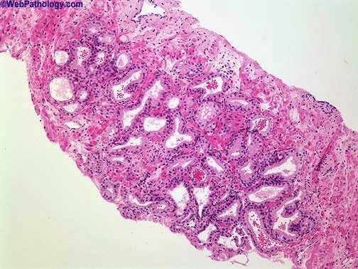
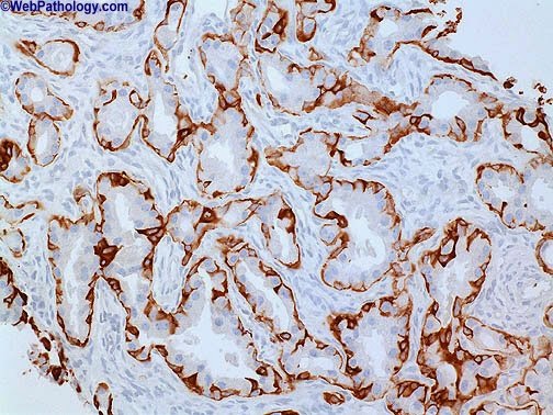
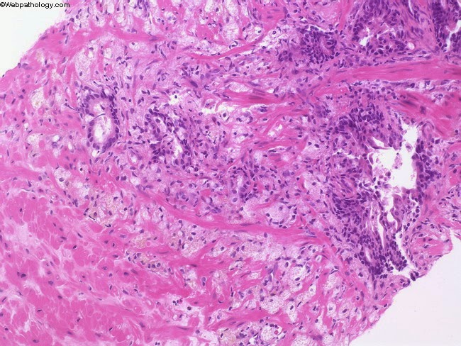
Mimics of Prostate Cancer
http://www.oncopathology.info.
Atrophy

- looks suspicious for adenocarcinoma at first glance.
- the nuclei are small and hyperchromatic.
- No prominent nucleoli are seen.
- Some glands are lined by obviously benign flattened atrophic epithelium.
- The immunostain for high molecular weight cytokeratin can be helpful in distinguishing between atrophy (fragmented basal cell layer) from atrophic variant of prostatic adenocarcinoma (no basal cell layer).
Atypical adenomatous hyperplasia
- It may show the infiltrative architecture of cancer,
- lacks the cytologic features such as prominent nucleoli.
- The immunostain for high mol. wt. Cytokeratin will show fragmented basal cell layer in most cases.
Post-Atrophic Hyperplasia
- Post-atrophic hyperplasia architecturally mimics adenocarcinoma
- lacks the cytologic features.
- In difficult cases, the immunostain for high mol. wt. cytokeratin can be performed which would show at least a few basal cells in post-atrophic hyperplasia.
Sclerosing Adenosis

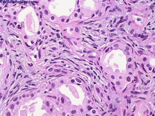
- small glands with infiltrative growth pattern in a cellular spindled stroma.
- The plump spindle cells in the stroma are nicely seen here.
- The lining acinar epithelial cells lack cytologic atypia – no significant nuclear or nucleolar enlargement is seen
- Myoepithelial differentiation in basal cells of the acini of Sclerosing adenosis is illustrated with the immunostain for muscle specific actin.
Cowper’s Glands


- They have a lobular configuration and are often associated with skeletal muscle fibers
- The glands are lined by goblet cells distended with mucin.
- The small hyperchromatic nuclei are pushed to the periphery.
- Sometimes ducts lined by cuboidal cells are present in the center of the lobules.
Mucinous Metaplasia

- Mucinous metaplasia is seen in about 1% of prostates.
- It may occasionally resemble prostatic adenocarcinoma. However, it lacks prominent nucleoli and the does not show immunoreactivity for PSA and PAP.
- The cells are positive for PAS, mucicarmine and Alcian blue.
Prostatic xanthoma

- Prostatic xanthoma is an uncommon benign lesion that may mimic high-grade prostatic adenocarcinoma.
- It consists of lipid-laden macrophages that may be arranged in small circumscribed nodules or infiltrating cords extending into the stroma
- diffusely positive for CD68 (shown here), and negative for CAM5.2, PSA, and PSAP.
Thanks to Dr.Dharam Ramani for the images.
i-Path Undergoes Corporate Rebrand to PathXL


i-Path Diagnostics will complete its global corporate rebrand to PathXL on 27th August 2011 following the PathXL launch in the USA earlier this summer.
PathXL is a global pioneer in the use of web-based solutions for digital pathology and provides innovative software for use in education, research and clinical sectors worldwide.
The rebrand reflects the company’s expansion on two fronts. Geographically PathXL has announced a number of strategic partnerships in recent months which strengthen its position to achieve rapid growth outside the UK and Europe - more announcements to follow. PathXL has also broadened the development and promotion of its workflow capabilities to create a comprehensive digital pathology platform that is web-based, open, and affordable. PathXL is committed to delivering digital workflow excellence.
The company will rebrand to PathXL at the European Congress of Pathologists in Helsinki on 27th August. If you are visiting the conference, please join us at our new PathXL stand 5C46.
Des Speed, CEO of PathXL commented:
“Our company is already recognised as being the leader in digital pathology education software, with an estimated 60% of UK trainee pathologists using PathXL’s innovative web-based platform each year. We recognise that digital pathology is moving from an era of point solutions to much greater maturity - transforming entire workflows in education, research and ultimately clinical use. We intend to become recognised as the visionaries and leaders in digital workflow excellence.”


Recent review article published on current state of whole slide imaging in pathology
A group of us recently summarized our thoughts from respective talks presented at the College of American Pathologists Companion Society meeting at this years' United States and Canadian Academy of Pathology.
I previously summarized who spoke at the actual meeting (see: Thoughts on CAP Companion Society Meeting at USCAP 2011).
The publication is avaialble free of charge from the Journal of Pathology Informatics (see links) or you can download the PDF here.
Review of the current state of whole slide imaging in pathology Liron Pantanowitz1, Paul N Valenstein2, Andrew J Evans3, Keith J Kaplan4, John D Pfeifer5, David C Wilbur6, Laura C Collins7, Terence J Colgan8
Correspondence Address: © 2011 Pantanowitz et al; This is an open-access article distributed under the terms of the Creative Commons Attribution License (http://creativecommons.org/licenses/by/2.0), which permits unrestricted use, distribution, and reproduction in any medium, provided the original work is properly cited. DOI: 10.4103/2153-3539.83746
Whole slide imaging (WSI), or "virtual" microscopy, involves the scanning (digitization) of glass slides to produce "digital slides". WSI has been advocated for diagnostic, educational and research purposes. When used for remote frozen section diagnosis, WSI requires a thorough implementation period coupled with trained support personnel. Adoption of WSI for rendering pathologic diagnoses on a routine basis has been shown to be successful in only a few "niche" applications. Wider adoption will most likely require full integration with the laboratory information system, continuous automated scanning, high-bandwidth connectivity, massive storage capacity, and more intuitive user interfaces. Nevertheless, WSI has been reported to enhance specific pathology practices, such as scanning slides received in consultation or of legal cases, of slides to be used for patient care conferences, for quality assurance purposes, to retain records of slides to be sent out or destroyed by ancillary testing, and for performing digital image analysis. In addition to technical issues, regulatory and validation requirements related to WSI have yet to be adequately addressed. Although limited validation studies have been published using WSI there are currently no standard guidelines for validating WSI for diagnostic use in the clinical laboratory. This review addresses the current status of WSI in pathology related to regulation and validation, the provision of remote and routine pathologic diagnoses, educational uses, implementation issues, and the cost-benefit analysis of adopting WSI in routine clinical practice. Keywords: Consultation, diagnosis, digital, education, frozen section, imaging, informatics, telepathology, validation, virtual microscopy, whole slide imaging
|


Learn the Latest About Telemedicine, Neuropathology, and Image Analysis of Biomarkers
Courtesy of the Digital Pathology Association.
I think this years' program will be among the best line up of speakers, topics (including update from the FDA on their thoughts), exhibitors, posters and networking opportunities this meeting has had. The talks and meeting come at an important time for digital pathology including new vendors in the space, regulatory issues and new business models, all of which will be discussed.
2011 PATHOLOGY VISIONS CONFERENCE
OCT. 30 – NOV. 2, San Diego, CA

A distinguished field of 41 speakers will highlight the hot topics in the world of digital pathology at the 2011 Pathology Visions Conference. Telemedicine, neuropathology, and the image analysis of biomarkers are just some of the subjects experts will discuss at the 7th annual Digital Pathology Association Conference.
Telemedicine and Neuropathology
In the clinical arena, Clayton Wiley, M.D., Ph.D., will discuss inter-institutional and interstate tele-neuropathology. Dr. Wiley will share his knowledge on telemedicine and its emersion as an efficient means of distributing professional medical expertise to geographically dispersed sites. He’ll also share his expertise on neuropathology and why it’s well-suited to utilize telepathology.
Image Analysis of Biomarkers
Tony Magliocco, M.D., will highlight the development of quantitative microscopy methods as prognostic and predictive cancer biomarkers. The digital pathology research expert will discuss three emerging analytical methods that hold promise for routine diagnostic pathology, including fractal analysis, analysis of DAB stained ki67 tissues with a focus on breast cancer, and the use of multiplex fluorescent imaging to study "functional tissue dynamics."
Guest Discount
The Pathology Visions Conference is also offering a special discounted rate for conference guests. For a reduced registration fee, your spouse or special guest can attend all meals, take part in the Sunday evening welcome reception and enjoy all that San Diego has to offer.
Poster Competition Extended Deadline Aug. 31
See contest details ?
Share your knowledge with others in a dynamic, interactive collegial environment and be rewarded! Submit your abstract for the poster competition at the Pathology Visions Conference. The deadline has been extended to Aug. 31.
"Access to sub-specialist pathologists using telepathology and image analysis of biomarkers are two of the strongest value propositions for why institutions adopt digital pathology," said Dirk Soenksen, President of the Digital Pathology Association and Chair of the Pathology Visions Program Committee. "Attendees at this year's conference will learn best practices for these and other applications of digital pathology, as well as how digital pathology can improve patient care and enable personalized medicine."
History Lesson
Did you know early pathologists started laying the groundwork for the digital pathology industry nearly a century and a half ago? Dr. Clive Taylor, M.D.,D. Phil., will give the audience a history lesson about the journey from microscopy to whole slide images. Dr. Taylor’s insight into how the past has shaped our industry will give attendees a firmer grasp on the important role they will play in the future of digital pathology.
These three speakers offer just a sneak peek at a line-up filled with distinguished presenters. The Pathology Visions Conference will host 38 additional research, education and clinical experts. Click here for a complete list of the speakers and details of their presentations.
Conference attendees will also have the opportunity to earn up to 13 CME credits, take part in an FDA panel discussion, and participate in cutting-edge industry workshops. DPA white paper and scientific poster sessions, roundtable discussions, networking events, and dozens of exhibitor displays will round out the three day conference.
| About the DPA The mission of the Digital Pathology Association is to facilitate education and awareness of digital pathology applications in health care. Members will be encouraged to share best practices and promote the use of the technology among colleagues in order to demonstrate efficiencies, awareness, and its ultimate benefits to patient care. |
|
|


OMNYX: Meet the Startup with Big Backing that is Digitizing Pathology
By Matt Pross, TEQ Staff Writer
Nice piece on Omnyx and their approach. Digital pathology is often cited as a $2 billion market as is mentioned a couple times in this story.
"Leave it to the big boys to go and carve out a $2 billion global market with the goal to redefine pathology worldwide. That’s exactly what GE Healthcare and the University of Pittsburgh Medical Center (UPMC) set out to do when they partnered to form Omnyx™. Focused on developing an entirely digital, integrated pathology solution, the joint venture has made significant progress toward this formidable goal since its founding in March 2008.

“The technology that was in use before OMNYX had a very niche focus and was used mainly in low volume/niche applications,” Tony Melanson, Vice President, Strategy & Marketing for OMNYX, said. “We saw the shortcomings of this technology and asked ourselves ‘what would it take to create a solution that would enable the high quality and high throughput necessary for pathology to evolve from an analog workflow into a digital practice?’ With its foundation in information technology systems for radiology, GE saw digitizing pathology as a natural progression and a great fit into the company’s existing portfolio.
We also knew pathology was uncharted territory and decided to solicit the help of Carnegie Mellon University and their leading Human Computer Interaction teams to help us design an efficient workflow and appealing user interface with the help of UPMC pathologists.""


