Decoding Human DNA

In a milestone for the understanding of human genetics, scientists just announced the results of five years of work in unraveling the secrets of how the genome operates.

The ENCODE project, as it is known, dispensed with the idea that our DNA is largely "junk," repeating sequences with no function, finding instead that at least 80 percent of the genome is important.

The new findings are the latest in a series of increasingly deep looks at the human genome. Here are some of the major milestones scientists have passed along the way.

1. An understanding of heredity, 1866

The realization that traits and certain diseases can be passed from parent to offspring stretches back at least to the ancient Greeks, well before any genome was actually decoded. The Greek physician Hippocrates theorized that "seeds" from different parts of the body were transmitted to newly conceived embryos, a theory known as pangenesis. Charles Darwin would later espouse similar ideas.

What exactly these "seeds" might be was destined to remain a mystery for centuries. But the first person to put heredity to the test was Gregor Mendel, who systematically tracked dominant and recessive traits in his famous pea plants. Mendel published his work on the statistics of genetic dominance in 1866 to little notice. [Genetics by the Numbers: 10 Tantalizing Tales]

2. Chromosomes come to light, 1902

But the painstaking work of cross-breeding pea plants wouldn't languish for long. In 1869, Swiss physician Johannes Friedrich Miescher became the first scientist to isolate nucleic acids, the active ingredient of DNA. Over the next several decades, scientists peering deeper into the cell discovered mitosis and meiosis, the two types of cell division, and chromosomes, the long strands of DNA and protein in cell nuclei.

In 1903, early geneticist Walter Sutton put two and two together, discovering through his work on grasshopper chromosomes that these mysterious filaments occur in pairs and separate during meiosis, providing a vehicle for mom and dad to pass on their genetic material.

"I may finally call attention to the probability that the associations of paternal and maternal chromosomes in pairs and their subsequent separation may constitute the physical basis of the Mendelian law of heredity," Sutton wrote in the journal The Biological Bulletin in 1902. He followed up with a more comprehensive paper, "The Chromosomes in Heredity" in 1903. (German biologist Theodor Boveri came to similar conclusions about chromosomes at the same time Sutton was working on his chromosome discovery.)

Originally posted here:
Decoding Human DNA

Posted in DNA

Biology in pictures: the science that could save the world

Another of the images, taken by Wong Chi Keung, shows fields of seaweed being grown in Fujian, china, where it is being harvested to produce biofuels.

Oil produced by the seaweed is collected and refined so it can be used in lawn mowers and cars.

A bumblebee collecting pollen from a wild flower, taken by William Richardson in Fulham, London, illustrates the threat posed to pollinators.

A graduate student overseeing the growth of algae in test tubes at the University of Tennessee in Knoxville, USA, also shows the striking green the blooms can create.

Taken by Mohammad Moniruzzaman, the project it illustrates is aimed at better understanding the organisms that make up these important ocean blooms, which are major contributors to the world's oxygen supply.

The winners are due to be announced in October at Charles Darwin House in London. The entire shortlist alongside those from the young photographers category will be put on display as part of Biology Week.

Read the original post:
Biology in pictures: the science that could save the world

Undergraduate Research Assistants

Dr. Raison (FSHD & College of Medicine, Psychiatry) is looking for Undergraduate Research Assistants to help on a research study that will examine the effect of exposure to infrared light on major depressive disorder (MDD). As an Undergraduate Research Assistant you will: - Gain invaluable research experience, which is important for those of you interested in applying to Graduate School - Be able to register for up to 3 units of Directed Research under Dr. Raison What is required from you: -Up to 10 hours per week (flexible) -1 year commitment (Fall 2012 and Spring 2013) *Undergraduates who have had a course in research methods are preferred. IF INTERESTED: please email Elizabeth Gleason (egleason@psychiartry.arizona.edu), Project Coordinator, with a current resume no later than Monday, September 10th. Please be sure to include your Undergraduate G.P.A. on your resume. In the email please: (1) describe any relevant research experience you may have and (2) why you are interested in joining the lab and gaining research experience.

Source:
http://physiologynews.blogspot.com/feeds/posts/default?alt=rss

Baylor’s Cancer Genetics Laboratory at CAP ’12

CGL Logo

The Cancer Genetics Laboratory (CGL) is a combined effort of multiple departments at Baylor College of Medicine. The CGL provides cancer genetic testing along with expert interpretations of the test results. Our extensive menu ranges from karyotyping and FISH analysis to next-generation sequencing and cancer chromosomal microarrays.

Please
look for Baylor College of Medicine’s Cancer Genetics Laboratory (CGL) at the
upcoming College of American Pathologists Annual Meeting in San Diego
(September 9-12 Booth #616).  CGL invites
all to visit the booth and learn more about their new services.  Members of the CGL clinical team will be
available to discuss CGL’s options for clinical pathology testing including:

  • Oncology
    Microarrays
  • Cancer
    Sequencing Panels Using Next-Generation Technology
  • Cancer
    Exome Sequencing

Dr.
Federico Monzon, CGL’s Director of Molecular Pathology will be available to
discuss current & future offerings.

To
schedule a meeting with Dr. Monzon please email Scott Johnson at scottj@bcm.edu.  

Search for tests by name
# A B C D E F G H I J K L M N O P Q R S T U V W X Y Z
Quick Links


Source:
http://feeds.feedburner.com/DigitalPathologyBlog

Translational Imaging Impacting Personalized Health. From Cell, To Whole Animal, To Tissue

The World Molecular Imaging Congress (WMIC) 2012 Annual Meeting in being held in Dublin, Ireland this week (September 5 - 8, 2012).


PerkinElmerLearn more about pathway characterization, therapeutic effect and treatment at the cellular, whole body and tissue level through PerkinElmer's translational imaging solutions. Our broad offering of imaging and analysis solutions enables you to see and understand more in every area of research — from cellular imaging, to in vivo imaging, to pathology. With intuitive, high-performance software, broad portfolio of in vivo imaging reagents and leading imaging systems, effectively translate your research from the bench top and beyond. Visit PerkinElmer at booth #404 at WMIC to experience our translational imaging technologies first-hand.

Hear from Kevin Hrusovsky, SVP & President, PerkinElmer on Translational Imaging Impacting Personalized Health-An Academic and Industry Partnership, September 7, 2012, 6:15 PM - 7:45 PM at WMIC.

Join the WMIC Gala Event on September 8th at the Guinness Storehouse, with music by PerkinElmer's own Molecular Groove. Return to the 80's with us including special guest appearances.

Register for the Gala Event

LEARN MORE AT IN VIVO UNIVERSITY

Visit http://www.perkinelmer.com/tissueimaging for PerkinElmer’s Digital Pathology tools.

Source:
http://feeds.feedburner.com/DigitalPathologyBlog

Journal of Pathology Informatics – New Articles Published


Cover Cover Cover
Utilization and utility of clinical laboratory reports with graphical elements

Brian H Shirts, Nichole Larsen, Brian R Jackson
J Pathol Inform 2012, 3:26 (25 August 2012)
[ABSTRACT]   [HTML FULL TEXT]   [PDF]   [Mobile HTML Full text ]   [EPub]

Diagnosis of dysplasia in upper gastro-intestinal tract biopsies through digital microscopy
Dorina Gui, Galen Cortina, Bita Naini, Steve Hart, Garrett Gerney, David Dawson, Sarah Dry
J Pathol Inform 2012, 3:27 (25 August 2012)
[ABSTRACT]   [HTML FULL TEXT]   [PDF]   [Mobile HTML Full text ]   [EPub]

Dynamic nonrobotic telemicroscopy via skype: A cost effective solution to teleconsultation
Sahussapont J Sirintrapun, Adela Cimic
J Pathol Inform 2012, 3:28 (25 August 2012)
[ABSTRACT]   [HTML FULL TEXT]   [PDF]   [Mobile HTML Full text ]   [EPub]

Experience with CellaVision DM96 for peripheral blood differentials in a large multi-center academic hospital system
Marian A Rollins-Raval, Jay S Raval, Lydia Contis
J Pathol Inform 2012, 3:29 (25 August 2012)
[ABSTRACT]   [HTML FULL TEXT]   [PDF]   [Mobile HTML Full text ]   [EPub]

Different tracks for pathology informatics fellowship training: Experiences of and input from trainees in a large multisite fellowship program
Bruce P Levy, David S McClintock, Roy E Lee, William J Lane, Veronica E Klepeis, Jason M Baron, Maristela L Onozato, JiYeon Kim, Victor Brodsky, Bruce Beckwith, Frank Kuo, John R Gilbertson
J Pathol Inform 2012, 3:30 (30 August 2012)
[ABSTRACT]   [HTML FULL TEXT]   [PDF]   [Mobile HTML Full text ]   [EPub]

A core curriculum for clinical fellowship training in pathology informatics
David S McClintock, Bruce P Levy, William J Lane, Roy E Lee, Jason M Baron, Veronica E Klepeis, Maristela L Onozato, JiYeon Kim, Anand S Dighe, Bruce A Beckwith, Frank Kuo, Stephen Black-Schaffer, John R Gilbertson
J Pathol Inform 2012, 3:31 (30 August 2012)
[ABSTRACT]   [HTML FULL TEXT]   [PDF]   [Mobile HTML Full text ]   [EPub]

Use of a wiki as an interactive teaching tool in pathology residency education: Experience with a genomics, research, and informatics inpathology course
Seung Park, Anil Parwani, Trevor MacPherson, Liron Pantanowitz
J Pathol Inform 2012, 3:32 (30 August 2012)
[ABSTRACT]   [HTML FULL TEXT]   [PDF]   [Mobile HTML Full text ]   [EPub]


Jpi_blogJpi_blogJpi_blog

Source:
http://feeds.feedburner.com/DigitalPathologyBlog

PerkinElmer and Massachusetts General Hospital Pathology Collaborate to Develop a Complete Sequencing Informatics System for Cancer Research

Global leader in life sciences to help leading research hospital advance innovation in cancer research

WALTHAM, Mass., Aug 28, 2012 (BUSINESS WIRE) -- PerkinElmer, Inc., a global leader focused on improving the health and safety of people and the environment, today announced that it has entered into a collaboration with Dr. John Iafrate and Dr. Long Le of the Massachusetts General Hospital (MGH) Pathology Department and Cancer Center to develop a sequencing informatics system for profiling the genetic changes in tumors for use in advanced cancer research.

Based on PerkinElmer's Geospiza(R) informatics platforms, the new system is expected to help guide the development of cancer treatments by enabling genotype analysis to define key genetic targets from which clinically relevant information can be leveraged. Under the terms of the agreement, PerkinElmer will develop a highly automated sample preparation and data analysis system suitable for cancer genotyping in clinical research at the MGH Pathology Department and Cancer Center. PerkinElmer's Geospiza GeneSifter(R) Laboratory and Analysis edition software platform will be enhanced with sophisticated variant detection algorithms. The new system will also integrate existing laboratory processes and equipment with enhanced LIMS and data analysis capabilities.

The informatics system will support the cancer research of Dr. Long Le and Dr. John Iafrate of the MGH Pathology Department and Cancer Center. "A key challenge presented by next generation sequencing applications in clinical research is not only maintaining high efficiency and throughput via automation solutions, but also ensuring that the informatics driving the instrumentation provides transparency and traceability for the entire process," said Dr. Le.

Kevin Hrusovsky, president of Life Sciences & Technology at PerkinElmer, said, "Working with the MGH Pathology Department and Cancer Center to support their critically important cancer research is a deeply rewarding collaboration and is consistent with our goal to help researchers eradicate this deadly disease. We are delighted to provide MGH with a complete next generation sequencing informatics and sample preparation solution to help advance their medical research."

The MGH Pathology Department and Cancer Center are currently using the high quality service and data from PerkinElmer's DNA Sequencing Services group and deploys PerkinElmer's Sciclone(R) automated liquid handling platform and the NGS Express(TM) Workstation for benchtop sequencers.

For more information on PerkinElmer's Geospiza informatics offering, please visit http://www.geospiza.com

About PerkinElmer, Inc.

PerkinElmer, Inc. is a global leader focused on improving the health and safety of people and the environment. The Company reported revenue of approximately $1.9 billion in 2011, has about 7,000 employees serving customers in more than 150 countries, and is a component of the S&P 500 Index. Additional information is available through 1-877-PKI-NYSE, or at http://www.perkinelmer.com .

SOURCE: PerkinElmer, Inc.

Source:
http://feeds.feedburner.com/DigitalPathologyBlog

More technology, better learning resources, better learning? Lessons from adopting virtual microscopy in undergraduate medical education.


Institution: Center for Learning Research, Department of Teacher Education, Faculty of Education, University of Turku, Turku, Finland. lhelle@utu.fi.

Abstract

The adoption of virtual microscopy at the University of Turku, Finland, created a unique real-world laboratory for exploring ways of reforming the learning environment. The purpose of this study was to evaluate the students' reactions and the impact of a set of measures designed to boost an experimental group's understanding of abnormal histology through an emphasis on knowledge of normal cells and tissues. The set of measures included (1) digital resources to review normal structures and an entrance examination for enforcement, (2) digital course slides highlighting normal and abnormal tissues, and (3) self-diagnostic quizzes. The performance of historical controls was used as a baseline, as previous students had never been exposed to the above-mentioned measures. The students' understanding of normal histology was assessed in the beginning of the module to determine the impact of the first set of measures, whereas that of abnormal histology was assessed at the end of the module to determine the impact of the whole set of measures. The students' reactions to the instructional measures were assessed by course evaluation data. Additionally, four students were interviewed. Results confirmed that the experimental group significantly outperformed the historical controls in understanding normal histology. The students held favorable opinions on the idea of emphasizing normal structures. However, with regards to abnormal histology, the historical controls outperformed the experimental group. In conclusion, allowing students access to high-quality digitized materials and boosting prerequisite skills are clearly not sufficient to boost final competence. Instead, the solution may lie in making students externally accountable for their learning throughout their training. Anat Sci Educ. © 2012 American Association of Anatomists.

Copyright © 2012 American Association of Anatomists.

Anat Sci Educ. 2012 Aug 28. doi: 10.1002/ase.1302. [Epub ahead of print]

Logo_en

Source:
http://feeds.feedburner.com/DigitalPathologyBlog

Morbid Anatomy Library Open Hours This Weekend, September 8th and 9th, 11-7, As Part of the Brooklyn Museum's "Go" Open Studio Project

This weekend--Saturday September 8th and Sunday September 9th--the Morbid Anatomy Library (seen above) will be open from 11-7 as part of the Brooklyn Museum's Go Open Studio Project. So please stop by for a perusal of the stacks, a turn through the drawers, and a conversation with the lovely and very clever Morbid Anatomy Library interns Kelsey Kephart and Dru Munsell.
The Morbid Anatomy Library is located at 543 Union Street at Nevins, Brooklyn, buzzer 1E. To view a map, click here. To For more about the Morbid Anatomy Library and for directions and other such information, click here. For more about the Go Open Studio Project--and to see a full list of participating artists--click here.
Photo of The Library by Shannon Taggart

Source:
http://morbidanatomy.blogspot.com/feeds/posts/default?alt=rss

Congress for Curious Peoples One-Day Symposium: London Edition, Last Tuesday Society, This Saturday, September 8

 This Saturday, September 8, you are cordially invited to join myself and a host of distinguished scholars, makers, and museum folk as we investigate, via a one day symposium termed "The Congress for Curious Peoples," some of the provocative intersections explored in the exhibition "Ecstatic Raptures and Immaculate Corpses: Visions of Death Made Beautiful in Italy," on view at the London-based Last Tuesday Society until the end of the month.

This first ever UK edition of The Congress for Curious Peoples will feature participants from The Wellcome Collection, The Wellcome Library, and The Gordon Museum of Pathology, as well as some of my very favorite artists, thinkers and scholars, and will take on such heady topics as enchantment and enlightenment, or the sublimation of the magical into the rational world; the secret life of objects, or the non-rational allure of objects and the psychology of collecting; and beautiful death and incorruptible bodies, or the shared drive to immortalize the human body and aestheticize death in both medicine and Catholicism, and will

Full info follows; hope very very much to see you there!

Congress for Curious Peoples: London Edition
Date: Saturday September 8

Time: 11am - 5:30 pm
Admission: £15.00 (Tickets here)
Location: The Last Tuesday Society
Address: 11 Mare Street, London, E8 4RP

Produced by Morbid Anatomy

11-12: Introduction by Morbid Anatomy's Joanna EbensteinKeynote panel: Enchantment and Enlightenment (20 minute presentations followed by moderated discussion)

12-1: Lunch

1-2:30 The Secret Life of Objects: The Allure of Objects and the Psychology of Collecting (20 minute presentations followed by moderated discussion)

2:30-3:00 Break

3:00-5:30 Beautiful Death and Incorruptible Bodies: Eternal Life and Aestheticized Death in Medicine and Catholicism (15 minute presentations followed by moderated discussion)

You can find out more by clicking here, and purchase tickets by clicking here.

Source:
http://morbidanatomy.blogspot.com/feeds/posts/default?alt=rss

"Ecstatic Raptures and Immaculate Corpses: Visions of Death Made Beautiful in Italy" Exhibition Opening Party, This Thursday, September 6


This Thursday, if you are in London or environs, please join Morbid Anatomy and The Last Tuesday Society for a free and gin-drenched opening party for my new exhibition "Ecstatic Raptures and Immaculate Corpses: Visions of Death Made Beautiful in Italy"! Full details follow. Hope very much to see you there!

"Ecstatic Raptures and Immaculate Corpses: Visions of Death Made Beautiful in Italy" Exhibition Opening Party
Date: Thursday September 6
Time: 6:00-8:00 PM
Location: The Last Tuesday Society
Address: ***Offsite at 11 Mare Street, London, E8 4RP

Admission: FREE
Produced by Morbid Anatomy
Click here to download Invitation

This Thursday, September 6, if you find yourself in London town, please join us for an opening party for an exhibition of photographs by Joanna Ebenstein of the Morbid Anatomy Blog, The Morbid Anatomy Library and Observatory with waxworks by Eleanor Crook and Sigrid Sarda.

In her many projects, ranging from photography to curation to writing, New York based Joanna Ebenstein utilizes a combination of art and scholarship to tease out the ways in which the pre-rational roots of modernity are sublimated into ostensibly "purely rational" cultural activities such as science and medicine.Much of her work uses this approach to investigate historical moments or artifacts where art and science, death and beauty, spectacle and edification, faith and empiricism meet in ways that trouble contemporary categorical expectations.In the exhibition "Ecstatic Raptures and Immaculate Corpses" Ebenstein turns this approach to an examination of the uncanny and powerfully resonant representations of the dead, martyred, and anatomized body in Italy, monuments to humankind's quest to eternally preserve the corporeal body and defeat death in arenas sacred and profane.The artifacts she finds in both the churches, charnel houeses and anatomical museums of Italy complicate our ideas of the proper roles of--and divisions between--science and religion, death and beauty; art and science; eros and thanatos; sacred and profane; body and soul.

In this exhibition, you will be introduced to tantalizing visions of death made beautiful, uncanny monuments to the human dream of life eternal. You will meet "Blessed Ismelda Lambertini," an adolescent who fell into a fatal swoon of overwhelming joy at the moment of her first communion with Jesus Christ, now commemorated in a chillingly beautiful wax effigy in a Bolognese church; The Slashed Beauty, swooning with a grace at once spiritual and worldly as she makes a solemn offering of her immaculate viscera; Saint Vittoria, with slashed neck and golden ringlets, her waxen form reliquary to her own powerful bones; and the magnificent and troubling Anatomical Venuses, rapturously ecstatic life-sized wax women reclining voluptuously on silk and velvet cushions, asleep in their crystal coffins, awaiting animation by inquisitive hands eager to dissect them into their dozens of demountable, exactingly anatomically correct, wax parts.

You can find out more about the show here, and view more images by clicking here.

Source:
http://morbidanatomy.blogspot.com/feeds/posts/default?alt=rss

Two Conferences on Death, Art and Culture: Calls for Papers

I have just been alerted to two fabulous looking death and culture conferences both of which are now soliciting papers! Full info for each follows. Apply away!

1) Art and  Death: A Series of Three Workshops
1 November 2012, 21 February and 23 May 2013

The Courtauld Institute of Art, Somerset House, Strand, London WC2R 0RN

Call for Papers
Submission by 20 September 2012 for workshop 1 (1 November 2012): Anticipation and Preparation

A series of three workshops will be held at the Courtauld Institute of Art in 2012-2013 to explore the inter-relationship between art and death. These workshops have arisen from an informal group of doctoral students with shared interests in funerary monuments. The workshops will be structured to recognize that the certainty of death is accompanied by the foreknowledge and uncertainty of what may come after, and that visual representations of these phases have varied over time and between countries. The first workshop will focus on the images and objects related to the impact that the certainty of death has on individuals and the community; the second on art in the context of dying, death and burial; and the final one on representations of the perceived fate of body and soul after death, as well as the continuation of a relationship (if only in memory) between the living and the dead.

Subjects for the workshops could include, but are not limited to:

Workshop 1 (1 November 2012): Anticipation and Preparation
•    Death insurance? Religious gifts and foundations
•    Protective objects and amulets
•    Tombs commissioned during a lifetime, testamentary desire and fulfilment
•    Contemplating images of death, warnings to the living
•    The cult of the macabre, images of illness and decay
•    Apocalyptic visions

Workshop 2: (21 February 2013): Death and Dying
•    A ‘good death’
•    War and violence
•    Funerals/Professional mourners
•    Funerary monuments, memorial architecture, cemetery design
•    Post-mortem portraits
•    Images of the corpse in painting, sculpture, film, photography, etc
•    Crucifixion imagery
•    Death in museum collections

Workshop 3 (23 May 2013) Life after Death
•    Images of the soul /resurrected or re-incarnated body
•    Depictions of the afterlife
•    The incorruptible body, saints, relics and reliquaries
•    Remembering the dead, commemoration in art and/or performance
•    The ‘immortality’ of the artist, post-mortem reputations

Format and Logistics:
•    Length of paper: 20 minutes
•    Four papers per workshop
•    Location: Research Forum, The Courtauld Institute of Art
•    Timing: 10am-midday
•    Expenses: funds are not available to cover participants’ expenses

We welcome proposals relating to all periods, media and regions (including non-European) and see this as an opportunity for doctoral and early post-doctoral students to share their research.

Please send proposals of no more than 250 words to: Jessica.Barker[at]courtauld.ac.uk and Ann.Adams[at]courtauld.ac.uk by the following dates:

•    20 September 2012 for workshop 1 (1 November 2012): Anticipation and Preparation
•    10 January 2013 for workshop 2 (21 February 2013): Death and Dying
•    11 April 2013 for workshop 3 (23 May 2013): Life after Death

For planning purposes, it would be helpful to have an indication of interest in the later workshops, in advance of submission of a proposal.

Organised by Jessica Barker and Ann Adams (The Courtauld Institute of Art)

2) Graduate Student Conference: “Death: the Cultural Meaning of the End of Life”
January 24–25, 2013
LUCAS (Leiden University Centre for Arts in Society)

This conference aims to explore how death has been represented and conceptualized, from classical antiquity to the modern age, and the extent to which our perceptions and understandings of death have changed (or remained the same) over time. The wide scope of this theme reflects the historical range of LUCAS’s (previously called LUICD) three research programs (Classics and Classical Civilization, Medieval and Early Modern Studies and Modern and Contemporary Studies), as well as the intercontinental and interdisciplinary focus of many of the institute’s research projects.

The LUCAS Graduate Conference welcomes papers from all disciplines within the humanities. The topic of your proposal may address the concept of death from a cultural, historical, classical, artistic, literary, cinematic, political, economic, or social viewpoint.

Questions that might be raised include: How have different cultures imagined the end of life? What is the role of art (literature, or cinema) in cultural conceptions of death? How might historical or contemporary conceptualizations of death be related to the construction of our subjectivity and cultural identity? What is the cultural meaning(s) of death? To what extent has modern warfare changed our perceptions of death? How is death presented in the media and how has this changed? In what ways has religion influenced our reflections on death and the afterlife?

Please send your proposal (max. 300 words) to present a 20-minute paper to lucasconference2013[at]gmail.com.

The deadline for submissions is November 15, 2012.

For further information on the first workshop, click here. For further information on the second conference, click here. Special thanks to Lisa Kereszi for turning me onto the latter!

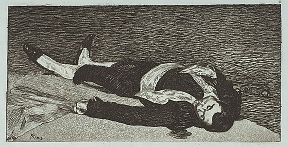
Image: Dead Toreador (Torero Mort). Édouard Manet (French, Paris 1832–1883 Paris)

Source:
http://morbidanatomy.blogspot.com/feeds/posts/default?alt=rss

Lilly Diabetes Launches Mobile Application for Those Who Support People with Type 1 Diabetes

App serves as a teaching tool and can provide guidance for treatment with Lilly Glucagon for Injection (rDNA origin) during severe hypoglycemic events


Lilly_Glucagon_mobile_app_screenINDIANAPOLIS – September 4, 2012 – Lilly Diabetes today announced the release of a new mobile application designed for caregivers and healthcare providers who support people with type 1 diabetes. The Lilly Glucagon Mobile App is a tool to teach how to use Glucagon for Injection, through simulated practice. Glucagon, 1 mg (1 unit), is indicated to treat severe hypoglycemia (low blood sugar). Severe hypoglycemia due to insulin may result in loss of consciousness (insulin coma). The app is designed to help people be more prepared, and also  provide an opportunity to store locations and expiration dates of their Lilly Glucagon Emergency Kits. The Lilly Glucagon Mobile App is now available on the iTunes® store as a free download for iPhone® or iPad® mobile devices.

People with type 1 diabetes who experience severe hypoglycemia during insulin treatment may require glucagon, a hormone produced in the pancreas to raise blood sugar levels. Glucagon. U.S. Library of National Medicine. NIH. Updated September 2010. http://www.ncbi.nlm.nih.gov/pubmedhealth/PMH0000691/.  Although Glucagon is rarely needed and only used during a severe hypoglycemic event, individuals in the person’s support network, such as family members, teachers, coaches, trusted friends and colleagues, should be trained to give the medicine, which is injected with a syringe. The app can also be used by diabetes educators and school nurses as a teaching tool.


GlucagonappIn type 1 diabetes, the body does not produce insulin, a hormone produced by the pancreas in response to an increase in blood sugar, such as after a meal. As many as three million Americans may have type 1 diabetes. Each year more than 15,000 people under age 20 are diagnosed with the disease. Glucagon should not be used in patients who have pheochromocytoma or patients who are allergic to Glucagon.  Patients must inform relatives or close friends that if they become unconscious, medical assistance must always be sought. If a patient is unconscious, Glucagon can be given while awaiting medical assistance.

“Lilly Diabetes is committed to developing personalized solutions to help people with diabetes achieve their treatment goals and improve their outcomes,” said Matt Caffrey, U.S. Product Brand Director, Marketing Specialty, Lilly Diabetes. “The Lilly Glucagon Mobile App leverages the power and reach of mobile technology, providing another opportunity to support people living with type 1 diabetes. Lilly Diabetes is constantly striving to create new and better tools to support the diabetes community in a variety of ways.” 

The Lilly Glucagon Mobile App is an interactive tool to help caregivers better understand Glucagon’s role in diabetes management. Its purpose is to educate and prepare the caregiver on how to use Glucagon in the event of an emergency. The app was developed with input from healthcare providers and people with diabetes.  

The Lilly Glucagon Mobile App includes:
·        Information about severe hypoglycemia and Glucagon 
·        Simulated practice demonstrating how to prepare and inject Glucagon
·        Visual and audio emergency instructions
·        Tools to keep track of kit locations and alerts for expiration dates 
·        Important safety information

Important Safety Information for Glucagon 
What is the most important information about Glucagon?
·        Glucagon should not be used in patients with pheochromocytoma or who may be allergic to glucagon.
·        Patients need to tell their healthcare provider if they have been diagnosed with or have been suspected of having an insulinoma as glucagon should be used cautiously in this situation.
·        Anyone who may need to help patients during an emergency should become familiar with how to use glucagon before an emergency arises. Read the Information for the User provided in the kit.
·        Patients need to make sure that relatives or close friends know that if they become unconscious, medical assistance must always be sought. If a patient is unconscious, glucagon can be given while awaiting medical assistance.
·        The kit must not be used after the date stamped on the bottle label.
·        Questions concerning the use of this product should be directed to a doctor, nurse or pharmacist.

WARNING: PATIENTS MAY BE IN A COMA FROM SEVERE HYPERGLYCEMIA (HIGH BLOOD GLUCOSE) RATHER THAN HYPOGLYCEMIA. IN SUCH A CASE, THE PATIENTS WILL NOT RESPOND TO GLUCAGON AND WILL REQUIRE IMMEDIATE MEDICAL ATTENTION.

Who should not use glucagon?
Glucagon should not be used in patients who have pheochromocytoma or who are allergic to glucagon.

What should patients tell their doctor before taking glucagon?
Patients should tell their doctor about all medical conditions and prescription and over-the-counter drugs. Patients should tell their doctor if they have been diagnosed with or have been suspected of having pheochromocytoma or an insulinoma.

How should glucagon be used?
·        It is important to act quickly. Prolonged unconsciousness may be harmful.
·        Family and friends need to know to turn the patient on their side to prevent choking if they are unconscious.
·        The contents of the syringe are inactive and must be mixed with the glucagon in the accompanying bottle immediately before giving injection. Glucagon for Injection must not be prepared until it is ready to be used.
·        Glucagon should not be used unless the solution is clear and of a water-like consistency.
·        The usual adult dose is 1 mg (1 unit). For children weighing less than 44 lbs (20 kg), 1/2 adult dose (0.5 mg) is used. For children, 1/2 of the solution from the bottle (0.5 mg mark on syringe) should be withdrawn. The unused portion should be discarded.
·        Patients should eat as soon as they awaken and are able to swallow. A doctor or emergency services must be informed immediately.

What is some important Information about Low Blood Sugar (Hypoglycemia)?
·        Early symptoms of low blood sugar include: sweating, drowsiness, dizziness, sleep disturbances, palpitation, anxiety, tremor, blurred vision, hunger, slurred speech, restlessness, depressed mood, tingling in the hands, feet, lips, or tongue, irritability, lightheadedness, abnormal behavior, inability to concentrate, unsteady movement, headache, and personality changes. These symptoms may be different for each person and can happen suddenly.
·        If low blood sugar is not treated, it may progress to severe low blood sugar that can include: disorientation, seizures, unconsciousness, and death
·        Low blood sugar symptoms should be treated with a quick source of sugar which should always be carried with the patient. If symptoms do not improve or if the patient is unable to take a quick source of sugar, they should be treated with glucagon or with intravenous glucose at a medical facility.

What are the possible side effects of glucagon? 
·        Severe side effects are very rare, although nausea and vomiting may occur occasionally.
·        A few people may be allergic to glucagon or to one of the inactive ingredients in glucagon, or may experience rapid heart beat for a short while.
·        Patients who experience any other reactions which are likely to have been caused by glucagon should contact their doctor.

Patients and caregivers are encouraged to report negative side effects of Prescription drugs to the FDA. Visit http://www.fda.gov/medwatchor call 1-800-FDA-1088.

How should glucagon be stored?
·        Before dissolving glucagon with diluting solution, the kit should be stored at controlled room temperature between 20° to 25°C (68° to 77°F).
·        After glucagon is dissolved with diluting solution, it should be used immediately. Any unused portion should be discarded. Glucagon should be clear and of a water-like consistency at time of use.

For more safety information, please access Information for the User and Information for the Physician.

HI GLUC PR ISI [17JUL12]

The glucagon design is a trademark of Eli Lilly and Company. Glucagon is available by prescription only.

About Lilly Diabetes
Lilly has been a global leader in diabetes care since 1923, when we introduced the world's first commercial insulin. Today we work to meet the diverse needs of people with diabetes through research and collaboration, a broad and growing product portfolio and a continued commitment to providing real solutions—from medicines to support programs and more—to make lives better. For more information, visit http://www.lillydiabetes.com

About Eli Lilly and Company 
Lilly, a leading innovation-driven corporation, is developing a growing portfolio of pharmaceutical products by applying the latest research from its own worldwide laboratories and from collaborations with eminent scientific organizations. Headquartered in Indianapolis, IN, Lilly provides answers — through medicines and information — for some of the world's most urgent medical needs. Additional information about Lilly is available at http://www.lilly.com


Source:
http://feeds.feedburner.com/DigitalPathologyBlog

NovoPath Showcases Smartphone App at CAP ‘12


PRINCETON, NJ, September 4, 2012 – NovoPath, Inc., a leader in Anatomic Pathology (AP) Best-of-Breed Software Solutions, announces the first smartphone apps for Android and Apple users, enabling secure on-the-go access to full-length AP reports.  The implementation of NovoNotifier Mobile means clinicians receiving AP reports from NovoPath partner laboratories now have a unique tool at their disposal, assisting in their quest to provide the very best care to their patients.  NovoNotifier Mobile will be featured on the NovoPath booth (#602) at the CAP ’12 Meeting, September 9-12, in San Diego.
 
According to a recent report from iPharma Connect 2012, over 80% of physicians are currently using smartphones, with adoption increasing at a tremendous rate.  Via NovoNotifier Mobile, Anatomic Pathology reports are now available in a continuous real-time portable format to many of these doctors.  NovoNotifier Mobile places results directly into the hands of the busy clinician, regardless of their location – be it in a point-of-care setting, at home, or anywhere in between.  This immediate access to lab generated anatomic pathology test results, enabled by mobile AP reporting, empowers the clinician to respond quicker – a significant improvement for the patient anxiously awaiting their diagnostic or prognostic outcome.
 
To access NovoNotifier Mobile, the clinician (working with a pathology lab using the NovoPath AP LIS Software Solution Version 8.0) is provided secure discreet user ID-based access to their patient’s anatomic pathology reports.  Following initial setup, AP reports are automatically sent via unattended delivery, with a “signal” sent to their smartphone notifying the physician that new reports are awaiting their review, at their convenience.  After viewing the report, the clinician has the option to preserve or delete the file – knowing it will be maintained on the lab’s website.  This affords clinicians the flexibility provided by immediate access, without tying up all the memory in their handheld.
 
Wally Soufi, NovoPath CEO, commented, “Mobile access means our partner lab’s clinicians no longer need to schedule time in their busy day to view web-based patient reports.  Via NovoNotifier Mobile, these same clinicians can now view AP reports in their entirety – in the same customized format developed by their pathology laboratory – anytime, anyplace.  The mobile view is 100% identical to the reports they have come to rely upon on their desktop. NovoPath is pleased to be the first to provide instant access to portable AP reports, and believes that this decrease in turn-around-time will translate to even better patient outcomes.”
About NovoPath, Inc.
 
NovoPath, Inc. develops and markets software solutions for the Anatomic Pathology Laboratory market segment that includes local, regional, national, in-house laboratories as well as community and university teaching hospitals and medical centers. Since the release of its flagship product in 1999, NovoPath, Inc. has focused exclusively on Anatomic Pathology. NovoPath's mission is to provide unique and unparalleled solutions and services to all aspects of the Anatomic Pathology sector in a way that improves workflow, reduces the probability of human error, ensures results accuracy for greater patient safety, protects patient confidentiality, and above all, produces more precise and informative diagnostic outcomes. More information is available at http://www.NovoPath.com.

Source:
http://feeds.feedburner.com/DigitalPathologyBlog

Anthropomorphic Mouse Taxidermy Class with Sue Jeiven, London, Last Tuesday Society, September 29-30, 1-5

I am so very very excited to announce that Sue Jeiven's is bringing her wonderful and ubiquitously sold out Observatory "Anthropomorphic Mouse Taxidermy Class" to the London as part of my month-long Morbid Anatomy Presents lineup at The Last Tuesday Society.
There will be two iterations of the class, one on Saturday the 29th and one on Sunday the 30th of September. No former taxidermy experience is required, and you need bring nothing; you will leave with your own taxidermied mouse set in a tableau, and the skills to create your own in the future; past student projects can be seen by clicking here. It must also be mentioned that Sue is a passionate and amazing teacher, and we have had nothing but excellent feedback about her class.
Class size is limited to 15, and, at least in Brooklyn, this class tends to sell out very quickly, so if interested, I suggest you purchase tickets straight away. 
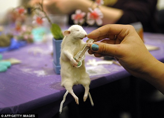
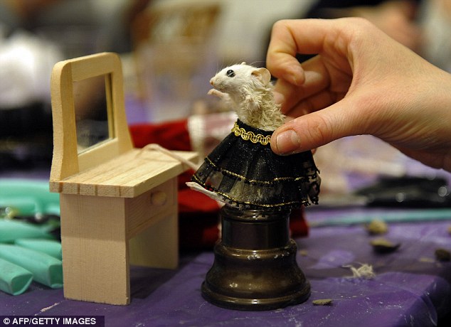
For the Brits among you, you might want to check out this writeup about the Brooklyn iteration of the class in--yes, you guessed it--The Daily Mail, from which the classroom photos above were drawn. You can also watch a brief featurette on Sue and her work in the episode of The Midnight Archive above.
Full details for the class follow; you can purchase tickets by clicking here. Hope very much to see you there!

Anthropomorphic Mouse Taxidermy Class  with Susan Jeiven
Dates: Saturday the 29th September 2012 and Sunday the 30th September 2012 
Cost: £60.00
Time: 1-5
Location: Last Tuesday Society, 11 Mare Street London E8 4RP

Anthropomorphic taxidermy–the practice of mounting and displaying taxidermied animals as if they were humans or engaged in human activities–was a popular art form during the Victorian and Edwardian eras. The best known practitioner of the art form is British taxidermist Walter Potter who displayed his pieces–which included such elaborate tableaux as The Death of Cock Robin, The Kitten Wedding, and The Kitten Tea Party–in his own museum of curiosities.

We invite you to join taxidermist, tattoo artist and educator Susan Jeiven for a beginners class in anthropomorphic taxidermy. All materials–including a mouse for each student–will be provided, and each class member will leave at the end of the day with their own anthropomorphic taxidermied mouse. Students are invited to bring any miniature items with which they might like to dress or decorate their new friend; some props and miniature clothing will also be provided by the teacher. A wide variety of sizes and colors of mice will be available.

No former taxidermy experience is required.

Also, some technical notes:

  • We use NO harsh or dangerous chemicals.
  • Everyone will be provided with gloves.
  • All animals are disease free.
  • Although there will not be a lot of blood or gore, a strong constitution is necessary; taxidermy is not for everyone.
  • All animals were already dead, nothing was killed for this class. All mice used are feeder animals for snakes and lizards and would literally be discarded if not sold.
  • Please do not bring any dead animals with you to the class

Source:
http://morbidanatomy.blogspot.com/feeds/posts/default?alt=rss

DNA leads to arrest in 1980 murder of Oxnard girl

More than three decades ago, 15-year-old Stacy Knappenberger was found fatally beaten and stabbed multiple times inside her Oxnard home. Investigators also suspected she been sexually assaulted in July 1980 attack.

Time and technology finally caught up with the man Oxnard police detectives say killed the A-grade student in the 5300 block of South J Street.

Thursday afternoon, Oxnard police detectives and members of the Ventura County Cold Case Task Force arrested Thomas Young, 65, in Fairfield, Ala., for the murder of Knappenberger. Young was connected to the crime via DNA evidence collected at the time of the killing.

Young lived in the Oxnard area at the time.

We know this is a very emotional day for the family and we hope that this helps in the healing process. We know that they have thought about Stacy every single day since she was killed in 1980," said Oxnard Police Chief Jeri Williams. "Its also a very rewarding day for law enforcement and a tribute to the good work that was put into this case over the past 32 years.

Despite an extensive investigation in 1980, Oxnard detectives developed no suspect information, but in 2000, due to advances in technology, the evidence in this case was reexamined by Oxnard detectives and the DNA evidence was submitted for testing by the Ventura County Sheriffs crime lab.

Williams said in 2010, a DNA hit was made on a suspect through the Combined DNA Index System (CODIS) that contains DNA profiles of arrested and convicted criminal offenders. That suspect was identified as Young.

Following the DNA hit, the case was assigned to the Ventura County Cold Case Task Force. Young was located with the assistance of the sheriffs office in Jefferson County, Ala., and arrested about 2:30 p.m. Thursday under the authority of a Ventura County murder warrant.

Young was booked into the Jefferson County Jail in Birmingham, Ala., for murder and is awaiting extradition to Ventura County.

-- Richard Winton

View post:
DNA leads to arrest in 1980 murder of Oxnard girl

Posted in DNA

DNA match leads to arrest in 1980 Oxnard slaying

When 15-year-old Stacy Knappenberger was killed in 1980, Oxnard investigators were at a loss.

The body of the Hueneme High School student was found in her home. She had been beaten and stabbed multiple times, and authorities suspected she had been sexually assaulted.

But there were no arrests until this week, when, guided by a DNA match, officers in Fairfield, Ala., converged on the home of Thomas Young Jr., 65, a Vietnam veteran described by a next-door neighbor as mild-mannered and religious.

"I am totally taken aback," said Eleanor Rogers, his neighbor in Fairfield. "He always appeared to be so very nice and respectful. We would plant flowers together."

Paul Knappenberger, Stacy's father, said Friday that he didn't know Young and doubted that his daughter had either.

"I believe he stalked her," he said.

Knappenberger, who has been in touch with detectives over the years, said he planned to be at Young's arraignment, which had not been scheduled as of Friday.

"I'm relieved that, after 32 years, this is finally coming to a conclusion," he said.

In a statement thanking Oxnard police and Ventura County prosecutors, the family said it was "hoping to have closure in the death of Stacy, and possibly give hope to others still waiting for justice for their loved ones."

Oxnard Police Chief Jeri Williams called the arrest "a very rewarding day for law enforcement and a tribute to the good work that was put into this case over the past 32 years."

Read the original post:
DNA match leads to arrest in 1980 Oxnard slaying

Posted in DNA

Digital Pathology Market – Global Trends, Developments & Forecasts (2012 – 2017)


M&mBy: marketsandmarkets.com

Publishing Date: October 2012

Report Code: MD 1307

Discount on Prebooks 

 
             Single User License :: US $ 4650             Corporate User License ::US $ 7150           purchase report
download pdf  request for customisation

Digital pathology includes scanning systems enabled in part by virtual microscopy, which is the practice of converting glass slides into digital slides that can be viewed, managed, and analyzed using an integrated system. This includes hardware as well as software systems enabling image management and workflow. Systems for research purpose also include RUO, CE-IVD and FDA-cleared 510(k) image analysis. It is a key driver to getting the right slide to the right pathologist (Telepathology) and enables diagnostic collaboration between specialists and researchers and the promise of workflow enhancement and efficiency.

These systems are useful for preclinical hisopathological studies by CROs and pharmaceutical companies, clinical diagnosis (this may not include image interpretation), pathological slides in clinical research. Within this space, cancer & toxicological pathology is of specific importance.

This report investigates the current digital pathology market, technological advancements, activities, trends observed in the market, explore potential opportunities for companies involved in this space, integrate stakeholder perception and quantify these findings into future market projections and geographic analysis.

request for customization

M&m

Source:
http://feeds.feedburner.com/DigitalPathologyBlog

Research and Markets: Encyclopedia of Human Nutrition, Four-Volume Set. Edition No. 2

DUBLIN--(BUSINESS WIRE)--

Research and Markets (http://www.researchandmarkets.com/research/tvq5w5/encyclopedia_of_hu) has announced the addition of Elsevier Science and Technology's new report "Encyclopedia of Human Nutrition, Four-Volume Set. Edition No. 2" to their offering.

This Second Edition is a thorough revision and 20% expansion of the 1998 release, reflecting the continuing scientific advances in the field of human nutrition. Now a four-volume set, nearly 300 articles with concise, up-to-date information are complemented by an award-winning indexing system. Included is expanded coverage of epidemiology of diet-related diseases, functional foods, food safety, clinical nutrition and gastrointestinal disorders. Virtually everyone will find the Encyclopedia of Human Nutrition an easy-to-use resource making it an ideal reference choice for both the professional and the non-professional alike.

Also available online via ScienceDirect - featuring extensive browsing, searching, and internal cross-referencing between articles in the work, plus dynamic linking to journal articles and abstract databases, making navigation flexible and easy. For more information, pricing options and availability visit http://www.info.sciencedirect.com.

FEATURES OF SECOND PRINT EDITION

- Now a four-volume set with over 250 articles

- Expanded coverage of epidemiology of diet-related diseases, functional foods, food safety, and gastrointestinal disorders, among other topics

ONLINE FEATURES AND FUNCTIONALITIES

- Browse the whole work by volume, authors or article titles

- Full and extensive subject index can be searched or browsed online, and takes you directly to the indexed paragraph, section, figure or table

Excerpt from:
Research and Markets: Encyclopedia of Human Nutrition, Four-Volume Set. Edition No. 2