Cutting Calories May Boost Aging Brains

(HealthDay News) -- Eating less to remember more might become a new prescription for some elderly people, German researchers say.

They found that memory and thinking skills improved among healthy, overweight subjects who cut their calorie intake by 30 percent over a three-month period.

If further research supports this conclusion, "from a public health point of view, you could actually do something for the prevention of cognitive decline from aging," said lead researcher Dr. Agnes Floel, assistant professor of neurology at the University of Munster.

The study suggests that the calorie restriction may boost memory and cognition by reducing insulin resistance and inflammation, which may be linked to age-related cognitive decline. Improvements in memory could be especially important, the study added, because memory losses are an early indication of Alzheimer's disease and its precursor, mild cognitive impairment.

The research also tested whether a dietary increase in unsaturated fatty acids, such as the omega-3 fatty acids found in some fish, would yield similar benefits. Although these healthy fats have spurred better cognitive performance in rats, the new study failed to find a similar effect in humans. Read more...

Aging system

Arizona Telemedicine Program Receives State Medical Association Award

Congratulations to Drs. Weinstein and Lopez and their colleagues at teh University of Arizona and Arizona Telemedicine Program. The scale of this program and the number served as part of a public health initiative is an example for other states and universities.

Ronald S. Weinstein, MD, FCAP, director, and Ana Maria López, MD, MPH, FACP, medical director, of the Arizona Telemedicine Program (ATP) at the University of Arizona College of Medicine, have been honored with Distinguished Service Awards by the Arizona Medical Association (ArMA).


Distingsvcawards The ArMA Distinguished Service Awards are given to individuals or organizations providing outstanding service to the community individually or collectively.


“In the last 15 years, Arizona has seen the development of a robust telemedicine program in our state, and these two physicians have been vital to its development and success,” notes ArMA president Gary R. Figge, MD, who presented the awards on June 4 at the ArMA 2010 President’s Banquet. “The work of these two physicians has indisputably demonstrated their service and dedication to the Arizona community.”


Telemedicine is the use of telecommunications technology to provide health-care services to patients who are geographically separated from a physician or other health-care providers. Since Drs. Weinstein and López began their work, the ATP has emerged as one of the largest telemedicine programs in the world and has received numerous national and international awards for its patient services, distance education programs, research and innovations. Patients in 70 communities statewide have received more than one million teleconsultations facilitated by the ATP. This year, 500 hours of continuing medical education and continuing education will be delivered to 34 communities using bi-directional video conferencing.


Dr. Weinstein is foundingdirector of the ATP and UA professor of pathology and public health. In 1996, he and Arizona state Rep. Robert “Bob” Burns (now state senate President Burns) established a pilot telemedicine program in Arizona. This initially consisted of eight pilot sites and included the establishment of telemedicine clinics in underserved rural communities, several Indian Health Service hospitals, and at an Arizona State Prison in Yuma. Since then, the ATP has created a large statewide broadband health-care telecommunications network, which it operates, linking dozens of health-care organizations in Arizona for the first time.


Dr. Weinstein also has been recognized for his innovations in the fields of pathology and telepathology and for creating a number of innovative education programs and courses. A Massachusetts General Hospital-trained pathologist and Harvard-trained cancer scientist, he served for 32 years as an academic pathology department chairman. While chairman of the Department of Pathology at Rush Medical College in Chicago (1975-1990), he developed robotic telepathology, introduced the word “telepathology” into the English language and authored a stream of scientific papers and books on telepathology. As chair of the Department of Pathology at the UA (1990-2007), he established an international telepathology diagnostic network and validated the diagnostic accuracy of telepathology. He is known as the “father of telepathology” and recently received the Eliphalet Nott Medal from Union College (Schenectady, New York), which recognizes “the perseverance of alumni who have attained great distinction in their field.” Previous Nott Medal Awardees have included a Nobel Laureate and the inventor of the laser. Dr. Weinstein has had a career-long interest in organized medicine and has had many leadership roles. He is past president of five national professional societies, including the United States and Canadian Academy of Pathology, the International Society for Urologic Pathology (ISUP) and the American Telemedicine Association (ATA). He has received Distinguished Service Awards from both the ATA and the ISUP. Dr. Weinstein also has been an innovator in other areas. He and Richard A. McNeely, former director of Biomedical Communications at the Arizona Health Sciences Center, co-designed the T-Health amphitheater in Phoenix. The T-Health amphitheater received the 21st Century Achievement Award, Education and Academia, from the Computerworld Honors Program. Dr. Weinstein is the author or co-author of more than 500 scientific articles, book chapters, monographs and published abstracts. A popular teacher, Dr. Weinstein is a recipient of the UA Basic Science Teacher-of-the-Year Lifetime Teaching Award and has been honored at five UA College of Medicine graduation ceremonies.


Dr. López is founding medical director for the ATP. She also is associate dean for outreach and multicultural affairs at the UA College of Medicine, UA professor of medicine and pathology, and a member of the Arizona Cancer Center and the UA BIO5 Institute. She serves on the board of directors of University Physicians Healthcare, the non-profit corporation created in 1985 as the medical practice of the physicians of the UA College of Medicine. She also serves as elected governor of the Arizona chapter of the American College of Physicians. In 2005, the National Library of Medicine recognized her as a Local Legend as part of a program highlighting the positive, enduring contributions of women physicians nationally to the health care of their communities.


Drs. Weinstein and López have continuously collaborated on developing and managing innovative academic programs since 1990, when Dr. López was chief resident in medicine at University Medical Center.


About the Arizona Telemedicine Program


Established in 1996, the Arizona Telemedicine Program (ATP) is a large, multidisciplinary, university-based program that provides telemedicine services, distance learning, informatics training and telemedicine technology assessment capabilities to communities throughout Arizona and in neighboring states. A division of the ATP, the Institute for Advanced Telemedicine and Telehealth (T-Health), is housed in the historic Phoenix Union High School building on the campus of the University of Arizona College of Medicine – Phoenix. One of the nation’s first regional demonstration learning centers, T-Health incorporates both telemedicine and telehealth – distance learning and health care delivery – using a wide range of technologies, including real-time videoconferencing, electronic transmission of digital medical images and data and the Internet. For more information, visit the website, http://www.telemedicine.arizona.edu


About the Arizona Medical Association


The Arizona Medical Association is a voluntary membership organization for Arizona physicians. The mission of the Arizona Medical Association is to promote and provide leadership in the art and science of medicine; to preserve and improve the health of all people in Arizona by developing and maintaining the highest standards; to represent the physician and the profession in the public forum; and to defend the freedom and ability of the physician to practice medicine in the best interests of the patient.


Original story link: http://opa.ahsc.arizona.edu/newsroom/news/2010/arizona-telemedicine-program%E2%80%99s-drs-weinstein-and-lopez-receive-distinguished-serv

Tonight at Observatory! "Morbid Ink: Field Notes on the Human Memorial Tattoo" with Dr. John Troyer


Morbid Anatomy presents at Observatory Tonight (!!!), July 20th. Hope to see you there!

Morbid Ink: Field Notes on the Human Memorial Tattoo
An Illustrated lecture with Dr. John Troyer, Deputy Director, Centre for Death and Society, University of Bath

Date: Tuesday July 20th

Time: 8:00

Admission: $5

Presented by Morbid Anatomy

In 1891, Samuel F. O’Reilly of New York, NY patented the first “…electromotor tattooing-machine,” a modern and innovative device that permanently inserted ink into the human skin. O’Reilly’s invention revolutionized tattooing and forever altered the underlying concept behind a human tattoo, i.e., the writing of history on the body. Tattooing of the body most certainly predates the O’Reilly machine (by several centuries) but one kind of human experience remains constant in this history: the memorial tattoo.

Memorial tattooing is, as Marita Sturken discusses the memorialization of the dead, a technology of memory. Yet the tattoo is more than just a representation of the dead. It is a historiographical practice in which the living person seeks to make death intelligible by permanently altering his or her own body. In this way, memorial tattooing not only establishes a new language of intelligibility between the living and the dead, it produces a historical text carried on the historian’s body. A memorial tattoo is an image but it is also (and most importantly) a narrative.

Human tattoos have been described over the centuries as speaking scars and/or the true writing of savages; cut from the body and then collected by Victorian era gentlemen. These intricately inked pieces of skin have been pressed between glass and then hidden away in museum collections, waiting to be re-discovered by the morbidly curious. The history of tattooing is the story of Homo sapiens’ self-invention and unavoidable ends.

Tattoo artists have a popular saying within their profession: Love lasts forever but a tattoo lasts six months longer.

And so too, I will add, does death

Dr. John Troyer is the Death and Dying Practices Associate and RCUK Fellow at the Centre for Death and Society at the University of Bath. He received his doctorate from the University of Minnesota in Comparative Studies in Discourse and Society in May 2006. From 2007-2008 he was a Visiting Assistant Professor in the Department of Comparative Studies at The Ohio State University teaching the cultural studies of science and technology. Within the field of Death Studies, he analyzes the global history of science and technology and its effects on the dead body. He is a co-founder of the Death Reference Desk website and his first book, Technologies of the Human Corpse, will appear in spring 2011.

You can find out more about this presentation here. You can get directions to Observatory--which is next door to the Morbid Anatomy Library (more on that here)--by clicking here. You can find out more about Observatory here, join our mailing list by clicking here, and join us on Facebook by clicking here.

Philips and Dako join forces in digital pathology

Eindhoven, the Netherland and Glostrup, Denmark – Royal Philips Electronics (NYSE: PHG, AEX: PHI) and Dako, yesterday announced that they have signed an agreement to integrate a selection of Dako’s image analysis applications into Philips’ future digital pathology solutions.

 http://www.research.philips.com/initiatives/digitalpathology/news/100721-dako.html

“Anatomic pathology is an essential element of virtually every cancer diagnosis and the demand for it is ever-increasing. Our goal is to develop integrated digital solutions that enhance the operational efficiency and productivity of pathology departments, as well as increasing diagnostic confidence,” says Bob van Gemen, General Manager of Philips Digital Pathology. “I am convinced that our partnership with Dako, with its leading market position and expert knowledge in detecting and quantifying specific biomarkers in cancer tissue, will significantly accelerate our clinical applications development program.”

“We are very pleased to announce this collaboration with Philips, a leading company in the healthcare industry that is committed to entering the digital pathology market,” says Lars Holmkvist, CEO of Dako. “By joining forces with Philips, we will be able to deliver highly competitive diagnostic tools based on Philips’ extensive clinical expertise and technology know-how and Dako’s expertise in advanced staining and image analysis in order to benefit pathology laboratories, pathologists and ultimately patients.”

Currently, anatomic pathology workflows to examine tissue samples are based on the microscope, through which pathologists examine tissue sections mounted on glass slides and treated with different stains. The staining enhances the contrast between, or reveals the presence of, cellular and molecular components such as cell nuclei or specific proteins. Accurate interpretation of the results is critical to the diagnosis and staging of each individual patient’s disease and requires a great deal of skill and experience.Digitizing the images that pathologists normally view through a microscope may enable the introduction of objective and quantitative image analysis tools.

Dr. Clive Taylor, MD, PhD, Professor at University of Southern California, USA, and a renowned expert in pathology, expresses about the collaboration: “Digital pathology has been long in gestation, in comparison to radiology, where images also are the currency of practice, and where image acquisition, transfer, interpretation and storage is almost entirely digital. In part, this lag is because acquisition of histopathology images is dependent upon a 100 year old technique of ‘tissue fixation’, sectioning and staining. In part, it is because, somewhat surprisingly, fully digitized histopathology images are much larger than CT files, and difficult to manage and analyze. Progress has been slow because there has been no single institution, or company, that embraces both of these areas. It is exciting that collaborations like that between Dako and Philips are now bringing diverse but appropriate expertise to bear on implementing a full digital pathology program.”

A fast pathology slide scanner and an associated image management system form the basis of Philips’ proposed integrated solutions for digitizing pathology workflows. The Philips-Dako collaboration will initially focus on leveraging Dako’s image analysis software for tissue-based breast cancer diagnosis using its reagents for staining HER2, Estrogen Receptor (ER), Progesterone Receptor (PR), p53 and Ki-67 proteins. The detection and quantification of these proteins in biopsy tissue are highly relevant for the classification of breast cancers and the selection of appropriate therapy. Philips and Dako will also explore the possibility of extending the collaboration to include image analysis software for immunohistology-based prostate and colon cancer diagnostics.

Pre-2005 Medical Residents Entitled to FICA Refund

I am late learning this apparently but a colleague recently told me if you were a resident between 1995 to 2005 you did not need to pay FICA. It was withheld in all cases, but most residencies/graduate medical education departments filed application for a refund based upon your status as a student.

Here is the IRS information on the issue:

http://www.irs.gov/newsroom/article/0,,id=219731,00.html

The Internal Revenue Service has made an administrative determination to accept the position that medical residents are excepted from FICA taxes based on the student exception for tax periods ending before April 1, 2005, when new IRS regulations went into effect.

The IRS has begun contacting hospitals and universities who filed FICA (social security and Medicare tax) refund claims for these periods with more information and procedures.

Medical residents with pending claims do not need to take any action at this time.  The IRS will send letters to medical residents after it it sends letters to all employers that filed claims.

Taxpayers with currently pending suits should contact the Department of Justice attorney assigned to the case.

Additional information:

Tonight! Morbid Anatomy in Conversation with Stephen Asma, Author of "On Monsters: An Unnatural History of Our Worst Fears," Bryant Park Reading Room

Tonight at Bryant Park Reading Room! Stephen Asma and I talk monsters, within and without, as investigated in his recent book On Monsters: An Unnatural History of Our Worst Fears. Full details follow; Hope to see you there!!

Word for Word Université at Bryant Park
In cooperation with Oxford University Press

Presents

Stephen Asma, author of On Monsters: An Unnatural History of Our Worst Fears

In conversation with

Joanna Ebenstein, Morbid Anatomy Blog and Library

“Real or imagined, literal or metaphorical, monsters have exerted a dread fascination on the human mind for many centuries. Using philosophical treatises, theological tracts, newspapers, films, and novels, author Stephen T. Asma unpacks traditional monster stories for the clues they offer about the inner logic of our fears and fascinations throughout the ages.” – Amazon.com review

Please join us for a fascinating discussion of the monsters in our lives and our need to classify them. Stephen Asma is the distinguished scholar and Professor of Philosophy at Columbia College Chicago. Joanna Ebenstein is the creator and writer of the Morbid Anatomy blog and the related Brooklyn-based Morbid Anatomy Library.

Place: Bryant Park Reading Room*
Date: July 21, 2010
Time: 7pm

This program is free to the public. For more details, visit http://www.bryantpark.org.

*The Bryant Park Reading Room is located on the 42nd Street side of Bryant Park, between 5th Avenue and Sixth Avenue. Look for the big burgundy/white umbrellas.

Directions to Bryant Park: Subways B, D, F, V to 6th Ave. @ 42nd St. 7 line to 5th Ave.@ 42nd St.; Bus M1, M2, M3, M4, Q32, to 5th Ave.@ 42nd St.; M5, M6, M7 to 6th Ave.@ 42nd St.

More information about the event and the venue can be found here. You can find more about Stephen Asma's books here and here and more about he and his work here.

Image: As used in Asma's book, and as seen in the Anatomical Theatre exhibition: Museum of Anatomical Waxes “Luigi Cattezneo” (Museo Delle Cere Anatomiche “Luigi Cattaneo”): Bologna, Italy "Iniope–conjoined twins" Wax anatomical model; Cesare Bettini, Early 19th Century

What is to become of the general surgical pathologist?

There was a story in the New York Times yesterday entitled "Prone to Error: Earliest Steps to Find Cancer" (Tuesday, July 20, 2010) written by Stephanie Saul, a Pulitzer Prize winning investigative journalist.  There is also a 7 minute video that accompanies the article produced by Shayla Harris, an award winning video producer with the New York Times.  

CANCER-3-popup In short, the article deals with examples of errors in breast cancer diagnoses and personal stories of women affected including a case involving a misdiagnosis lawsuit.  

The video mentions the "American College of Pathology" I think referring to the College of American Pathologists, refers to pathologists "helping doctors diagnose cancer" while we "work in a lab rather than seeing patients".  It also says that the biopsy samples may be the size of a "few grains of salt".  

There are some excellent comments by Dr. Shahla Masood (note the clipboard and seeing a patient in her web profile picture) and reference to a certification program for breast pathology credited to Dr. James Connolly citing possible financial implications for referrals to "breast pathologists".

There is no doubt this is a confusing area of pathology, even among experts as the story points out, and no one likes to hear about adverse outcomes and second and third opinions in some cases may be warranted.

This brings up a number of complex issues and the story is not favorable to pathology or pathologists.  The "American College of Pathology" and a "few grains of salt" and call out a pathologist who did obtain a second opinion?  Come on.  

Nonetheless, the story highlights errors made out of "hundreds of thousands of breast biopsies performed in this country a year", presumably from imaging guided biopsies detected from screening mammograms.  The debates continue about appropriate screening.

One mistake is too many but in my opinion the story puts pathology in a bad light and will only further undermine patients' confidence in the accuracy of their testing and diagnoses.  

I don't know which is worse the public thinking all pathologists are like "Quincy" or worse, like Dr. Kevorkian or have anything at all to do with the movie "Pathology" or this.   

Today, the CAP (aka "American College of Pathologists) sent a letter to the editor of the New York Times addressing pathologists’ commitment to continually improve the accuracy of diagnosing DCIS.

Having spent enough time in small community hospitals by myself for periods of time as a part-timer in doing so, I have a lot of respect for those folks who do it as a career without immediate "next door experts" or having renown "experts" in your department as I have enjoyed.  Add to that problems with classification and reproducibility and a drive towards one having their own "breast radiologist", "breast oncologist", "breast surgeon" and "breast pathologist" - what is the general pathologist to do?  And what happens when your "breast pathologist" is on vacation or at a "breast meeting" and these cases come across your microscope?

It is a delicate balance with significant push-pull involved with sub-specialty designations.  

And what are we as patients to do?  Go to cancer centers or specialty hospitals and seek out internal medical sub-specialists or surgical sub-specialists bypassing generalists and community hospitals? 

Answer to Case of the Week 55

Answer: Rhinosporidiosis (infection with Rhinosporidium seeberi)

Congratulations to Anonymous (x 3!), Kenneth, Chris, Victor, and Santoshpath who all got this correct! The keys to the diagnosis is the clinical history (location in nasal mucosa), exposure history (India), and histopathologic features of a polypoid mass containing mature sporangia (large, thick-walled spherical structures) and smaller internal sporangiospores (daughter cells). The sporangia stain with the fungal stains Gomori methenamine silver (GMS) and periodic acid-Schiff (PAS), and may mimic the appearance of similar appearing fungi such as Coccidiodes immitis (as one viewer suggested) and Chrysosporium spp. (the agent of adiaspiromycosis). Myospherulosis, a non-infectious entity, is also in the differential diagnosis. The difference between these entities is in the size of the spherule/sporangia and the internal structures.

This organism was previously considered to be a fungus, but it is now considered to be an aquatic protistan parasite (classified under Mesomycetozoea, which includes parasites of fish and amphibians). Rhinosporidiosis is endemic in India, Sri Lanka, South America, and Africa.

Although the disease most commonly involves the mucosa of the nose, mouth, and conjunctiva, involvement of the skin, ear, genitals, and rectum has also been described. Disseminated infection has been described in 3 individuals.

Thank you all for viewing and writing in!

Arizona Telemedicine Program Receive State Medical Association Award

Congratulations to Drs. Weinstein and Lopez and their colleagues at teh University of Arizona and Arizona Telemedicine Program. The scale of this program and the number served as part of a public health initiative is an example for other states and universities.

Ronald S. Weinstein, MD, FCAP, director, and Ana Maria López, MD, MPH, FACP, medical director, of the Arizona Telemedicine Program (ATP) at the University of Arizona College of Medicine, have been honored with Distinguished Service Awards by the Arizona Medical Association (ArMA).

 
Distingsvcawards The ArMA Distinguished Service Awards are given to individuals or organizations providing outstanding service to the community individually or collectively.
 
“In the last 15 years, Arizona has seen the development of a robust telemedicine program in our state, and these two physicians have been vital to its development and success,” notes ArMA president Gary R. Figge, MD, who presented the awards on June 4 at the ArMA 2010 President’s Banquet. “The work of these two physicians has indisputably demonstrated their service and dedication to the Arizona community.”
 
Telemedicine is the use of telecommunications technology to provide health-care services to patients who are geographically separated from a physician or other health-care providers. Since Drs. Weinstein and López began their work, the ATP has emerged as one of the largest telemedicine programs in the world and has received numerous national and international awards for its patient services, distance education programs, research and innovations. Patients in 70 communities statewide have received more than one million teleconsultations facilitated by the ATP. This year, 500 hours of continuing medical education and continuing education will be delivered to 34 communities using bi-directional video conferencing.
 
Dr. Weinstein is foundingdirector of the ATP and UA professor of pathology and public health. In 1996, he and Arizona state Rep. Robert “Bob” Burns (now state senate President Burns) established a pilot telemedicine program in Arizona. This initially consisted of eight pilot sites and included the establishment of telemedicine clinics in underserved rural communities, several Indian Health Service hospitals, and at an Arizona State Prison in Yuma. Since then, the ATP has created a large statewide broadband health-care telecommunications network, which it operates, linking dozens of health-care organizations in Arizona for the first time.
 
Dr. Weinstein also has been recognized for his innovations in the fields of pathology and telepathology and for creating a number of innovative education programs and courses. A Massachusetts General Hospital-trained pathologist and Harvard-trained cancer scientist, he served for 32 years as an academic pathology department chairman. While chairman of the Department of Pathology at Rush Medical College in Chicago (1975-1990), he developed robotic telepathology, introduced the word “telepathology” into the English language and authored a stream of scientific papers and books on telepathology. As chair of the Department of Pathology at the UA (1990-2007), he established an international telepathology diagnostic network and validated the diagnostic accuracy of telepathology. He is known as the “father of telepathology” and recently received the Eliphalet Nott Medal from Union College (Schenectady, New York), which recognizes “the perseverance of alumni who have attained great distinction in their field.” Previous Nott Medal Awardees have included a Nobel Laureate and the inventor of the laser. Dr. Weinstein has had a career-long interest in organized medicine and has had many leadership roles. He is past president of five national professional societies, including the United States and Canadian Academy of Pathology, the International Society for Urologic Pathology (ISUP) and the American Telemedicine Association (ATA). He has received Distinguished Service Awards from both the ATA and the ISUP. Dr. Weinstein also has been an innovator in other areas. He and Richard A. McNeely, former director of Biomedical Communications at the Arizona Health Sciences Center, co-designed the T-Health amphitheater in Phoenix. The T-Health amphitheater received the 21st Century Achievement Award, Education and Academia, from the Computerworld Honors Program. Dr. Weinstein is the author or co-author of more than 500 scientific articles, book chapters, monographs and published abstracts. A popular teacher, Dr. Weinstein is a recipient of the UA Basic Science Teacher-of-the-Year Lifetime Teaching Award and has been honored at five UA College of Medicine graduation ceremonies.
 
Dr. López is founding medical director for the ATP. She also is associate dean for outreach and multicultural affairs at the UA College of Medicine, UA professor of medicine and pathology, and a member of the Arizona Cancer Center and the UA BIO5 Institute. She serves on the board of directors of University Physicians Healthcare, the non-profit corporation created in 1985 as the medical practice of the physicians of the UA College of Medicine. She also serves as elected governor of the Arizona chapter of the American College of Physicians. In 2005, the National Library of Medicine recognized her as a Local Legend as part of a program highlighting the positive, enduring contributions of women physicians nationally to the health care of their communities.
 
Drs. Weinstein and López have continuously collaborated on developing and managing innovative academic programs since 1990, when Dr. López was chief resident in medicine at University Medical Center.
 
About the Arizona Telemedicine Program
 
Established in 1996, the Arizona Telemedicine Program (ATP) is a large, multidisciplinary, university-based program that provides telemedicine services, distance learning, informatics training and telemedicine technology assessment capabilities to communities throughout Arizona and in neighboring states. A division of the ATP, the Institute for Advanced Telemedicine and Telehealth (T-Health), is housed in the historic Phoenix Union High School building on the campus of the University of Arizona College of Medicine – Phoenix. One of the nation’s first regional demonstration learning centers, T-Health incorporates both telemedicine and telehealth – distance learning and health care delivery – using a wide range of technologies, including real-time videoconferencing, electronic transmission of digital medical images and data and the Internet. For more information, visit the website, http://www.telemedicine.arizona.edu
 
About the Arizona Medical Association
 
The Arizona Medical Association is a voluntary membership organization for Arizona physicians. The mission of the Arizona Medical Association is to promote and provide leadership in the art and science of medicine; to preserve and improve the health of all people in Arizona by developing and maintaining the highest standards; to represent the physician and the profession in the public forum; and to defend the freedom and ability of the physician to practice medicine in the best interests of the patient.
 

Sale of Incredible Private Collection of Taxidermy and Oddities, July 16-18 (Today through Sunday), San Francisco, CA


This just in! Tia, proprietor of the wonderful Case of Curiosities website, is selling off a variety of curiosities from her incredible personal collection (as seen above) today, tomorrow and Sunday in San Fransisco, California. I have seen this woman's collection and, I promise you, it will have your mind reeling in wonder and delight. If I lived in California, I would be there in a flash!

Full details follow:

3 DAY PRIVATE COLLECTION SALE!
?July 16,17,18 (Fri-Sun) 10am-3pm??
3 Phoenix Terrace, San Francisco CA 94133?
Off of Pacific, between Jones & Taylor, last house on right, light blue.
415-563-7705

PLEASE, No early birds, loitering or loud conversation, this is a private street.
Victorian & Edwardian taxidermy (some pets),Outsider taxidermy, osteological and pickled specimens, antique insects, glass display domes, various objects of natural curiosity, antique and vintage medical tools, supplies, prosthetics, images, charts and models.

Artifacts & Ephemera.

Mission/Arts & Crafts/Craftsman furniture, pottery, copper. Early Victorian & Eastlake arm chairs, antique picture frames. Vintage and antique oil paintings & lighting.

Handmade vintage amusement park cabinet, painted wood, shaped like a CLOWN, c.1940’s? Vintage Paint by Number paintings.

Cash or Paypal.
Sorry, no email photos or shipping.

Sign up for my e-newsletter for sale updates and link to photos when available.

You can find out more by visiting A Case of Curiosities by clicking here. Thanks, Ronni, for the photos and the reportage!

Next Tuesday at Observatory! "Morbid Ink: Field Notes on the Human Memorial Tattoo" with Dr. John Troyer


Morbid Anatomy presents at Observatory next Tuesday, July 20th. Hope to see you there!

Morbid Ink: Field Notes on the Human Memorial Tattoo
An Illustrated lecture with Dr. John Troyer, Deputy Director, Centre for Death and Society, University of Bath

Date: Tuesday July 20th

Time: 8:00

Admission: $5

Presented by Morbid Anatomy

In 1891, Samuel F. O’Reilly of New York, NY patented the first “…electromotor tattooing-machine,” a modern and innovative device that permanently inserted ink into the human skin. O’Reilly’s invention revolutionized tattooing and forever altered the underlying concept behind a human tattoo, i.e., the writing of history on the body. Tattooing of the body most certainly predates the O’Reilly machine (by several centuries) but one kind of human experience remains constant in this history: the memorial tattoo.

Memorial tattooing is, as Marita Sturken discusses the memorialization of the dead, a technology of memory. Yet the tattoo is more than just a representation of the dead. It is a historiographical practice in which the living person seeks to make death intelligible by permanently altering his or her own body. In this way, memorial tattooing not only establishes a new language of intelligibility between the living and the dead, it produces a historical text carried on the historian’s body. A memorial tattoo is an image but it is also (and most importantly) a narrative.

Human tattoos have been described over the centuries as speaking scars and/or the true writing of savages; cut from the body and then collected by Victorian era gentlemen. These intricately inked pieces of skin have been pressed between glass and then hidden away in museum collections, waiting to be re-discovered by the morbidly curious. The history of tattooing is the story of Homo sapiens’ self-invention and unavoidable ends.

Tattoo artists have a popular saying within their profession: Love lasts forever but a tattoo lasts six months longer.

And so too, I will add, does death

Dr. John Troyer is the Death and Dying Practices Associate and RCUK Fellow at the Centre for Death and Society at the University of Bath. He received his doctorate from the University of Minnesota in Comparative Studies in Discourse and Society in May 2006. From 2007-2008 he was a Visiting Assistant Professor in the Department of Comparative Studies at The Ohio State University teaching the cultural studies of science and technology. Within the field of Death Studies, he analyzes the global history of science and technology and its effects on the dead body. He is a co-founder of the Death Reference Desk website and his first book, Technologies of the Human Corpse, will appear in spring 2011.

You can find out more about this presentation here. You can get directions to Observatory--which is next door to the Morbid Anatomy Library (more on that here)--by clicking here. You can find out more about Observatory here, join our mailing list by clicking here, and join us on Facebook by clicking here.

"Betty Boop's Snow White," The Fleischer Brothers Starring Cab Calloway, 1933

"Betty Boop's Snow White" (1933) is undoubtedly Fleischer's masterpiece as lapse, particularily its final sequence in an underworld--both an Orphean journey (i.e. the myth of Orpheus), and an Orphic journey (a silly dance of death set to music.)...the evil queen turns Koko the Clown into a shapeshifting ghost, while her mirror keeps sprouting hands; and a blackface to tell her who is the fairest of them all. At the same time, Koko as ghost is rotoscoped from a clip of Cab Calloway...

Koko sings "St. James Infirmary," while turning into a twenty dollar gold piece, then into a "shot of that of booze." At the same time,. to illustrate the line of "crap shootin' pallbearers," the walls behind him is lined with murals of skulls and cows together, gambling. That bears scrutiny, usually requires a few viewings: it is intentionally traced like the wall of a Coney Island Mystery Cave Ride. It is also traced out of a collective imaginary (at least the collective of animators). The skulls of African Americans reenact the greasy underworld of back-alley saloon life and in Harlem. But not Harlem as blacks knew it--this is Harlem as the white male Fleischer animators saw it....
--The Vatican to Vegas: A History of Special Effects, Norman Klein, 2004

You can find out more about this wonderful book--which contains countless gems such as this--by clicking here. Thanks to my good friend Ben for introducing me to this book, which has been captivating me for the past few weeks.

Digital Pathology Consultants Launches a Digital Pathology Webinar Series

Digital Pathology Consultants, a leader in digital pathology consulting services has launched a free webinar series provided by http://www.thedigitalpathologywiki.com and its sponsors; established to arm adopters and users with the information necessary to be successful with digital pathology.

Broomfield, CO (PRWEB) July 12, 2010

Digital Pathology Consultants, a leader in digital pathology consulting services announced today the start of a free webinar series provided byhttp://www.thedigitalpathologywiki.com and its sponsors; established to arm adopters and users with the information necessary to be successful with digital pathology.

The series will kick off with "Digital Pathology: How to Put The Pieces Together" presented by Amanda Lowe, President of Digital Pathology Consultants on July 20, 2010 at 12 PM EST. Amanda will provide an overview of the key pieces of digital pathology including acquisition, integration, data management, and interpretation and how to efficiently put the pieces together. This is an excellent webinar for individuals who are planning to adopt a digital pathology solution, or who have a solution and want to know how to maximize the investment and use the technology to its full potential.

The series will continue with a monthly one-hour webinar presented by industry leaders to educate the community, share best practices, keep current on industry advancements, and to cultivate healthy discussions about digital pathology.

Future webinars include "Pathology 2.0" presented by Keith J. Kaplan, MD a surgical pathologist and CIO at Carolinas Pathology Group, on August 10, 2010 at 12 PM EST and "Image Analysis In A Regulated Environment" by Steve Potts, PhD, CEO of Flagship Biosciences on September 23, 2010 at 12 PM EST.

The webinar series is free to all registered attendees, however space is limited. Register today for this unique opportunity where the information you learn can be applied immediately.

About Digital Pathology Consultants, LLC

Digital Pathology Consultants, founded by Amanda Lowe, is the leading provider of digital pathology consulting services for the biopharma and healthcare industries. Our services help clients research, plan, select, implement, and validate digital pathology solutions. Knowledge, trust, and an open exchange forum shape the foundation of Digital Pathology Consultants. Delivering results is our promise, exceeding your expectations is our guarantee.

Tattoo Collection, Department of Forensic Medicine at Jagiellonian University, Krakow, Poland


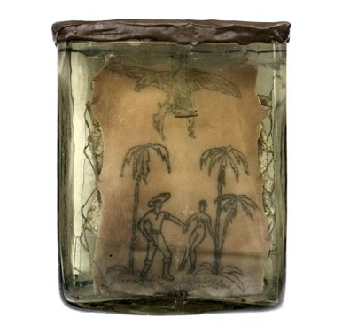
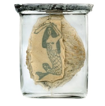
I just stumbled upon a pretty incredible "photo story" documenting a collection of tattoos found in the Department of Forensic Medicine at Jagiellonian University, Krakow, Poland. The images above are all drawn from the photo essay; below is an excerpt from the very interesting article which accompanies the images:

Preserving the Criminal Code
Photo Stories
Katarzyna Mirczak

The tattoo collection at the Department of Forensic Medicine at Jagiellonian University in Krakow, Poland consists of 60 objects preserved in formaldehyde, a method devised by one of the experts employed by the Department at the turn of 20th century.

The tattoos were collected from the prisoners of the nearby state penitentiary on Montelupich Street as well as from the deceased on whom autopsies were performed.

The majority of the prison tattoos represent connections between the convicts. Besides gestures and mimics it is a kind of secret code – revealing why 'informative' tattoos appeared on uncovered body parts: face, neck or arms.

The collection was created with a view to deciphering the code – among prisoners known as a 'pattern language'. By looking closely at the prisoners' tattoos, their traits, temper, past, place of residence or the criminal group in which they were involved could be determined.

In Poland, tattoos are common among criminals. Traditionally, they could be found on people who exhibited a tendency towards perverse behaviour: such as burglars, thieves, rapists and pimps. It was noticed that a significant percentage of tattooed people showed signs of personality disorders and aggressive behaviour. In the 1960s in Poland, getting a prison tattoo required special skills and criminal ambition – it was a kind of ennoblement, each tattoo in the criminal world was meaningful...

The entire photo story, with the full article and image collection (highly recommended!), can be found by clicking here; text and images by Katarzyna Mirczak as published on the Foto8 website.

PS: If you are interested in this topic, then you certainly won't want to miss our upcoming Observatory lecture "Morbid Ink: Field Notes on the Human Memorial Tattoo" with Dr. John Troyer, Deputy Director, Centre for Death and Society, University of Bath next Tuesday, July 20th. More on that event here.

Leica Microsystems Wins European Scanner Contest

First Prizes for Scan Speed and Best-Focused Images

Leica Microsystems‘ Slide Scanner Leica SCN400 and Autoloader Leica SL801 have come first in two categories of the first European Scanner Contest (ESC). The prizes were awarded in the “Scan Speed and Best-Focused Images” category, each at 20x and 40x magnification.

The jury was convinced by the digital pathology system by Leica Microsystems: Thanks to the patented “Dynamic Focus”, the Slide Scanner Leica SCN400 produces virtual images of exceptional quality at unprecedented speed. Another product designed for high throughput in the pathology lab is the Autoloader Leica SL801, which is capable of scanning up to 384 samples at a time. “By winning first prize, Leica Microsystems has again asserted its market position. We will continue to play a key role in shaping the future of virtual microscopy and digital pathology,” said Dr. Bodo Falke, Sales Manager Central Europe for Leica Microsystems, at the award presentation in Vilnius, which hosted the 10th European Congress on Telepathology and the 4th International Conference on Virtual Microscopy last week.

The European Scanner Contest was held within the framework of “Pathology Week“, a joint event of the German Society of Pathology and the Association of German Pathologists. With this competition, the organizers have created the first opportunity to compare the speed and imaging quality of different scanners using identical samples. The contest is also intended to promote the development of virtual microscopy products.

Source: Leica Microsystems

http://www.leica-microsystems.com/news-media/press-releases/press-releases-details/article/leica-microsystems-wins-european-scanner-contest/


Diabetes Medicines

Diabetes (http://www.dreddyclinic.com/findinformation/dd/diabetes.htm) means your blood glucose, or blood sugar, is too high. If you can't control your diabetes with wise food choices and physical activity, you may need diabetes medicines. The kind of medicine you take depends on your type of diabetes, your schedule, and your other health conditions.
With Type 1 diabetes, your pancreas does not make insulin. Insulin is a hormone that helps glucose get into your cells to give them energy. Without insulin, too much glucose stays in your blood. If you have type 1 diabetes, you will need to take insulin.
Type 2 diabetes, the most common type, can start when the body doesn't use insulin as it should. If your body can't keep up with the need for insulin, you may need to take pills. Read more...

Healthy blood

Upcoming Observatory Presentation: "Nature as Miniaturist: An Illustrated Survey of the Bogs of Southern New Jersey," Lord Whimsy, August 5


On Thursday, August 5th, please join Morbid Anatomy in welcoming good friend and fellow traveler Lord Whimsy at Observatory as he delivers an illustrated lecture on "the botanical oddities found in the ancient Ice Age bogs of the New Jersey Pine Barrens."

Whimsy will augment his lecture with the display of a variety of live plant specimens illustrating the diversity of this region, a demonstration on building and and maintaining your own container gardens, and plant care sheets to guide you in cultivating a variety of indigenous carnivorous plants. Copies of his book, The Affected Provincial’s Companion, Volume One will also be available for sale and signing.

Herr Whimsy is a wonderful speaker and a fascinating thinker. I promise, this is one event you won't want to miss! Full details follow; hope to see you there!

Nature as Miniaturist: An Illustrated Survey of the Bogs of Southern New Jersey
An Illustrated lecture and specimen demonstration with author, artist, and Gentleman Naturalist Lord Breaulove Swells Whimsy

Date: Thursday, August 5th

Time: 8:00

Admission: $5
Presented by Morbid Anatomy

Tonight, author, artist and Gentleman Naturalist Lord Breaulove Swells Whimsy will be giving an illustrated lecture on the botanical oddities found in the ancient, Ice Age bogs of the New Jersey Pine Barrens. These tiny, alien worlds are home to rare orchids, carnivorous plants, and bizarre species of plants and animals–some of which are found nowhere else on Earth. Whimsy, a lifelong resident of the Pine Barrens, will also give a demonstration of how to build and maintain your own container garden for these strange, wonderful plants. Live specimens of these plants will be on display, and care sheets for carnivorous plants like Venus Flytrap will also be made available. Whimsy’s book The Affected Provincial’s Companion, Volume One will also be available for sale and signing.

Lord Breaulove Swells Whimsy (aka V. Allen Crawford III) is an artist, designer, author, failed dandy, bushwhacking aesthete, and middle-aged dilettante. Whimsy is the author of The Affected Provincial’s Companion, Volume One (Bloomsbury), which has been optioned for film by Johnny Depp’s production company, Infinitum Nihil. He and his wife are proprietors of Plankton Art Co., an illustration and design studio. Their most notable project to date is the collection of 400 species identification illustrations that are on permanent display at the American Museum of Natural History’s Hall of Ocean Life.

You can find out more about this presentation here. You can visit Lord Whimsy's website by clicking here, and find out more about his book by clicking here. You can get directions to Observatory--which is next door to the Morbid Anatomy Library (more on that here)--by clicking here. You can find out more about Observatory here, join our mailing list by clicking here, and join us on Facebook by clicking here.

Olympus America Licenses Digital Pathology Patents to BioImagene

Olympus continues to enter into nonexclusive licensing agreements with other whole slide vendors.  The latest announcement is with BioImagene.  While the terms were not made public and often are not clearly one can see the value of this rich suite of patents that several vendors have entered into agreements with Olympus for imaging and data handling.  I gather more companies will also follow given what Olympus owns in order to continue to move the field. 


CENTER VALLEY, Pa., July 12 /PRNewswire/ -- Olympus America Inc. has signed a nonexclusive worldwide licensing agreement with BioImagene, Inc. of Sunnyvale, California, allowing BioImagene to access an extensive portfolio of Olympus patents in the field of digital pathology and virtual microscopy. This is the most recent of several licenses that Olympus has granted for the technologies, which are considered critical for developing future digital imaging and data handling systems for pathology.  

Digital pathology technology has the potential to change the way doctors review and manage millions of biopsy slides and other pathology specimens now handled using glass slides and traditional optical microscopes.  The patents included in the licensing deal cover methods and equipment for creating, storing and delivering virtual microscopy slides. The technology enables pathologists to view and share high-resolution virtual microscopy images over the Internet.

"The digital pathology field is evolving rapidly," said F. Mark Gumz, President and CEO of Olympus Corporation of the Americas, the parent company of Olympus America Inc., "and allowing other companies in this field like BioImagene to license these important patents will help advance the field of pathology, and ultimately promote better access to healthcare for patients around the world."

"Digital pathology is at an important inflexion point, moving very rapidly towards broad-based adoption. We believe that our relationship with Olympus will be a catalyst for this trend," said Ajit Singh, Ph.D., President and CEO of BioImagene. "Olympus has been a pioneer in this field, and their patent portfolio will complement BioImagene's innovations in end-to-end digital pathology."

In the United States alone, $13.2 billion was spent in anatomic pathology testing during 2008, according to a 2010 Laboratory Economics report. The Olympus portfolio enables development of solutions that can streamline the review of pathology slides and improve the speed of reporting, while saving costs and enhancing patient care. Virtual microscope slide technology also has the potential to aid hospitals in moving to comprehensive patient Electronic Medical Record (EMR) systems – a transition that is already beginning in some parts of the world.

The companies are not making specific terms and conditions of the agreement public.

About Olympus Scientific Equipment Group

Olympus America Scientific Equipment Group provides innovative microscope imaging solutions for researchers, doctors, clinicians and educators. Olympus microscope systems offer unsurpassed optics, superior construction and system versatility to meet the ever-changing needs of microscopists, paving the way for future advances in life science.

About Olympus

Olympus is a precision technology leader, designing and delivering innovative solutions in its core business areas: Medical and Surgical Products, Life Science Imaging Systems, Industrial Testing Instruments and Cameras and Audio Products.

Olympus works collaboratively with its customers and affiliates worldwide to leverage R&D investment in precision technology and manufacturing processes across diverse business lines. These include:

  • Gastrointestinal endoscopes, accessories, and minimally invasive surgical products;
  • Advanced research, clinical and educational microscopes and research and educational digital imaging systems;
  • Industrial research, engineering, test, inspection and measuring instruments; and
  • Digital cameras and voice recorders.

Olympus serves the healthcare field with integrated product solutions and financial, educational and consulting services that help customers to efficiently, reliably and more easily achieve exceptional results. Olympus develops breakthrough technologies with revolutionary product design and functionality for the consumer and professional photography markets, and also is the leader in gastrointestinal endoscopy and clinical and educational microscopes.  For more information, visit http://www.olympusamerica.com.

About BioImagene

BioImagene (http://www.bioimagene.com) is the leading provider of innovative and scalable digital pathology solutions for diagnostics. The company's total digital pathology solution is comprised of the Virtuoso™ digital pathology application software, iScan™ family of automated digital slide scanners, and a rich menu of Companion Algorithms™. BioImagene's innovative product line includes a unique image viewing input device called the iSlide™, and a high-performance pathology workstation called Crescendo™. BioImagene is also the platinum sponsor of PathXchange™, a vendor neutral, not-for-profit website for the pathology community.

SOURCE Olympus America Inc.

Auction Report: "The Gallery of Creation, a Museum of Natural History, Created by Joseph Hurt Studio, Inc."


\
Video: The Gallery of Creation Adventure of Lion & Lamb
Watch robotic animals discuss the creation of the world from a biblical prospective. Found here.

Regular Morbid Anatomy readers might recall a recent blog post about the a Georgia-based creationist natural history (sic) museum--"The Gallery of Creation, a Museum of Natural History"--being disseminated at public auction. Morbid Anatomy reader Sarah of the blog A Discourse on the Arts and Sciences was curious to know the outcome of that auction; Following is her exclusive investigative report, for your reading pleasure:

While the contemporary art market has struggled over recent months in terms of desirable material entering the market, the antiques market has been blessed. In particular, the genres of natural history, taxidermy and, surprisingly, kitsch have been a virtual goldmine in recent weeks, which have been bolstered even further by interesting provenances as well.

The Australian sale of the Owsten Collection (see report here) sold extremely well, with numerous pieces of highly significant natural history selling well above high estimates. I was particularly in love with the collector’s cabinets, myself.

Another captivating sale which I heard of, via Morbid Anatomy (thank you), was the de-accessioning of the collection of "The Gallery of Creation, A Museum of Natural History" on June 25th and 26th in Social Circle, Georgia (more on that here).

This Creationist natural history museum was founded by William Hurt Studios and catered to church groups, school classes and family outings. I wish I had gotten a chance to see it before it closed.

I have to admit, my first reaction was one of sheer captivation. What a great combo, natural history and that special kind of kitsch which only seems to happen in the South. Maybe my family stopped at too many tourist destinations during my childhood, but I shamelessly confess to a love of this kind of nostalgia. I envisioned lots of big blonde hair waving paddles for--quite simply--one of the oddest assortment of items I have ever come across available for sale in one place.

This particular sale was one of those examples of the sometimes odd attractions collectors can have. It certainly wasn’t about the ‘finest’, or art--it was about the sometimes most determined aspect of our attractions --nostalgia --as well as an occasional appreciation for good old fashioned camp.

In following up to the sale, I found that many of the pieces did sell quite reasonably. Prices do not include buyer’s premiums.


Oil Painting on Canvas of the Ark
8’6” wide x 5’ high
$600


Model of a Velosoraptor
$425


Vitrine Display of Various Skeletons & Skulls
14 feet wide x 9 feet high x 4 feet deep
$4,250


Vitrine Display of Seascape with Animation Features
12 feet wide x 9 feet high x 4 feet deep
$2,600


Vitrine of Mounted Birds, including Peacock, Toucan,
numerous parrots (Sphinx Macaw) and others
13 feet wide x 9 feet high x 4 feet deep
$4,100


Large Animated Vitrine of Lion & Lamb Conversing
$3,750

Another upcoming auction item worthy of pangs of nostalgia, as well as camp, is none other than Trigger himself, the trusted companion of Roy Rogers to be sold on July 14th in New York. The dispersal of the Roy Rogers & Dale Evans Museum Collection is being handled entirely by Christies and will take place on July 14th and 15th; more on that here.


TRIGGER (1934-1965)
Estimated $100,000 - $200,000.
Roy Rogers & Dale Evans Museum Collection

Entertaining the masses so thoroughly for over two decades, Roy Rogers and Trigger were one of America's most recognizable duos, becoming instant classics in people's eyes, hearts and imaginations. Trigger also reached legendary status in his own right, and is undeniably one of the most memorable horses that ever lived. Trigger was apparently purchased for $2,500 back in the day.

Thanks so much, Sarah, for the excellent report. More information of the Museum of Creation can be found here. For backstory on the auction, click here. Click here to find out more about the Roy Rogers & Dale Evans Museum Collection auction. Click here to check out guest-poster Sarah's lovely blog A Discourse on the Arts and Sciences.

This Friday at Observatory: Live Human Dissection, Lecture and Book Launch by The Hollow Earth Society


This Friday! At Observatory! Live human dissection, lecture and Book Launch by The Hollow Earth Society; full details follow. Hope to see you there!

Suspicious Anatomy: Lecture, Live Human Dissection, and Book Launch The Hollow Earth Society presents SUSPICIOUS ANATOMY Workbook No. 15: The Human Cranius Lecture, Live Human Dissection and Book Launch Date: Friday, July 16th Time: 8:00 PM Admission: $5

"Not since Galen’s De Elementis has been set in ink a single compendium of medicological knowledge so extensive & practicably useful as SUSPICIOUS ANATOMY Workbook No. 15: The Human Cranius. Having intrinsic value to all persons—piratical, mysterious, upright, or otherwise—The Human Cranius is a PEERLESS GEM of uncanny truth. If you are a living human, you should make frequent, unabashed forays into this field guide to your hideous secondary body—the cranius, an organ-matrix & carnival of fangs which is trying to destroy you even as you read this sentence…"

From the genre-chainsawing minds of the Hollow Earth Society (Ethan Gould and Wythe Marschall) comes “the definitive guide to the horrifying world inside you”—finally available in lush, illustrated paperback!

In the tradition of John Hodgman, David Cronenberg, and H. P. Lovecraft, The Human Cranius explores an alternative anatomy at once mesmerizing and deeply unsettling. Gould and Marschall ask: What do we know about our own bodies? The answer: Very little…

In many ways, the art and human studies of modernity have given us the keys to our unconscious minds, but have left entirely to dry science (fixing plumbing, testing drugs) the workings of our bodies. What does it feel like to have guts? To face disease, age, mutation—in short, a self that is not only not whole but not even on its own side?

The SUSPICIOUS ANATOMY series seeks to address these physio–psychomological imbalances by producing, for your benefit, the entire unconscious of the body, the shadow-self, in words and elaborate images.

The official Human Cranius book launch features a lecture, medicological film snippets, and a live human dissection. Join us!

About the Hollow Earth Society: For over one hundred years, the Hollow Earth Society has probed the world’s most bizarre and pertinent mysteries via an ever-mutating set of handbooks, rogue histories, and practical manuals. The Society is currently led by Colonels Ethan Gould and Wythe Marschall.

Ethan Gould is a Brooklyn-based artist working in drawing, puppetry, writing, and video to exploit the moments when the formerly robust process of perceptual categorization snaps like the fragilest of dry twigs. A graduate of the University of Rochester, he helped to create several development programs at the American Folk Art Museum. His work has appeared in such disparate places as the Ontological-Hysteric Theater, The Assembly Theater Company, The Brooklyn Review, Pomp & Circumstance, and ABC’s Wife Swap.

Wythe Marschall is writer. A graduate of Bennington College and the MFA fiction program at Brooklyn College, where he teaches undergraduate literature, Wythe has published stories and essays in McSweeney’s Quarterly Concern, Ninth Letter, and elsewhere. He is the senior editor of the Atlas Obscura and an editor of Pomp & Circumstance, as well as a frequent reader for Electric Literature. His thoughts on letters, postmodernity, and hip hop can be found on his website, chronolect.com.

You can find out more about these presentation here. You can get directions to Observatory--which is next door to the Morbid Anatomy Library (more on that here)--by clicking here. You can find out more about Observatory here, join our mailing list by clicking here, and join us on Facebook by clicking here.