The NEW ClockWorks Theater, Grand Opening Party, Saturday April 30th, 7:30 PM


Exciting news! Good friend and brilliant puppeteer/manifester of alternate worlds Jonny Clockworks is opening a new incarnation of his infamous ClockWorks Theatre! Judging from Clockworks' fantastic former productions, this new venture is sure to become the center for innovative and beautiful puppet works in the New York area.

The theater celebrate its opening this Saturday, April 30th, with an exciting evening's programming; full details follow. Very much hope to see you there!

The Cosmic Bicycle Theatre reopens
The ClockWorks Theatre in it’s new incarnation
The Universal ClockWorks, Inc.
in Carroll Gardens ~ West
the Columbia Street Waterfront
Brooklyn , USA

April 30th Walpurgisnacht!
7:30 PM ~ The Blessing of the Wheel ~ Sidewalk Spectacle ~ Free
8:30 PM ~ Theatre Opens
9:00 PM ~ Der WunderKammer Puppet Kabaret ~ Admission $20

The new ClockWorks Theatre is located on Columbia Street in the Carroll Gardens neighborhood of Brooklyn, New York. More on the theatre here; more on the opening event here.

The History of Skin Grafts with Paul Craddock, Observatory, Monday May 9


Come learn about skin grafts, from ancient history to modern controversy, with London-based Paul Craddock, Monday May 9th! Hope to see you there.

Knowing Your Ass from Your Elbow; or, Thinking About Skin Grafts
An Illustrated Lecture with Paul Craddock, Ph.D. candidate with the London Consortium
Date: Monday, May 9
Time: 8:00 PM
Admission: $5
Presented by Morbid Anatomy and The Hollow Earth Society

Late in 2010, The Economist ran a series of advertisements about transplant surgery on the London Underground, putting pressing questions to subterranean commuters like Paul Craddock. Ethics, black markets, identity, and the gap between organ supply and demand were all covered, and both sides of the arguments given, urging us to take a view on weighty questions while waiting for a train. The adverts presented The Economist as being at the cutting edge of debate in light of recent advances in transplant technologies, face transplants, and so forth but these questions are nothing new.

No. Transplants did not even appear in the 1960s with the first heart transplant, or at any point in the twentieth century – or even the nineteenth. They were first widely addressed in the media and Literature of the eighteenth century but medical transplantation is actually an ancient skill. Skin grafts go back around 5,000 years, and were simple but bloody affairs.

This illustrated lecture is about the ‘re-discovery’ of skin grafting from India in the early nineteenth century and the questions that seemed to raise. In what ways is it important that we can move skin? How did we find out that it was possible? What does it tell us about ourselves? If it is possible to have skin that’s not our own, what does this do to our sense of self? And what about identity? Why is this important to you, now? By thinking about how these questions were presented in the past, we may view and wear our own skins differently.

Paul Craddock is a second-year Ph.D. candidate with the London Consortium holding the Science Museum's Science and Humanities Scholarship. He is working on The Poetics of Bodily Transplantation, 1702 - 1902, a cultural history of the transplant procedure in medicine covering the two hundred years prior to the recognized 'beginning' of organ transplant history. In a past life, he was a performance and sonic arts scholar, researching and teaching a phenomenology of sound and the experience of it as part of an environment. Currently based in London, Paul is also the Director of London Consortium TV, the London Consortium's televisual arm, and the Publicity and Guests' Secretary for the University of London's Extra Mural Literary Association. He recently found joy in producing and presenting films, of which he has made two and a third to date.

For more, click here. You can sign up for the Observatory mailing list by clicking here, or join us our Facebook group by clicking here. For more about Observatory, click here. To contact organizers with questions or suggestions, click here.

Image: Cowasjee from the Gentleman's Magazine of 1794

Announcing New Observatory Series: "Out of the Cabinet: Tales of Strange Objects and the People Who Love Them," Co-Curated by Evan Michelson


What, exactly, is it that motivates the collector? Is it primarily an obsession, an addiction or a compulsion? Is the urge to collect benign or malignantly invasive? No one has yet provided a compelling answer, and any collector would be hard-pressed to articulate what exactly is driving the need to hunt down and acquire that next essential piece. The symptoms of the collecting impulse are as varied as the collectors themselves: some collect categorically, some collect socially, and some are driven by aesthetic considerations above all, but most collectors agree that the thrill of that next find is one of life's greatest pleasures, and the love of certain objects can last a lifetime. --Evan Michelson, Collector, Morbid Anatomy Library Scholar in Residence, Star of TV's "Oddities"

I am very pleased to announce "Out of the Cabinet: Tales of Strange Objects and the People Who Love Them," a new collector "show and tell" and lecture series taking place at Observatory and organized as a collaboration between Morbid Anatomy and Evan Michelson, our Library Scholar in Residence and, more recently, star of TV's "Oddities."

"Out of the Cabinet" will take artifacts as a launching off point for exploring the non-rational power of objects and the alluring mysteries of collecting. For the duration of the series, a variety of collectors will be invited to bring in an object or objects from their own collections and use these artifacts as a departure point for a response of some sort, from an illustrated historical lecture to a loose meditation to a debate with another collector.

A few events have already been scheduled (see below), but we continue to seek private or institutional collectors living in--or with plans to visit--the New York City area. If interested, please email me at morbidanatomy [at] gmail.com; And please, feel free to send this post along to any other interesting collector types you may know.

Full details follow. Looking forward to hearing from all you collectors out there!

Out of the Cabinet: Tales of Strange Objects and the People Who Love Them: A Collector "Show and Tell" and Lecture Series
Presented by Morbid Anatomy and Morbid Anatomy Library Scholar in Residence/Star of TV's "Oddities" Evan Michelson

What, exactly, is it that motivates the collector? Is it primarily an obsession, an addiction or a compulsion? Is the urge to collect benign or malignantly invasive? No one has yet provided a compelling answer, and any collector would be hard-pressed to articulate what exactly is driving the need to hunt down and acquire that next essential piece. The symptoms of the collecting impulse are as varied as the collectors themselves: some collect categorically, some collect socially, and some are driven by aesthetic considerations above all, but most collectors agree that the thrill of that next find is one of life's greatest pleasures, and the love of certain objects can last a lifetime.

Above all, it is our own personal history that imbues an object with meaning, and gives it resonance beyond its intended life. Collectors tell their inner stories through their acquisitions, and we can suss a collector's personality through his or her items without a word being spoken; a collection is a physical manifestation of the perpetually unseen, inner life.

Whether it's the pursuit of beauty, a sense of stewardship, the creation of a personal narrative, a love of science and history or acquisitiveness run amok, the objects a collector lives with speak to an undeniable drive to possess something rare, beautiful or personally significant. In this series, collectors will present some choice objects from their collections and discuss what it means to be possessed by a possession, what layers of meaning an object can hold beyond price or rarity, and what shadowed corners of the psyche are illuminated by the things that hold us in their spell.

Out of the Cabinet Events scheduled thus far:

  • April 11, 2011: A Gathering of Bones: An Illustrated lecture by Collector Evan Michelson
  • May 20, 2011: The Witch’s Dungeon: An illustrated lecture and show and tell with proprietor of “The Witch’s Dungeon” Cortlandt Hull
  • June 2, 2011: The Bell Jar: An Artifact-Based Lecture with Collector John Whiteknight
  • June 23, 2011: Home-Made Visual Albums: An Artifact-Based Lecture with Collector David Freund

For more on this series, click here. To keep abreast of newly announced events as part of this series, sign up for the Observatory mailing list by clicking here, or join us our Facebook group by clicking here. For more about Observatory, click here. To contact organizers with questions or suggestions, click here.

Images: Top to bottom:

  1. The Artist in His Museum, Charles Willson Peale, Pennsylvania Academy of the Fine Arts, Philadelphia; found here.
  2. Early 20th Century Department Store Mannequin, From the Home Collection of Evan Michelson, Antiques Dealer, New Jersey, as seen in the Private Cabinets Series, Joanna Ebenstein, 2009
  3. From the Home Collection of Evan Michelson, Antiques Dealer, New Jersey, as seen in the Private Cabinets Series, Joanna Ebenstein, 2009
  4. Kitten Princess of Winter by Fine Art Taxidermist Tia Resleure of A Case of Curiosities, From the Collection of Ronni Ascagni, Art Director, New York City as seen in the Private Cabinets Series, Joanna Ebenstein, 2009
  5. Tim Knox and Todd Longstaffe-Gowan Collection, Private Collection, London, England, as seen in the Private Cabinets Series, Joanna Ebenstein, 2009
  6. Tim Knox and Todd Longstaffe-Gowan Collection, Private Collection, London, England, as seen in the Private Cabinets Series, Joanna Ebenstein, 2009

The Great Coney Island Spectacularium, Obscura Day Oddities Party, 2011

Fancy a peek at drunk people cavorting within our recently launched Great Coney Island Spectacularium? The video above was shot at our Obscura Day "Oddities" party a few weeks ago. The sharp-eyed among you might spot yourself, the cast of TV's "Oddities" and their spouses, friends, and my mother.

You can find out more about the Spectacularium in these recent articles from Time Out New York, New York Press, Boing Boing and Brooklyn Based. Or, of course, on the Spectacularium website, which can be accessed here.

Thanks, G. F. Newland, for alerting me to the existence of this troubling video.

2011 Annual American Association for the History of Medicine (AAHM) Meeting, Phildadelphia, April 28-May 1, 2011


If anyone has plans to be in the Philadelphia area this week, you might consider popping over to the 2011 Annual American Association for the History of Medicine (AAHM) Meeting, taking place this week at the Philadelphia Society Hill Sheraton Hotel from April 28-May 1, 2011.

There are about a dozen interesting looking panels, but the two panels that look the most unmissable to me are the following, both taking place on Friday April 29th: "Museum Practice and the Making of Medical Science: Specimen Collections, Networks, and Institutions in the Late 19th and Early 20th Centuries," which will include two of my favorite scholars, Eva Ahren and Lisa O'Sullivan; and "Digital Media and New Audiences for the History of Medicine," where I will be delivering a short paper, along with such esteemed luminaries as Michael Sappol and Lisa Rosner.

Details for these two panels follow; to view the full schedule, click here.

Very much hope to see you there!

FRIDAY, APRIL 29
10:15 - 11:45 AM
Museum Practice and the Making of Medical Science: Specimen Collections, Networks, and Institutions in the Late 19th and Early 20th Centuries
Ballroom E
MODERATOR: Susan Lawrence (UniversityofNebraska,Lincoln)

  • Eva Ahren (University of Uppsala) ?Making Space for Specimens?: Medical Museums and Institution-Building at the Karolinska Institute, 1860-1910
  • Ross Jones (University of Sydney) ?No interest in human anatomy as such?: Frederic Wood Jones Dissects Anatomical Investigation in the United States in the 1920s
  • Lisa O‘Sullivan (University of Sydney) Creating Medical Specimens and Meanings: Frederic Wood Jones and the Work of a ?Good? Anatomical Specimen

12:00 - 1:15 PM
Digital Media and New Audiences for the History of Medicine
Ballroom B

  • Joanna Ebenstein (Morbid Anatomy Library, New York City)
  • Susan Reverby (Wellesley College)
  • Lisa Rosner (Stockton College)
  • Michael Sappol (National Library of Medicine)
  • Karie Youngdahl (College of Physicians of Philadelphia)
  • Laura Zucconi (Stockton College)

Click here to download a PDF program containing the complete and very extensive schedule. More information about the conference can be found here.

Image source: Rhizome.org; no citation found. Click on image to see larger, more detailed version.

"Hunting Trophies," Unknown Artist, British, Albumen silver print, ca. 1870


"Hunting Trophies," Unknown Artist, British, Albumen silver print, ca. 1870. Full description from The Metropolitan Museum of Art website:

Shikar, or big game hunting, was an immensely popular pastime for the ruling class in India prior to British rule. When the British came into power, elaborate hunting ceremonies were used by Indians and British alike to display their prowess and status to each other. The British influence also brought improvements in hunting technology, which spurred an increase in the capture of game. Dozens of animals were killed in a single day's hunt and the trophies decorated the halls of the princes' extravagant hunting lodges. By the late 1870s, the population of many of these rare species had been severely depleted and a government-implemented system for conservation had begun to take hold.

From The Metropolitan Museum of Art website. Found via Wunderkammer blog.

Click in image to see much larger, more detailed version.

Aperio Awarded Patent for Innovation that Improves Digital Pathology Workflow

Aperio continues to add to its IP portfolio with streamlined workflow patented technology.

Creation of complete digital slide image file - during scanning - results in fastest scanners, streamlines workflow and simplifies IT

VISTA, Calif.--(BUSINESS WIRE)--Aperio, the global leader in providing digital pathology solutions that improve patient care, announced today that the United States Patent and Trademark Office has issued the company patent No. 7,826,649 entitled, “Data Management in a Linear-array-based Microscope Slide Scanner.”

The ‘649 patent describes techniques invented by Aperio for efficiently processing large image files — during the scanning process — to create a single high-quality digital slide image. The ability to create a single digital slide, including image compression, during the scanning process results in faster overall scanning and streamlines digital pathology workflow.

“This patent highlights our ability to create a single digital slide image during the scanning process, which is why our scanners continue to be the fastest on the market,” commented Dirk G. Soenksen, CEO of Aperio. “Our sub-95 second scan time includes all steps required to create a single digital slide image file by the scanning instrument. This patent requires others to divide the scanning process between the scanning instrument and a dedicated server, and to post-compress the imagery data on the server. Doing so takes much longer than their advertised scanning time, complicates the workflow, and requires moving and storing gigabytes of imagery data.”

Aperio has the highest throughput scanners available today. The company’s family of ScanScope® scanners are based on line-scanning, which has become the standard for scanning performance and image quality in the digital pathology industry. Line scanning rapidly produces seamless digital slide images with unsurpassed image quality, and is critical to a pathologist’s ability to make informed diagnoses and prognoses of diseases.

The patent strengthens Aperio’s comprehensive intellectual property portfolio comprising more than 80 issued and pending patents world-wide, and provides the company with ownership of key innovations that address critical pathology imaging needs.

With more system installations than all other digital pathology vendors combined, Aperio is the true global leader and reliable choice for world class digital pathology solutions. The company boasts a global installed base of more than 800 systems in over 30 countries, including more than 500 systems in hospitals and reference laboratories, the 13 largest pharmaceutical companies and a multitude of biotechnology and government organizations.

About Aperio

Aperio is the leading provider of digital pathology solutions in hospitals, reference labs, and pharmaceutical and research institutions across the world. Today, our affordable and complete product portfolio improves patient care by enhancing quality assurance, delivering more efficient workflows, facilitating access to new and more targeted therapies, and improving pathologists’ skills via lifelong education. Our comprehensive product line features our ScanScope® scanners, Spectrum™ image management (PACS) software, SecondSlide® slide sharing service for pathology, and image analysis tools and services. Aperio’s products are FDA cleared for specific clinical applications, and are intended for research and education use for other applications. For clearance updates and more information please visit http://www.aperio.com.


VA awards $1.38 billion in telehealth contracts

Courtesy of FierceMobileHealthcare:

The Department of Veterans Affairs this week awarded contracts to six IT vendors to run its massive (and growing) telehealth program for the next five years. The winners: Authentidate, American Telecare, Cardiocom, HealthHero Network, Visual Telecommunication Network/ViTelCare, and Viterion TeleHealthcare.

The prize: About $1.38 billion in VA telehealth contracts. The individual vendor contracts run anywhere from $150 million to $372 million over the five-year period, VA officials tellFierceMobileHealthcare. That total isn't guaranteed, though. Each vendor starts out with a single-year contract, which is renewable in each of the following four years.

What's really interesting: The VA's clear leap into telehealth. The smallest of the new contracts--$150 million for Viterion Tele-Healthcare--is just shy of the agency's entire telehealth budget of$163 million last year. It is somewhat on trend, though, given that the 2010 budget was a 50 percent jump over 2009 spending.

The contracts do cover both traditional video-based telehealth systems, according to Authentidate officials. They also cover sensor-based remote monitoring systems in patient homes.

To learn more:
- read Authentidate's press release
- check out MobileHealthWatch's coverage

Related Articles:
VA's telehealth budget may double, but strategic questions linger
Veterans using mobile devices for follow-up care
Telehealth prevents ICU deaths, VA research shows


The Essential Guide to Achieving the Perfect Pathology Fellowship: Useful Advice and “Must-Do’s” for First-, Second- and Third-Year Residents

How to build teamwork, boost productivity, and enhance harmony
in your lab’s multi-generational workforce

Jared Gardner, MDRobert Petras,  MD

YOUR PRESENTERS:

Jerad M Gardner, M.D., Chair, CAP Resident Forum and Fellow, Soft Tissue

Robert E. Petras, M.D., FCAP, FACG, Managing Director, Ameripath, Inc.

Justin M. Clark (Moderator), Director of Operations, THE DARK REPORT


LIVE Webinar April 28 – Register Now!

Early in their four-year residency programs, pathology residents should start searching for the perfect fellowship, and employers should emphasize the need for residents to acquire subspecialty expertise before they begin a career in pathology and laboratory medicine.

On Thursday, April 28, 2011, the Pathology Residents Education Program (PREP) has a timely learning opportunity designed to help pathology residents identify, apply, and interview for the perfect pathology fellowship. During this 50-minute webinar, you’ll learn precisely what it takes to demonstrate your skills and competencies—and then nail down an offer for the ideal pathology fellowship.

Our first speaker, Robert Petras, M.D., is internationally known for his subspecialist expertise in gastrointestinal pathology. Dr. Petras accepts fellows and will give you an insider’s perspective on all aspects of the fellowship experience. He has been active in promoting fellowship programs at the national level and has invaluable insights into how you can stand out as an applicant.

You’ll also hear from Jerad M. Gardner, M.D., Chair of the CAP Resident Forum and Fellow, Soft Tissue Pathology at Emory University. From the pathology resident’s perspective, Dr. Gardner offers practical information about the do’s and don’ts of submitting fellowship applications, handling the interview process, and responding appropriately to post-interview communications.

This webinar is perfect for first-, second- and third-year pathology residents who want to identify and obtain the ideal pathology fellowship. Pathology residency program directors and chairs of pathology will also find this information invaluable, and will want to make it available to their residents.

Pathology fellowships aren’t easy to get. To get a fellowship offer from your first choice, it’s essential for you to understand how to distinguish yourself from the other applicants. Find out what it takes to get a fellowship in either an academic or non-academic setting, including how to select a subspecialty, how to become a competitive candidate, and how to navigate the fellowship application process.

You’ll also learn about the fellowship interview and selection criteria and what you can do to prove you’re the best candidate for the position. We’ll even prepare you for handling an offer of acceptance—and what to do if you’re turned down.

If you’re a resident looking for a fellowship opportunity, this session will give you a head start by taking you through the application process and clearing up some of the more confusing aspects before you begin. Drs. Petras and Gardner offer insights, advice, and recommendations for conducting a search to find the best pathology fellowship in a non-academic setting.

Ask your chief resident or residency program director to register your residency program for this information-packed 50-minute webinar, “The Essential Guide to Achieving the Perfect Pathology Fellowship: Useful Advice and “Must Do’s” for First-, Second- and Third-Year Residents,” on Thursday, April 28, 2011.

And don’t forget the question-and-answer session at the end of this conference. It’s your opportunity to get answers to specific questions about how to apply for a academic or non-academic pathology fellowship.


THE DARK REPORT WEBINAR AT A GLANCE

PLACE: Your computer or your residency program’s conference room

COST: $249 per download *Add 30 days of post-conference online access to the recording of this event for $75

TO REGISTER: Click here or call Suzanne Galloway toll-free at 1-800-560-6363


For one low price—just $249—you and your entire team can take part in this fast-paced, insightful webinar. Best of all, you’ll be able to hear answers to your questions with the panelists when you here the Q and A session answers.

 Here’s just some of what you’ll learn during this in-depth 50-minute webinar:

  • How to select the right subspecialty.
  • Where to find out about the best pathology fellowship programs.
  • Easy steps for establishing yourself as the top applicant.
  • The best time to apply for pathology fellowships.
  • Why each fellowship program has different selection criteria.
  • Interviewing do’s and don’ts.
  • Advantages and disadvantages of the academic and non-academic experience.
  • Strategies for navigating the oft-confusing fellowship application process.
  • Must-do steps to take once you’ve been accepted into a fellowship program.
  • Why you need a backup plan—and what to do if you’re not accepted.

…and much more!

Individuals who benefit from this webinar download:

  • Pathology chief residents, residents and fellows
  • Academic pathology chairs
  • Residency program directors
  • Pathology department administrators
  • Undergraduate and graduate medical educators
  • Pathology and laboratory professionals

How to Register:

1. Online
2. Call toll free: 800-560-6363

Your registration order includes:

  • A site license to attend the webinar (invite as many people as you can fit around your computer at no extra charge)
  • A downloadable PowerPoint presentations from our speaker
  • A full transcript emailed to you soon after the conference

Register Now! Or for more information, call us toll-free at 800-560-6363.

Distinguished Experts:

Jerad M. Gardner, M.D. is Chair of the CAP Residents Forum and has served on the Residents Forum Executive Committee for the past three years. He is the soft tissue pathology fellow studying under Sharon Weiss at Emory University. This will be followed by a dermatopathology fellowship, also at Emory, beginning later this year. Dr. Gardner received his M.D. from Tulane University School of Medicine in 2006. He completed his residency in anatomic and clinical pathology at The Methodist Hospital. During residency, Dr. Gardner founded the Pathology Resident Wiki, a website that can be freely edited by any user. The main intent of the Wiki is to provide a comprehensive list of pathology fellowship and residency programs, including feedback about those programs from current and past trainees. He is passionate about helping residents understand and navigate the seemingly treacherous waters of the fellowship selection and application process.

Robert E. Petras, M.D. has been the National Director for Gastrointestinal Pathology Services at AmeriPath Inc. since 2002. He is also Managing Director of AmeriPath Cleveland, Medical Director and GI Pathology Fellowship Training Program, Director at AmeriPath’s Institute of Gastrointestinal Pathology and Digestive Disease Chairman of AmeriPath’s Gastrointestinal Pathology Resource Committee and an Associate Clinical Professor of Pathology at Northeastern Ohio Universities College of Medicine. Prior to 2002, Dr. Petras chaired the Department of Anatomic Pathology, was Vice Chairman of the Division of Pathology and Laboratory Medicine, and directed the Pathology Residency Training Program at the Cleveland Clinic Foundation. He received his doctorate of medicine from The Ohio State University College of Medicine and completed his residency at the Cleveland Clinic. Afterwards, he received subspecialty training in gastrointestinal pathology at St. Mark’s Hospital in London. He has been active in the American Society for Clinical Pathology (ASCP), the College of American Pathologists (CAP), and the United States and Canadian Academy of Pathology (USCAP). He frequently presents as faculty at various courses and workshops. He has received numerous awards including the 1997 ASCP Distinguished Service Award and the 2001 Impact Paper of the Year Award from the American Society of Colon and Rectal Surgeons. He has co-authored more than 200 publications in gastrointestinal pathology including Tumors of the Intestines, part of the AFIP Atlas of Tumor Pathology Fascicle series.

The Daily Scan on iPhone

The Daily Scan - Aperio's daily news and views about Digital Pathology and many other services has an iPhone App that keeps you connected to the site.  Click on image below for larger size.  App free of charge from iTunes store.

2011-04-18_22-03-13

iPhone 5 Rumor Mill

When Apple announced that it would be holding its annual Worldwide Developers Conference in June, the rumor mill started to heat up. From every corner of the world, so-called sources were saying what Apple will and won’t announce at the show. Many of those rumors surrounded the iPhone 5. Historically, Apple has announced details about the new iPhone at a keynote address to kick off the event. However, some say it won’t do that this year. Other reports, of course, say it will. In either case, it’s clear that there are many question marks surrounding the iPhone 5 that Apple is unwilling to answer. But that doesn’t mean that those questions should be ignored. Apple’s iPhone is integral to its success. And it relies upon consumer excitement to help maintain its increasing revenue and profits. Simply put, the iPhone 4 and the upcoming iPhone 5 are integral to Apple’s operation. And the sooner it answers the many questions consumers have, the better it will be for the company. Read more.

Tonight and Tomorrow at Observatory: Extreme Taxidermy and Dubiously Sourced Bodies and Anatomical Learning!

Tonight and tomorrow night at Observatory; hope to see you there!

Kitten Tea Parties, Auto Icons, and Habitat Groups: A Brief History of Taxidermy
An Illustrated Lecture by Dr. Pat Morris, Royal Holloway, University of London

Date: TONIGHT! Thursday, April 21
Time: 8:00 PM
Admission: $5

Tonight, taxidermy scholar and collector Pat Morris will discuss the fascinating and sometimes bizarre history of taxidermy as explored in his new book A History of Taxidermy: Art, Science and Bad Taste. Along the way, Morris will discuss anthropomorphic taxidermy of the sort made famous by Victorian museologist and taxidermist Walter Potter, "extreme taxidermy" (ie. human taxidermy), and the role of taxidermy in the history of scientific display and popular culture. He will also detail the development of taxidermy as an art form, tracing its development from the stiff rudimentary mounts which characterized its beginnings to the artistic triumphs of the late 19th and early 20th Centuries.

Copies of his new book History of Taxidermy: Art, Science and Bad Taste will also be available for sale and signing.

Dr. Pat Morris is a retired staff member of Royal Holloway College (University of London), where he taught biology undergraduates and supervised research on mammal ecology. In that capacity he has published many books and scientific papers and featured regularly in radio and TV broadcasts. The history of taxidermy has been a lifelong hobby interest and he has published academic papers and several books on the subject. With his wife Mary he has travelled widely, including most of Europe and the USA, seeking interesting taxidermy specimens and stories. They live in England where their house is home to the largest collection and archive of historical taxidermy in Britain.


Ill-gotten Brains: The Grisly History of Sourcing Bodies for Anatomical Learning
An Illustrated Lecture with Megan Curran, Norris Medical Library, USC
Date: TOMORROW, Friday, April 22
Time: 8:00 PM
Admission: $5

The idea of donating your body to science is actually a very new concept. There wasn’t even a national law governing the process until the late 1960s. How, then, did medical illustrators, going back hundreds of years, acquire bodies to draw? Many bodies were “donated” alright, but the dead people didn’t know they were being so generous. Prisoners, the indigent, robbed graves, and even murders helped supply medical schools and doctors for centuries.

This wild history of sourcing human bodies spans from the dawn of modern anatomy in the Renaissance with Vesalius (and even artists like Da Vinci and Michelangelo), through the 19th century's institutionalized medical school body snatchings, and up to Nazi medicine and the controversy over plastinated bodies in exhibits like Body Worlds.

For tonight's lecture, join Megan Curran of USC's Norris Medical Library for fascinating accounts of how bodies were procured for the advance of science, often through less than ethical means, accompanied by images from USC's rare medical books.

Megan Curran is the Head of Metadata & Content Management for the Norris Medical Library of the University of Southern California in Los Angeles, CA. Megan manages the history of medicine and rare books collections at USC and has been working to promote that collection. Megan serves on the editorial board of the Journal of the Medical Library Association, and is on the board of the Archivists & Librarians in the History of the Health Sciences. Megan originally hails from Philadelphia and is a dyed-in-the-wool rare book nerd.

You can find out more about these events on the Observatory website by clicking here and here; You can get directions to Observatory--which is next door to the Morbid Anatomy Library (more on that here)--by clicking here. You can find out more about Observatory here, join our mailing list by clicking here, and join us on Facebook by clicking here.

Images: Top: The Auto-Icon of Jeremy Bentham. Mr. Bentham requested in his will that his body be preserved and stored in a wooden cabinet. It has been on display since 1850 at University College London. Photo by Joanna Ebenstein.
Bottom: Opera omnia anatomica & chirurgica, Andreas Vesalius

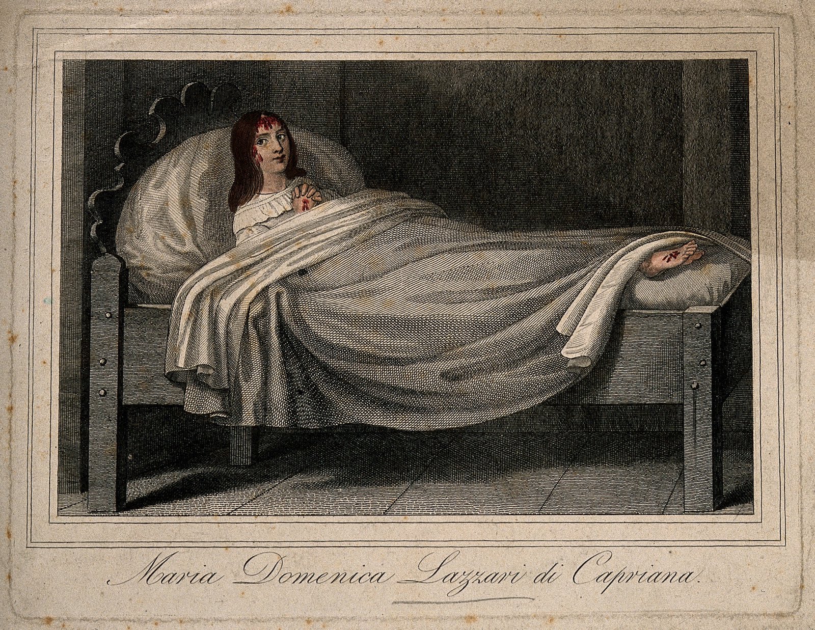
Visiting the Stigmatics of South Tyrol on Good Friday via The Wellcome Library, Circa 1840


...In the 1840s there was a steady flow of foreign tourists and pilgrims to the idyllic valleys of [South Tyrol] ... solely to visit two women who were said to have received spontaneously bleeding wounds (stigmata) on their hands, feet, or head like those caused to Jesus Christ when he was nailed to the cross and forced to wear the crown of thorns. One of the two women was Maria Domenica Lazzari (sometimes spelled Lazzeri), and the other was Maria von Moehrl (also called Mörl). The former was known as L'Addolorata (the woman of pain), the latter as L'Estatica (the woman of ecstasy), for reasons which will become clear.

On this Good Friday -- the holiday commemorating Jesus Christ's death by crucification -- why not take a moment to consider the medio-religious condition of stigmata, ie. spontaneous bleeding (mostly found in the female persuasion) on the hands, feet, and/or head, mimicking the wounds caused to Jesus Christ when he was nailed to the cross and forced to wear the crown of thorns?

All of the above text and images are drawn from two recent fascinating posts on the Wellcome Library blog; To read the full articles, click here and here.

Happy Good Friday!

Images, top to bottom (please click images to view larger, more detailed versions):

  1. Maria Domenica Lazzari. Coloured engraving, ca 1840. Wellcome Library no. 260i
  2. Maria von Moehrl. Watercolour by L. Giuditti after L.G. de Ségur, 1846. Wellcome Library no. 708243i
  3. Maria Domenica Lazzari. Watercolour by L. Giuditti after L.G. de Ségur, 1846. Wellcome Library no. 708242i

Italian Devil Automaton, 15th or 16th Century, From the Wunderkammer of Ludovico Settala


Automata were... theologically and culturally familiar, things with which one could be on easy terms. They were funny, sometimes bawdy, and they were everywhere... Mechanical devils were...rife. Poised in sacristies, they made horrible faces, howled and stuck out their tongues to instill fear in the hearts of sinners. The Satan-machines rolled their eyes and flailed their arms and wings; some even had moveable horns and crowns. A muscular, crank-operated devil with sharply pointed ears and wild eyes remains in residence at the Castello Sforzesco in Milan.
--Jessica Riskin, “Machines in the Garden.” Republics of Letters: A Journal for the Study of Knowledge, Politics, and the Arts 1, no. 2 (April 3, 2010)

In her article "Machines in the Garden," Jessica Riskin discusses the use of automatons for religious purposes. Some of my favorite examples include mechanical devils intended to frighten church visitors into obedience (as shown and quoted above); mechanical passion plays; and a mechanical Christ on the crucifix popular in the 15th Century, which was able

to bow down and lifte up it selfe, to shake and stirre the handes and feete, to nod the head, to rolle the eies, to wag the chaps, to bende the browes, ... and gathering a frowning, forward, and disdainful face, when it would pretend offence: and shewing a most milde, amiable, and smyling cheere and countenaunce, when it woulde seeme to be well pleased. --William Lambarde, A Perambulation of Kent: Conteining the Description, Hystorie, and Customes of that Shire. Written in the Yeere 1570. by William Lambarde of Lincolns Inne Gent

I highly encourage you to read this fascinating article in its entirety; you can do so by clicking here.

Images: Italian Automaton (The Devil), carved in wood, 15th and 16th centuries, from the Wunderkammer owned by Ludovico Settala. It could, in the words of the Cosmodromium Blog, "roll its eyes and move its tongue, emit a noise and spit smoke from the mouth." Images sourced here.

All images and ideas sourced from the Cosmodromium Blog; read whole post--and see additional images--by clicking here.

Recent MLO Articles on Digital Pathology

When I received my copy of MLO Medical Laboratory Observer this past week it reminded me there were a few articles of interest last month in the journal.  Both provide value propositions for digital pathology with use cases illustrated by Dr. Schwartz of Aperio.

Digital Pathology and imaging — past, present, and future

By Karen Lynn 

The first focus of digital pathology was to automate the microscope. “The ultimate goal was to begin the migration from a physical slide to a digital image and ensure users’ comfort during the transition,” says Jason Christiansen, PhD, senior director of Operations at HistoRx. In its earlier days, image-analysis applications were produced but limited to existing testing paradigms which had less impact than the leap to slide digitization. Fast-forward five years. Today, image quality is virtually identical to viewing a glass slide under the microscope. In fact, pathologists are willing to make diagnoses based on an image versus actual glass.

Viewing slides digitally gives numerous advantages that glass slides do not provide; for example, tumors and areas suspicious for disease can be measured more precisely; images can be manipulated and utilized for consultation and teaching purposes; images can be viewed by more than 100 people simultaneously from anywhere in the world; and automated, quantifying algorithms for estrogen receptors, progesterone receptors, and HER2 have since been developed and FDA-cleared, reports Joon Yim, MD, director of digital pathology at Acupath Laboratories. In addition, digital pathology supports rapid-assessment turnaround time for frozen sections which are critical to surgical protocols, and those slides can be digitally imaged and stored in a central repository.

Full text
Expanding the lab’s reach with digital pathology

By Jared Schwartz, MD, PhD 

Advances in digital-pathology systems, including rapid slide creation, data management, and image-visualization techniques are transforming the practice of pathology. A powerful tool in anatomic pathology, advancements in digital pathology continue to enhance efficiency and accuracy, resulting in lower costs, significant workflow efficiencies, and improved patient care.

Digital slides are a complete representation of the entire glass slide, viewable on a computer monitor at any magnification. Web-based pathology picture archiving and communication system, familiarly known as PACS, allow pathologists to work remotely anytime, anywhere, to deliver accurate results faster than traditional methods.

As digital pathology becomes more accessible for the average lab, the ability to work digitally provides opportunities to offer new services and tests, attract new customers, and create new business lines. Implementing an outreach business enabled by digital pathology is an emerging strategy that is helping many labs maintain market share and increase growth and profitability.

Full text

Molecular Pathology Laboratory Network, Inc. Selects mScope Clinical For Its Pathology Imaging Communications & Integration Platform


This new partnership signifies a major breakthrough with a market 
leader in Comprehensive, Personalized Testing Services

MONTREAL (CANADA), April 13, 2011 – Aurora Interactive Ltd., a world leader in digital pathology communications announced today that the company has signed a license agreement for its mScope Clinical communications and imaging modalities integration platform with Molecular Pathology Laboratory Network, Inc. (MPLN), a leader in specialized reference laboratory services dedicated to providing superior diagnostic testing and complementary technologies, including molecular diagnostics, flow cytometry, fluorescence in situ hybridization,immunohistochemistry and cytogenetics.

“We chose Aurora for their cost efficient plug-and-play and proven integration solution. Given the variety of imaging modalities we use, we considered Aurora’s universality key to maintaining our strategic flexibility and creating a common user interface for our internal and external clients,” said Steve Olsen, MS, HTL (ASCP), Chief Operating Officer of MPLN.

“We are very excited about MPLN’s decision to partner with Aurora. MPLN is a cutting edge provider who is dedicated to client service and making a difference in patient care. We are proud they chose us as their communications and integration partner” stated Pierre Le Fèvre, President and Chief Executive Officer, Aurora Interactive.

About Aurora Interactive Ltd.

Aurora Interactive has developed the leading web based software platform (mScope) for simplification, productivity and ease of communications in digital pathology. mScope’s Universal Web Viewer has collaborative tools to view medical slides and images anytime, anywhere, regardless of file format. The software has four applications to meet digital pathology communications needs: mScope Education, mScope Clinical, mScope Research and mScope Universal Viewer. Aurora’s mission is to improve patient outcomes and help members of the medical community achieve their full potential by eliminating the learning, diagnostic and collaborative restrictions imposed by time and space. http://www.aurorainteractive.com.

About Molecular Pathology Laboratory Network, Inc.

Molecular Pathology Laboratory Network, Inc. (MPLN) is a privately-owned regional reference laboratory founded in 1989 with headquarters in Maryville, Tenn. and a satellite laboratory in Richmond, Va. A leader in personalized laboratory medicine, MPLN specializes in oncology and women’s health testing and has extensive expertise in chromosome analysis, flow cytometry, fluorescence in situ hybridization (FISH), real-time and quantitative polymerase chain reaction technologies, anatomic pathology, immunohistochemistry and gene sequencing. The laboratory also offers research and development services through its clinical trial division, Geneuity Clinical Research Services. MPLN is certified by the Clinical Laboratory Improvement Amendments (CLIA), accredited by the College of American Pathologists (CAP), and licensed by the states of Tennessee, Florida, New York and Maryland. For more information about MPLN, visit http://www.mplnet.com.


University of Minnesota licenses multiplexing software to Flagship Biosciences

Flagship receives exclusive license to software that pairs quantitative techniques with pathology expertise

MINNEAPOLIS / ST. PAUL (04/14/2011) —The University of Minnesota has finalized a license agreement with Flagship Biosciences for software that provides quantitative analyses of protein expression levels in human tissue samples. The software, IHC Map, improves a pathologist’s ability to characterize diseased tissues and assists researchers with clinical trials for pharmaceuticals and medical devices.

The exclusive license agreement for the university-developed software has also forged a partnership between BioNet, a central tissue procurement and research histology lab on the university’s Twin Cities campus, and Flagship Biosciences, a private company based in Flagstaff, Ariz. Flagship performs histopathology services for biotech, pharmaceutical, medical device and cosmetic companies. Flagship offers IHC Map to its customers and also refers organizations to BioNet for additional tissue procurement and research services that it does not offer.

“Flagship provides digital pathology services, primarily to the pharmaceutical industry,” said Dr. Steve Schmechel, director of BioNet, assistant professor of laboratory medicine and pathology and co-inventor of IHC Map. “For many of those functions, companies require both academic surgical pathologist skills and access to biospecimens or techniques that they may not have internally.”

BioNet's tissue procurement and histology services are available to researchers in both the public and private sector.  The lab procures biospecimens, such as tissue and blood samples, from subjects who have given consent, stores the specimens and associated annotation data, and reports the data, from which patient-identifying information has been removed, to the researcher. 

The instrumental tool in this partnership between industry and academia is IHC Map, software that analyzes tissue samples and identifies protein markers for disease. Without the software, a pathologist must analyze multiple slides individually, and make a qualitative judgment on the results. IHC Map allows protein or gene signatures taken on multiple sections to be aligned and visualized in a heatmap-like fashion. The technology improves the multiplexing of biomarkers on a tissue section, and allows the computation of expression to be reported as a comparison to other more stable proteins in the sample. The patent-pending software was developed at University of Minnesota and will be refined in Flagship’s pharma services by both veterinary and medical anatomic pathologists.Flagship Biosciences helps pharmaceutical and medical device clients develop tissue-based diagnostics. “Seventy to ninety million glass slides are analyzed each year in pharmaceutical drug development, and maybe another ten million in medical device development,” said Steven Potts, CEO of Flagship.  “We think this collaboration will make it easier for these clients to have specialized medical pathology expertise combined with quantitative techniques from Flagship.”

“Most diseases are due to alterations of genes that manifest themselves at the level of protein expression,” said Schmechel. “To characterize disease it is very useful to look at protein expression. Often looking at the expression of one protein is not sufficient, or not powerful enough to optimally characterize the disease. It’s better to look at multiple proteins simultaneously.”

Identifying protein expression across multiple genes can answer key questions for researchers: What is the nature of the disease? How is it likely to behave in the patient? Will the disease be responsive to a given therapy? “It’s been found that combining information from multiple genes to characterize tissue is a powerful way to identify aggressive cancers,” said Dr. Greg Metzger, co-inventor of IHC Map and associate professor of radiology at the U of M. In addition to quantifying expression of these proteins in a quantifiable measurement, the software also allows for greater standardization across tissue samples, and requires less time for the pathologist to analyze slides.

IHC Map was invented by Schmechel and Metzger; Stephen Dankbar, a software programmer from the department of radiology; and Jonathan Henriksen, an information technology specialist from BioNet. The research was funded by the National Institutes of Health and the American Recovery and Reinvestment Act.

The mission of the University of Minnesota’s Office for Technology Commercialization is to translate University research into new products and services that provide growth opportunities for its licensees, benefit the public good, improve the quality of life, and generate revenue to support the University’s research and education goals.


George W. Moore, prominent pathologist, dead at 65

Dr. Moore was one of the smartest informaticians I have had the pleasure of knowing.  I first met him at an APIII meeting in 2001 where he embraced "E-posters" and made sure to look at yours.  His website, http://www.netautopsy.org lists over 400 publications, book chapters and abstracts and he was a mentor to many.  His interests in surgical pathology and autopsy pathology were broad and given his combined interest and expertise in medical and pathology informatics, allowed Dr. Moore to refine data mining techniques and create mathematical models of disease and disease processes.   

April 10, 2011|By Nick Madigan, The Baltimore Sun

A pioneer in medical informatics, he held appointments at Johns Hopkins Hospital and other institutions

Dr. George William Moore, who had practiced pathology in Baltimore since 1976 and was a pioneer in the field of medical informatics, died April 4 after a long illness. He was 65.

During his career, Dr. Moore published hundreds of articles on pathology and computational medicine. His work in the closely related fields of medical informatics and pathology informatics, both of which emerged in the 1970s, was groundbreaking. Medical informatics covers the broad field of hospital computerization, while pathology informatics deals with the organization, retrieval and analysis of clinical laboratory data collected in medical centers.

60797418 Dr. Moore, celebrated for a fine singing voice as well as a prodigious beard, held appointments at Johns Hopkins Hospital, the University of Maryland School of Medicine and the Baltimore Veterans Affairs Medical Center. He developed a wide range of computational techniques for collecting and indexing the data contained in pathology reports. He also established novel statistical methods for analyzing the collected data.

He was born in Detroit and grew up in the suburb of Highland Park. He was educated in public schools and graduated at the top of his class at Highland Park High School in 1963.

He attended the University of Michigan at Ann Arbor, where he majored in cellular biology and earned a bachelor's degree in science. He received his doctorate in biomathematics from North Carolina State University at Raleigh. After a year of postdoctoral training at the University of Freiburg in Germany, Dr. Moore returned to Detroit, where he earned a medical degree from Wayne State University School of Medicine in 1976.

Five years earlier, he had married the former Barbara Lynne Struble. They had two sons, Geoffrey Walter Moore, who was born in 1974 and died three months ago, and Gregory Vincent Wayne Moore, born in 1980, who survives him.

Dr. Moore and his family moved to Baltimore in 1976, and lived most recently in Cedarcroft. In 1981, he completed an internship and residency in the pathology department at Hopkins, and stayed on as an assistant professor in pathology until 1989. That year, he accepted an appointment as associate professor at the University of Maryland School Of Medicine and continued to teach at Hopkins' various institutions. Also in 1989, Dr. Moore began practicing as a full-time pathologist at the VA Medical Center, a position he held until his death.

In 1967, Dr. Moore was named a Woodrow Wilson Fellow, and in 1980 received the Ludwig Aschoff Medal from the Medical Society of Freiburg. In 2007, he was named an honorary fellow by the Association for Pathology Informatics.

From 1957 until shortly before he died, Dr. Moore sang tenor in various chorus societies. He was particularly proud to have been a chorus member in a performance by the Detroit Symphony Orchestra in 1959, under the baton of Paul Paray, as well as a performance in 1966 of the University of Michigan Choral Union conducted by the Russian-born composer Igor Stravinsky. Most recently, Dr. Moore sang in the choir for the Episcopal Church of Christ the King, in Woodlawn, where he was a member of the congregation.

In addition to his wife and son, Dr. Moore is survived by a brother, James Michael Moore, and a sister, Kathleen Elizabeth Moore.


Tonight at The Coney Island Museum: Acep Hale on Con Men and Sideshow Legends Ward Hall and Bobby Reynolds!


Tonight, as night 7 of the Congress of Curious Peoples, we have a fantastic double header: magician and scholar Acep Hale lecturing on ""Legerdemain and Larceny'--a history of the con man--followed by a performance by sideshow talker legends Ward Hall and Bobby Reynolds onstage for the first time ever in Coney Island.

This is sure to be an incredible night! Full details follow; very much hope to see you there.

Acep Hale, "Legerdemain and Larceny"
Tonight, FRIDAY, APRIL 15th
CONEY ISLAND MUSEUM, 7:30 pm - $5

Coney Island, like most fairs, amusement parks, and midways has always had a reputation for hucksterism and the con job. Join magician and scholar, Acep Hale, as he explores the history of the con, taking the audience on a guided tour of the clowns, contrarians, murmurers and mystics that have held fast to lives of wandering wonder throughout the ages. Acep Hale is a street performing magician, musician, traveler, and rogue gentleman scholar. Driven by the 19th century belief in propaganda by deed he performs daily on street corners everywhere to prove that magic still lives around every bend, you don’t need a nine to five to stay alive, and hope springs eternal between the cracks of every sidewalk.”.

Sideshow Legends Ward Hall and Bobby Reynolds
Tonight, FRIDAY, APRIL 15th
SIDESHOWS BY THE SEASHORE, 8:30 pm - $15

Ward Hall (born 1930) has been around longer than anyone in the business and runs The World of Wonders Sideshow. He is "a modern-day P.T. Barnum and the last of the sideshow promoters. He's a national treasure who is loved and revered by showfolk, sideshow historians, and fans the world over."

Bobby Reynolds (born 1932) got his start at Hubert's Museum in Times Square and has been talking, performing in, and operating sideshows ...ever since. He still comes out of "retirement" every year or so to run shows all over the world. "With his two-headed babies, all frog band, and giant rat, Bobby has perfected the art of giving people the art of giving people what they didn't know they wanted."

Tonight, join Ward Hall, King of the Sideshow, and legendary side showman Bobby Reynolds as they perform

For more info--and to purchase tickets--click here and here.

Tomorrow Night: "A Gathering of Bones" Lecture with Evan Michelson, Morbid Anatomy Scholar in Residence and star of TV's Oddities, Coney Island Museum


Tomorrow night, why not consider joining me and Morbid Anatomy scholar in residence (and star of TV's "Oddities") Evan Michelson at Coney Island for her new lecture "A Gathering of Bones?" If her former lectures are any indication, this is sure to be a great one!

The event--which will take place within the newly opened Great Coney Island Spectacularium!--begins at 7:30. Drinks are half price at the bar until 8:00. Hope very very much to see you there!

"A Gathering of Bones," an Illustrated lecture by Evan Michelson
Date: Monday, April 11
Time: 7:30 PM
Admission: $5 (or free with Congressional Pass)

Location: The Coney Island Museum (1208 Surf Avenue)

Human bone: one of the most common materials on the planet. And yet, at one time the remains of certain individuals were prized more highly than the rarest, most precious metals and gems. The cult of the saints, the backbone of the early Christian Church, gave rise to an institutional fetishization of human remains that produced objects still unsurpassed in craftsmanship and opulence.

The aesthetic of the most humble and commo...n organic remains coupled with gold, silver, gems and textiles has for centuries proved irresistible to secular collectors and religious institutions alike. The ultimate collectible, the constituent parts of each and every human on the planet were once the object of obsession, veneration and murderous desire. As a collector myself, Christian relics provided my earliest exposure to the realm of transcendently beautiful, perverse and venerated objects.

The collection and categorization of human remains underwent a drastic change with the enlightenment, but the unquenchable human thirst for knowledge and comfort in the face of our own mortality has ensured that the corpus remains at the center of an unending human fascination with and confrontation of the greatest mystery of all. The gathering of bones continues to this day, still controversial, decadent and utterly essential to the human narrative.

This event is part of The Morbid Anatomy Library Collector Series.

Click here to purchase tickets ($5 each)

This event is made possible, in part, by a grant from the Andy Warhol Foundation.

Image: Galileo's finger mounted on a marble base and encased in a crystal jar, as on view at the Museum of History and Science in Florence, Italy. More on that--and image source--here. Click on image to see much larger, more detailed version.