New Book (In English!!!) About Honoré Fragonard's Incredible 18th Century Anatomical Ecorchés!


If the Founding Fathers wanted to visit Body Worlds they could have. Or pretty darn close, at least - they just needed to visit one of the many European cabinets of anatomical curiosities, to see the work of anatomists like Honore Fragonard.

Fragonard's eighteenth-century ecorches were the clear precursors to Gunther von Hagens' "Body Worlds" exhibits: preserved, injected, partially dissected bodies in lifelike, dramatic poses, with ragged strips of muscle draped like primitive clothing over exposed vessels and nerves. The effect is eerie - like a Vesalius illustration sprung to (half-)life... --Bioephemera, "If the Founding Fathers wanted to visit Body Worlds..."

Much has been said--and rightfully!--about the "uncanny similarity" (as one might charitably say) between the anatomical works of Body Worlds' impresario Gunther von Hagens and the 18th Century allegorical anatomical ecorchés of Honoré Fragonard, cousin of well-known rococo painter Jean-Honoré Fragonard. Bioephemera says it very very well today--as quoted above--and follows with a really nice, extensive review of the new, wonderful, lavishly illustrated Blast Books publication Fragonard Museum: The Ecorches.

To read the entire post on the Bioephemera website--very much recommended!--click here. To order a copy of the book for your very own--also highly recommended!--click here.

The specimens you see above -- and more! -- are housed in the amazing Le Musée de l’Ecole Nationale Vétérinaire d’Alfort (née Musée Fragonard) right outside of Paris; to find out more about that museum, click here. Images as credited; to see more images from the Musée, click here. To visit the museum website, click here.

Image credits and captions:

  • Top image: Fragonard's Horseman, found here.
  • Second image: Fragonard's man with the mandible, inspired by Samson smiting the Philistines with an ass's jaw, found here.

All other images from the Bioephemera post. Captions, top to bottom:

  • Nilgai/Doe of the Indies
  • Human Bust
  • Human Bust
  • Installation view of The Musee Fragonard

This Week At Observatory! Obscure Anatomical Collections! Behind the Scenes at The Museum of Natural History! Gender and Medical Illustration!

To celebrate (or mourn, depending on your point of view) yesterday's anti-climactic non-rapture, Morbid Anatomy has put together a very exciting week of illustrated lectures to take place at Brooklyn's Observatory! On Tuesday, join Dr. Rolf ter Sluis for a virtual tour of the underknown European anatomical collection he curates; on Thursday, author Jay Kirk will tell us the story of Carl Akeley, that "brooding genius who revolutionized taxidermy and created the famed African Hall we visit today at New York's Museum of Natural History;" and on Saturday, practicing medical illustrator Shelley Wall will discuss "how sexual anatomy, gendered bodies, and dimorphic sex have been represented in the visual discourse of medicine."

Heady stuff! Full details for all events follow; hope very much to see you there!

A Virtual Tour of the Anatomical Collections of the University of Groningen
An illustrated lecture with Dr. Rolf ter Sluis, Curator and Director of the Groningen University Museum
Date: This Tuesday, May 24th
Time: 8:00 PM
Admission: $5

Tonight, join Dr. Rolf ter Sluis--curator and director of the Netherlands based Groningen University Museum--for a virtual tour of the museum's historic and amazing anatomy and pathology collections. The majority of the collection consists of preparations in spirit, but also includes dry preparations where the veins have been injected with coloured wax, wax and Papier-mâché models, skeletons and skulls, preserved tattooed skin, and much more.

The core of the museum collection is drawn from the private collections of two important 18th century medical scientists, Petrus Camper and Pieter de Riemer. The collection of Camper, professor of medicine from 1763 - 1774, and his son Adriaan Gilles Camper consisted of anatomical, comparing anatomical and biological preparations, fossils, minerals and instruments. The collection was donated to the museum after Camper’s death in 1820 and there are still around 200 of his preparations in the museum collection. Another important part is the collection of the medical scientist Pieter de Riemer (1769 - 1831). He was especially interested in anatomy, surgery and obstetrics. The De Riemer collection, containing more than 900 preparations, came into the hands of the university in 1831.

Dr. Rolf ter Sluis is the Curator and Director of the Groningen University Museum. He also studied history and worked for 25 years as a registered nurse in Anaesthetics before taking on his role as curator and director of the collection.

Kingdom Under Glass: A Tale of Obsession, Adventure, and One Man’s Quest to Preserve the World’s Great Animals
An illustrated lecture and book signing with author Jay Kirk
Date: This Thursday, May 26th
Time: 8:00 PM
Admission: $5
***Books will be available for sale and signing

During the golden age of safaris in the early twentieth century, one man set out to preserve Africa's great beasts. In his new book Kingdom Under Glass: A Tale of Obsession, Adventure, and One Man's Quest to Preserve the World's Great Animals, Jay Kirk details the life and adventures of naturalist and taxidermist Carl Akeley, the brooding genius who revolutionized taxidermy and created the famed African Hall we visit today at New York's Museum of Natural History. The Gilded Age was drawing to a close, and with it came the realization that men may have hunted certain species into oblivion. Renowned taxidermist Carl Akeley joined the hunters rushing to Africa, where he risked death time and again as he stalked animals for his dioramas and hobnobbed with outsized personalities of the era such as Theodore Roosevelt and P. T. Barnum. In a tale of art, science, courage, and romance, Jay Kirk resurrects a legend and illuminates a fateful turning point when Americans had to decide whether to save nature, to destroy it, or to just stare at it under glass.

Tonight, join author Jay Kirk for an illustrated lecture based on his new book Kingdom Under Glass. Books will be available for sale and signing after the event.

Jay Kirk's nonfiction has been published in Harper's, GQ, The New York Times Magazine, and The Nation. His work has been anthologized in Best American Crime Writing 2003 and 2004, and Best American Travel Writing 2009 (edited by Simon Winchester). He is a recipient of a 2005 Pew Fellowship in the Arts and is a MacDowell Fellow. He teaches in the Creative Writing Program at the University of Pennsylvania.


Pub(l)ic Identities: Reading Medical Representations of Sex
An illustrated lecture with medical artist Shelley Wall
Date: This Saturday, May 28th
Time: 8:00 PM
Admission: $5

"It's a girl!" "It's a boy!"... The genitals, those body parts conventionally expected to remain most hidden, are also the first and most powerful shapers of our public identity. In this illustrated talk, medical artist Shelley Wall considers how sexual anatomy, gendered bodies, and dimorphic sex have been represented in the visual discourse of medicine. From early anatomical atlases through to present-day clinical illustrations and the Visible Human datasets, medical imagery has influenced ideas about sexual identity and what it means to be "normal".

Shelley Wall is a medical artist and professor in the Biomedical Communications graduate program, University of Toronto. Her research interests include biomedical representations of sex and gender, conventions in visualizing the embryology of sexual differentiation and intersex conditions, contemporary and historical visual practices in relation to women's health, and medical humanities.

You can fin
d out more about these events on the Observatory website by clicking here; you can access these events on Facebook here. You can get directions to Observatory--which is next door to the Morbid Anatomy Library (more on that here)--by clicking here. You can find out more about Observatory here, join our mailing list by clicking here, and join us on Facebook by clicking here.

Ventana looking for Pathologist/CMO at BioImagene location

Job Description

The Principal Pathologist is responsible for managing the clinical operations and the medical office of the company. The Principal Pathologist oversees the clinical division involved in validation of imaging applications, including oversight of studies leading to FDA 510(k) and PMA filings. The Principal Pathologist is a member of the Sunnyvale Management Team and will be jointly responsible for managing clinical collaborations for the company, including those with academic medical centers and reference laboratories. The Principal Pathologist develops research programs with key opinion leaders at these sites and works with internal business development along with R&D to execute these programs. The Principal Pathologist works closely with multiple other functions and groups, including Marketing, R&D, Quality and Regulatory to develop and review new products and applications as well as enhancing and refining existing products and applications and customer support for pathologist-related issues. The Principal Pathologist supervises the pathology requirements of the company, including image/slide review, test scoring, algorithm testing, algorithm validation, and application testing. Importantly, he or she will also represent Ventanas vision from a medical value and digital pathology perspective via preparation of abstracts and review articles, in meetings with external visiting pathologists, attend relevant scientific symposia, external pathology meetings and conferences.

ESSENTIAL FUNCTIONS:
- Oversight of FDA studies and filings
- Management of clinical collaborations
- Product/Application development and review
- Pathology supervision, including slide review/validation, tissue procurement, and application testing
- Presentations and Conference attendance
- Remain up to date on pathology, oncology and molecular biology literature related to Ventana's assay and imaging businesses
- Preparation of abstracts and peer reviewed publications
- Sales and Marketing support
- Customer support for pathologist-related issues

MINIMUM QUALIFICATIONS:

Formal Training/Education:
- M.D. Pathologist with American Board of pathology anatomic pathology board certification

Experience:
- 5+ years of pathology training and practice
- 5+ years experience in biomedical research (PhD or other degree) or biotech/pharma industry

Knowledge, Skills, and Abilities:
- Diagnostic pathology
- Advanced staining and diagnostic methods including immunohistochemistry and in situ hybridization
- Familiarity with, and interest in, personalized medicine and companion diagnostics.
- Biomedical research and development
- Clinical study design
- FDA filings
- Demonstrated record of accomplishments (e.g. publications in scientific journals, issued US patents).
- Scientific writing and presentations
- Management experience
- Medical Informatics

Contact Information

Please apply online at https://careers.ventana.roche.com, IRC20656, or contact allison.bacalia@ventana.roche.com for more information.


Laboratories as gate keepers and health maintenance providers

Laboratories engaged in direct patient care have at their disposal huge amounts of health care information related to both screening and diagnostic tests. 

When it is time for your 6 month dental check up you receive a card or more recently electronic reminder that your appointment you made 6 months ago is approaching or you need to call the office and schedule one.  My ophthalmologist provides a similar service with reminders for routine visits.  

Imagine the laboratory notifying you that "Mrs. Jones it has been 1 year since your last pap smear according to our records.  It may be time to visit your health care provider (like this universal term now) for your annual examination and pap smear (this may not go along with recommendations the provider follows -- need to think this one through). The intent of course, much like the dentist or ophthalmologist, that you choose the same provider and he/she chooses your laboratory.  

What about the pathology laboratory sending you a letter about 12 months after you had some atypical nevi removed that were examined by your laboratory that it may be time for another "skin check" by of course "your health care provider".  It won't necessarily say that it may be time to see Dr. Jones at Jones Dermatology but why not?  Particularly if Dr. Jones is still a client of yours.  Presumably Dr. Jones sent out a reminder as well but you know what could happen to those small cards that get mixed in with all the other mail.  

Ditto for cholesterol or glucose tests.  For example. "Ms. Smith, Our laboratory performed blood tests for you about three years ago.  We noticed that you have not had any subsequent testing.  May we suggest it may be an appropriate time to consult with your health care provider for a full physical examination and routine blood work that may be required".  

It is starting to sound like a drug ad -- "Call your doctor".  

By the way, have you ever tried to call your doctor.  I spend some of my days calling back results to referring physicians.  "Call the doctor" or "Notify the referring physician of abnormal results" is not as easy as it sounds.  Without fail, you call the wrong office as they are at the "other" office.  Again without fail, the office staff knows they are not at that office, but can't find the number for the other office.  Or a pager.  Or cell phone number.  The directory of physician listings has not been updated since President Bush's administration (the first Bush) and the other office isn't even in the listings. Once you make contact with the correct office you find out they have left for the hospital or other office...

Anyways, laboratories have at their disposal huge amounts of health maintenance data that is one of the keys being discussed with electronic medical records (EMRs), health information exchanges (HIEs) and accountable care organizations (ACOs). 

Laboratories could provide one of the necessary portals and sources of key data for continuity of care whether it is a screening colonoscopy or mammography (assuming their is biopsy material to record), pap smears or serum chemistries or cholesterol testing.  Ditto for drug levels for organ transplant patients lost to follow up.

The lab has the data to mine. Issues about self-referral, lack of patient-physician (health care provider) relationship and potentially unnecessary testing abuses would have to be worked out.  

I think with EMRs, HIEs and ACOs on the horizon, the laboratory should be one of the key organizations considered as potential gatekeepers and data keepers for patient management and outcomes.

In doing so we may be able to lose the term "lost to follow-up".


Managed Image Analysis – A new model for busy researchers

With our unique managed service model, the experts at i-Path take care of all your digital pathology needs.

Image2 i-Path has devised a model which will allow anyone to take the first step to using digital pathology quickly, easily and cost efficiently.

This managed service model works on a project by project basis, you are not tied into an annual contract, simply call us as a project comes up. There is no need for large capital investment or staff training.

Send your slides to i-Path where experts will scan your slides at your desired magnification and QC all slides.


  • Analysis of the whole digital slide is performed using our proprietary tissue measurement or tumour measurement algorithms.
  • i-Path can also work with you to create bespoke algorithms which can be used to analyse the whole digital slide or regions defined by you.
  • A comprehensive report will be generated giving full results of the study.

Simply Contact Us with your needs and we can get your project started.

AMA Changes Criteria for CME Credits

Courtesy of CAP:

Effective July 1, 2011, the American Medical Association (AMA) requires all continuing medical education (CME) qualifying for AMA PRA Category 1 CME Credit™ to have a scored assessment that all participants must pass before claiming CME credit. Access information about AMA PRA Category 1 Credit™ and requirements at ama-assn.org/resources/doc/cme/pra-booklet.pdf or contact the AMA directly at AMA-assn.org.

Learn what the CAP is doing to help our members affected by the upcoming AMA criteria change for CME credit:

  • CAP Education has added assessments to its AMA PRA Category 1 Credit™ CME qualifying education programs to comply with this requirement, and it offers participants up to three attempts to pass.
  • The new requirement applies to all enduring educational materials, such as print and online programs. It applies only to education programs released after July 1, 2011, and it is not retroactive to completed programs or those in progress that are released before July 1, 2011.
  • CAP Education has placed a notice on the registration page of each education course subject to this change, as well as a statement of this change on the information page of each program that is subject to this requirement.

The College is making great efforts to notify CAP members of this change prior to July 1, 2011.

For more information, please visit the AMA site describing AMA PRA Category 1 CME Credit™ and actual requirements at ama-assn.org/resources/doc/cme/pra-booklet.pdf.

CAP@YourService – June

The New World of Laboratory Medicine

Digital Pathology Consultants introduces a new webinar "The New World of Laboratory Medicine" presented by Richard Friedberg MD, PhD on May 23, 2011 at 3 PM EST / 12 PM PST, supported by our sponsors at http://www.thedigitalpathologywiki.com.

Description

To thrive in highly competitive markets, clinical laboratories need to look beyond traditional models of service delivery and begin to think outside the box. This may mean integrating with other diagnostic services and delivering different types of results, from laboratory to imaging, to physicians and other clients. This webinar, presented by Richard Friedberg MD, PhD, will explore how lab medicine has changed and is likely to evolve in the coming years and how you can position yourself for the future.

About Richard Friedberg MD, PhD

Chairman, Department of Pathology, Baystate Health, Springfield, MA

Medical Director at Baystate Reference Laboratories

Professor and Deputy Chairman, Department of Pathology, Tufts University School of Medicine

In 2007 and again in 2010, Dr. Friedberg was elected to the Board of Governors of the College of American Pathologists, where he also serves on the Council on Accreditation (vice-Chair) and Council of Government & Professional Affairs (Executive Committee), as well as CAP committees such as Strategy Management (Chair) and Finance. Dr. Friedberg holds a BS with Honors from Stanford University, an MD from Duke University, a PhD in coagulation biochemistry from Duke University, and a Master’s (SM) in Health Care Management from Harvard University. He is a Certified Physician Executive (CPE) by the American College of Physician Executives.

To register visit: https://www1.gotomeeting.com/register/557090784

Indica Labs, Inc. Formed To Meet Growing Demand For Digital Pathology Software

Albuquerque, New Mexico 5/12/2011 – Indica Labs, Inc. receives an initial round of seed funding to begin product development of a series of image analysis tools for digital pathology. The venture backed company will immediately begin development operations in the Albuquerque, New Mexico area.

Traditional pathology methods typically involve manual assessment of glass slides under a microscope. But the recent emergence of new technologies such as whole-slide scanners and image management systems has made digitization of glass slides commonplace. This digital transformation has led to an increasing need for whole-slide image analysis software and visualization tools.

“Our software will address the growing demand for sophisticated image processing algorithms that will aid pathologists in reaching an accurate assessment more efficiently and effectively as well as assist researchers and scientists in generating highly reproducible and quantitative data” says CEO, Steven Hashagen. “The number of institutions that have invested in digital pathology hardware and software systems is rapidly growing, and our tools will allow these institutions to leverage more value out of their investments”.

Indica Labs’ initial product offering will include a set of digital pathology software tools that will be used by pathologists in academic medical centers, government research institutions, global pharmaceutical companies, and small biotech firms.

For more information, please visit http://www.indicalab.com.


About Indica Labs, Inc.

Indica Labs provides sophisticated image analysis software for digital pathology. Indica Labs' software tools seamlessly integrate into leading digital pathology platforms to provide reproducible and highly quantitative data to pathologists in hospitals, academic medical centers, government research institutions, global pharmaceutical companies, and small biotech firms.

Contact

Indica Labs, Inc.

info@indicalab.com

(505) 573-5167

AI-Powered Pathology


Aperio Launches State-of-the-Art Digital Slide Scanning Instrument Featuring Highest Throughput, Compact Design and Superior Image Quality

Sixth Generation ScanScope® AT Highest-Capacity Scanner Available Today 

Image002  

VISTA, Calif.-- Aperio, the global leader in providing digital pathology solutions that improve patient care, today announced the launch of the ScanScope® AT, the latest addition to Aperio’s industry-leading, patented line of ScanScope slide scanning instruments.

For more than a decade, Aperio has been the leader in producing high-quality lab instruments for digitizing microscope slides. The new ScanScope AT incorporates the very latest technology to deliver the most compact, highest throughput, lowest cost-per-slide, highest-capacity scanner on the market today with superior image quality and highest first scan success rate for high-throughput applications.

“The ScanScope AT underscores Aperio’s commitment to the continued development and evolution of our already industry-leading digital pathology technology,” said Dirk G. Soenksen, CEO of Aperio. “The AT has a small footprint for easier installation, yet produces the same high-resolution, superior quality images that Aperio is known for.”

Made possible through a combination of improved efficiencies in scan speed, robotic slide loading and handling, and user interface software design combined with high image quality to minimize rescans, the ScanScope AT is a groundbreaking development for the digital pathology market.

With just a 16-inch by 24-inch footprint, the ScanScope AT features a 400-slide capacity with sustained throughput of 30 slides per hour. Utilizing patented line scanning and inline compression technology, the AT can scan a 20x slide in an all-inclusive time of 95 seconds, producing a fully prepared image ready for immediate viewing.

In addition, automated tissue finding, focusing, and calibration processes yield the highest first-scan success rates, resulting in reduced cost per slide and lower total cost of ownership due to low re-scan rates and high-quality images.

The ScanScope AT comes equipped with an elegant, extensively tested, next generation autoloader designed to minimize mechanical complexity. The new autoloader substantially reduces travel distance for the slide, improving overall scan time and reliability. The system interoperates with Sakura slide racks, allowing slides to be transferred into Aperio slide racks 20 at a time, further saving time and improving throughput.

Greg Crandall, Aperio’s chief engineer, stated, “The design of the AT benefits tremendously from Aperio’s 11 years of experience building scanners. Not only is it our fastest scanner to date, it is also our most reliable in terms of slide handling. We have created a horizontal slide loader that holds almost four times as many slides as our previous model while fully supporting the slide throughout a very short travel distance, thus maximizing the safety of even imperfectly prepared slides.”


This Friday at Observatory: Collector Cortland Hull With and on His Collection of All Things Classic Monster Movie!


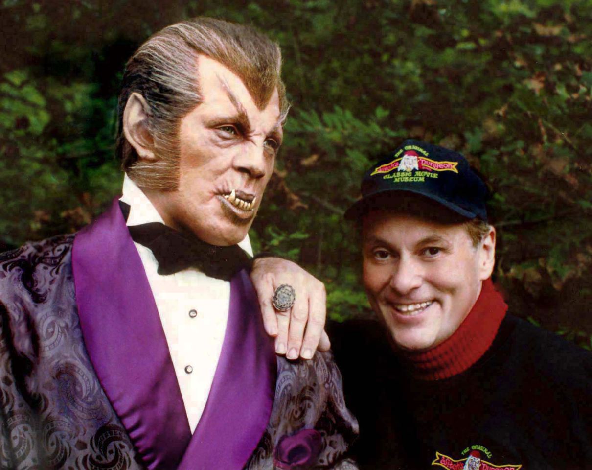
This Friday at Observatory, please join Morbid Anatomy in welcoming collector and museologist Cortland Hull as he shares some artifacts from his private museum of classic movie monster artifacts, shows some film clips, and provides a visual history of the actors & makeup artists behind the classic monsters. This event is part of the new Out of the Cabinet: Tales of Strange Objects and the People Who Love Them series.

Full details follow; hope to see you there!

The Witch's Dungeon
An illustrated lecture and show and tell with collector, artist, and proprietor of "The Witch's Dungeon" Cortlandt Hull
Date: Friday, May 20th
Time: 8:00 PM
Admission: $5
Part of
Out of the Cabinet: Tales of Strange Objects and the People Who Love Them, presented by Morbid Anatomy and Evan Michelson

Friday, May 20th may be a dark and stormy night. Brave souls normally catch the coach at midnight from the Borgo Pass to access the lawless and far off lands of Bristol, CT, spoken about in hushed tones as the home of the Witch's Dungeon. But on this rare occasion the stars have aligned and like the Baba Yaga's chicken-footed cabin, the Witch's Dungeon is coming to Observatory!

Tonight, Cortlandt Hull will be speaking about his life's work: the creation and evolution of The Witch's Dungeon, a museum consisting of life size reproductions of classic film monsters. Growing up during the 1960's monster boom, Cortlandt began construction of the Witch's Dungeon 45 years ago in the back yard of his parent's house. Over its near half century in existence, the Witch's Dungeon has continually creaked open its doors, striking chords with patrons, becoming a true piece of Americana, and attracting many of the actors and filmmakers commemorated in the museum.

Cortlandt will also be screening clips of his multiple documentary films, providing a visual history of the actors & makeup artists who created the classic films. Original head props from fantasy films will be on display along with samples of Cortlandt's work from the Witch's Dungeon.

Cortlandt Hull--artist, museologist, and film historian--began "THE WITCH'S DUNGEON CLASSIC MOVIE MUSEUM" when just 13. in 1966. It is now considered the longest running tribute to the makeup artists & actors from classic horror films. Featuring accurate life-size figures of Boris Karloff, Vincent Price, Lon Chaney, Bela Lugosi, and many others. Many of the figures are made from the actual life casts of the actor's faces Cortlandt has produced documentaries on the history of classic horror & fantasy films. Actor, Henry Hull ("Werewolf of London") was Cortlandt's great uncle, and Josephine Hull ("Arsenic & Old Lace") was his great aunt, so fantasy & horror is "in the blood"! He has lectured at universities, and film festivals, across the country, and has written for books and magazines.

You can find out more about this event on the Observatory website by clicking here; you can access this event on Facebook here. You can get directions to Observatory--which is next door to the Morbid Anatomy Library (more on that here)--by clicking here. You can find out more about Observatory here, join our mailing list by clicking here, and join us on Facebook by clicking here.

Image: Cortlandt Hull with figure of his great uncle, Henry Hull, "The Werewolf Of London"

Celebrate the Centenary of The Great Dreamland Fire of 1911 with A Brand-New Disaster Spectacle, Period Amusements, Free Hendrick's Gin, and More!


Unveiling of a brand new 19th Century style disaster amusement! Free Hendrick's Gin! Disaster tunes of yester-year! Lord Whimsy! Stars of TV's Oddities! Vintage Coney Island films curated by Zoe Beloff! Rare appearance of the old Dreamland Bell!

All this and more await you next Friday at our Centennial Celebration of the Great Dreamland Fire. Please, come celebrate the end of an era with us!

Full invite below. Hope very much to see you there!!!

Centennial Celebration of the Great Dreamland Fire Featuring the Opening of Coney Island’s Newest Cosmorama
Presented by The Coney Island Museum, The Morbid Anatomy Library, and Atlas Obscura
Date: Friday May 27, 2011
Time: 7:00 PM
Admission: $25 (Tickets at the door, or purchase here)
Location: The Coney Island Museum, 1208 Surf Avenue, Brooklyn (map here)

Next Friday, May 27th, you are cordially invited to a party commemorating the "awful splendor" of The great Dreamland fire of May 27, 1911, the most devastating disaster to hit New York City in the pre-9-11 era, a fire which devastated a never-to-be-rebuilt-Dreamland 100 years ago on this day.

This event will mark the premiere of the Cosmorama of the Great Dreamland Fire, a 360 degree immersive cosmorama telling the story of the great fire in pictures, sound, and light. Based on Coney Island’s great immersive disaster spectacles, the cosmorama is the product of months of labor, thousands of dollars, and the expertise of artists and artisans from the Metropolitan Opera, and uses real boards from the original Coney Island boardwalk in its construction.

The party will also feature a complementary gin bar with custom cocktails, disaster tunes of yester-year curated by The Foppinton Brothers, vintage Coney Island films, a rare appearance of the old Dreamland Bell, celebrity appearances, anatomical give-aways, myriad performances, and much more!

Full line-up:

Tickets are available by clicking here or purchasing at the door. See you there!

Nicotine Raises Blood Sugar Levels in Lab

(HealthDay News) -- Smoking is damaging to everyone's health, but the nicotine in cigarettes may be even more deadly for people who have diabetes.

In lab experiments, researchers discovered that nicotine raised blood sugar levels, and the more nicotine that was present, the higher the blood sugar levels were. Higher blood sugar levels are linked to an increased risk of complications from diabetes, such as eye and kidney disease.

"Smoking is really harmful for diabetics. It's even more harmful to them than to a non-diabetic," said study author Xiao-Chuan Liu, an associate professor in the department of chemistry at California State Polytechnic University in Pomona. "This study should encourage diabetics to quit smoking completely, and to realize that it's the nicotine that's raising [blood sugar levels]."

For that reason, it's also important to limit the use of nicotine replacement products, such as nicotine patches, Liu said.

"If you're using them for a short period of time to quit smoking, that's OK. But, if you still have this addiction to nicotine and are using this product long-term, it will do harm. Don't use electronic cigarettes or nicotine gum for a long time. You need to stop nicotine intake," he advised. Read more...

AyurGold for Healthy Blood

Volunteer Training for Hospice Family Care

Hospice Family Care will be providing a free hospice volunteer training beginning this week. See dates and times below:

Tuesday May 10 5:30 – 8:30 p.m.
Thursday May 12 5:30 – 8:30 p.m.
Saturday May 14 9:00 a.m. – 4:00 p.m.
Tuesday May 17 5:30 – 8:30 p.m.
Thursday May 19 5:30 – 8:30 p.m.
Tuesday May 24 5:30 – 8:30 p.m.
Thursday May 26 5:30 – 8:30 p.m.
Tuesday May 31 5:30 – 8:30 p.m.
Thursday June 2 5:30 – 8:30 p.m.

Hospice Family Care is certified by Medicare to care for people facing any end-stage disease or terminal illness. Patients and families dealing with End-Stage Heart or Lung Disease, Diabetes, Cancer, and many other illnesses can benefit. Our team addresses physical and practical needs, emotional and spiritual needs, and all quality of life issues. Pain and other symptoms are treated with priority.
Hospice care is available to anyone, of any age, or any financial status, and is a Medicare benefit. Many seniors are not aware they already have this coverage or how to take advantage of this special care. As part of this benefit, Medicare covers a variety of services, carried out by our special hospice team, and pays necessary additional benefits such as related prescriptions and disposable supplies, all at 100%
Volunteers are always needed! Our volunteers are very special people from all walks of life who are sensitive to the needs of our patients and families and who support our philosophy of supportive care. Our volunteers are individuals who respect and revere life and, in the performance of their volunteering, bring their energy and support to our patients, family members, and our care team. Volunteers attend a comprehensive 30-hour orientation encompassing various aspects of patient and family needs, and the volunteer role in the hospice team.
Any person sixteen years of age or older who has successfully completed our free orientation program can volunteer with us.
Our next training will be in May. There is an initial interview and you are then signed up for the next class.
How can you join the Hospice Family Care team? If you are interested in joining our team of volunteers please contact Rosy Cunningham at:

Rosy Cunningham
Volunteer Coordinator
Hospice Family Care
6300 E. El Dorado Plaza, Ste. A-100
Tucson, AZ 85712
(520) 790-9299 Office
(520) 790-9211 Fax
(520) 307-0330 Cell

Thank you so much,

Rosy

FREE White Paper – Connecting Labs to Physicians’ Electronic Health Records: Effective Strategies for Laboratories to Add Value

FREE Special Edition White Paper

download your report now!

Download Your FREE Special Report Today!
Simply Complete the Form Below


Connecting Labs to Physicians’ Electronic Health Records: Effective Strategies for Laboratories to Add ValueConnecting Labs to Physicians’ Electronic Health Records: Effective Strategies for Laboratories to Add ValueThe American healthcare system is in the process of significant change. Healthcare represents the fastest growing segment of our GDP, accounting for 17.3% of expenditures in 2009 and 19.3% projected for 20191, with Medicare and Medicaid representing 35% of that segment. Most metrics and outcome measures indicate that our healthcare system is operating inefficiently and that we are not getting our fair share from our investment. There is a broad consensus that these trends are unsustainable and that a change is mandated. It is generally agreed that health information technology (HIT) must play a significant role in effecting this change.

Health information technologies have been slow to trickle down to physician offices in the United States, and the adoption of electronic health records (EHR) in the U.S. lags significantly when compared to other nations. In the last several years, we have witnessed a major change. Ongoing government initiatives have actively endorsed the use of HIT across the continuum of care.

The Dark Report is happy to offer our readers a chance to download our recently published FREEWhite Paper “Connecting Labs to Physicians’ Electronic Health Records: Effective Strategies for Laboratories to Add Value” at absolutely no charge. This free download will provide readers with a detailed overview adding value connecting labs to physicians’ EHRs.  This report  will help laboratories to implement solutions that deliver the levels of service and connectivity needed to remain competitive and increase market share.


download your report now!

—

Among other topics, this white paper specifically addresses:

  1. HITECH Initiatives
  2. Meaningful Use
  3. CPOE
  4. Clean Orders
  5. LOINC
  6. For more about value adding techniques, please CLICK HERE

download your report now!

Table of Contents

Chapter 1. Introduction — Page 4

Chapter 2.
 Market Pressures Changing the Business of Healthcare — Page 6

Chapter 3.
 Stimulus Funding Synopsis  — Page 8

Chapter 4.
 The Path to Meaningful Use — Page 11

Chapter 5.
 Importance of Laboratory Data — Page 14

Chapter 6.
 Laboratory Competitive Pressures — Page 17

Chapter 7.
 Lab-EHR Connectivity Challenges— Page 19

Chapter 8. Advantages of a Partnering Strategy— Page 23

Chapter 9. The Clinical Data Exchange Solution — Page 26

Chapter 10. References — Page 31

Appendices

A-1 About Gai Elhanan, M.D., M.A. — Page 33
A-2 
About Mitch Fry — Page 34
A-3 Halfpenny Technologies, Inc.— Page 35
A-4 About DARK DAILY— Page 36
A-5 About The Dark Intelligence Group, Inc., and The Dark Report— Page 37
A-6 About Executive War College on Laboratory and Pathology Management— Page 38
A-7 About Kathleen N. Bowley— Page 40   

Terms of Use — Page 44

download your report now!


Alcatel-Lucent, Pittsburgh Hospital Team On Telemedicine Platform

The joint venture will combine live, store and forward, and home monitoring audio- and video-communications services for the University of Pittsburgh Medical Center.

By Nicole Lewis,  InformationWeek 
May 11, 2011
URL: http://www.informationweek.com/news/healthcare/interoperability/229500026 

In an effort to push further into the telemedicine market, telecommunications provider Alcatel-Lucent has announced that it will collaborate with the University of Pittsburgh Medical Center (UPMC) to develop a telemedicine platform and suite of applications.

The company says the new system will offer real-time clinical encounters in a virtual "exam room." By using a Web portal, patients will have access to both scheduled and emergency care from any location at any time through a variety of mobile devices using real-time video and audio communication among multiple participants in multiple locations.

Learn the security essentials of mobility

Business-Ready Mobile Infrastructure

At the same time, the system will generate, retrieve, and store patient data, and will make use of advanced technologies such as immersive and cloud-based communications.

Sid Ahuja, VP of Alcatel-Lucent Ventures, said the platform will be an important component of Alcatel-Lucent's connected hospital strategy that will allow hospitals to extend their reach and care beyond their physical walls. He also said the UPMC health delivery network, which has more than 20 hospitals, 400 outpatient sites, a health plan, and operates medical facilities in Italy and Ireland, will help Alcatel-Lucent test, validate, and build a better solution that will be offered to other healthcare organizations.

"The healthcare sector requires specific customizations of IT systems for reasons of privacy, quality of care, and reachability," Ahuja said in an interview. "Further, acceptance and usage of new technologies takes significant usage trials and commitment from healthcare organizations. We have decided to not just introduce new technology but to make sure it is vetted in the 'care process' practiced today." According to Ahuja, the plan is to have real UPMC trials with patient usage by end of this year. Alcatel-Lucent will also showcase the platform to other healthcare organizations and develop plans for other trials next year.

Currently, UPMC is using telepresence technology from Polycom in several areas of patient care including pathology, dermatology, stroke, psychiatry, trauma, and wound care. The next-generation telemedicine platform will upgrade the service with the necessary clinical input to ensure a richer and more robust product that fits the workflow of caregivers.

"The new platform will help to coordinate all three types of telehealth (live, store and forward, and home monitoring) into a single offering that is coupled with education and administration, all riding on an integrated telehealth network," said Andrew Watson, VP of UPMC's international and commercial services division. "We believe that almost all clinical services can be positively impacted by telemedicine, which can be used to treat chronic diseases, acute medical issues such as stroke, and for specialist care such as cardiology, endocrinology, and surgery."

Watson also said telemedicine provides UPMC with significant cost savings through increased home care, fewer readmissions, and greater provider efficiency.

The new telemedicine platform will build on the more than $1.3 billion that UPMC has invested over the last five years in health IT. This includes significant investments in electronic medical records and interoperability, a patient Web portal, primary and secondary data centers, and connectivity among UPMC's more than 20 hospitals both regionally and internationally. The telemedicine platform is being developed to leverage and complement these investments.

It is being developed along with Bell Labs, Alcatel-Lucent's research arm, and expected to be ready for commercial launch in early 2013.


Aperio Partners with Definiens to Market Definiens Tissue Studio for Advanced Image Analysis

2011-05-10_062029 Vista, CA and Munich, Germany – May 10, 2011 – Aperio, the global leader in providing digital pathology solutions that improve patient care, and Definiens, the leading Health Image Intelligence™ company, today announced a partnership in which Aperio will market Definiens Tissue Studio™, the company’s leading image analysis software for quantitative digital pathology, on a worldwide, non-exclusive basis.

Image analysis is a cutting-edge capability that provides automated quantitative analytical techniques to help pathologists interpret pathology slides, which informs treatment decisions and improves patient care.

Definiens Tissue Studio will be integrated into Aperio’s applications framework and accessible through its Spectrum™ information management system to expand the existing suite of image analysis tools that include Area Quantification, Cell Quantification, Microvessel Analysis, Rare Event Detection and Genie Histology Pattern Recognition.  The Spectrum system includes viewing tools and enables the execution of analysis applications in a geographically distributed environment for maximum throughput.

“This alliance furthers our ability to significantly improve intra- and inter-observer agreement and patient care by providing the most scalable and highest performance digital pathology solutions available,” said Dirk G. Soenksen, CEO of Aperio. “We are excited that Definiens recognizes the value of leveraging their leading image analysis software on the industry’s most robust digital pathology platform.”

Definiens Tissue Studio automatically detects regions of interest within a tissue sample and quantifies these regions on a cell-by-cell basis. The detailed quantification enables a large number of standard and user-defined measurements. In addition to classifications such as staining intensity and cell counts, the software quantifies individual cellular morphological characteristics such as circularity, elliptical fit, area, length, width and distance to tumor border to help accurately measure nuclear, cytoplasmic, and membrane biomarkers. Additionally, it allows users to create completely new features offering virtually unlimited quantification related to biomarker expression and tissue morphology.

Aperio users in both healthcare and life sciences will benefit from Definiens Tissue Studio’s fluorescence image analysis capabilities and morphometry tools. Morphometry can provide pathologists with a means to make more educated decisions about a course of treatment, and for  determining the relative effect of drug candidates on cells and tissues.

“Digital pathology is becoming an essential technology for increasing productivity and improving treatment decisions,” said Thomas Heydler, CEO of Definiens. “Automated image analysis is the next step in digital pathology, providing clinicians with greater insight into patients’ disease states. Definiens Tissue Studio is a natural fit for Aperio’s platform, and we expect to reach a wide customer base as a result of this partnership.”

Aperio’s digital pathology platform provides a unique combination of instruments, image management software and a patent-protected architecture that allows image analysis partners such as Definiens to offer their applications to the largest and fastest growing installed base (800 systems, including 500 in hospitals and reference labs) of digital pathology customers. Aperio plans to continually expand its capabilities with tools from other suppliers.

Graduating Physicians Opt for Jobs in Hospital-Owned Practices over Private Practices

Courtesy of Dark Daily

Trend could prove unfavorable to independent clinical laboratory companies

More physicians now join hospital-owned practices than any other type of practice. That’s one conclusion reported in a survey conducted by the Medical Group Management Association (MGMA). This is a trend that may have negative implications for independent clinical laboratory companies and pathology groups that provide medical laboratory testing to office-based physicians. 

In the MGMA survey, higher compensation packages offered by hospital-owned practices were cited as one reason why growing numbers of physicians choose a hospital-owned practice. The survey also determined that physicians believe they will have a better chance for reimbursements in hospital-owned practice settings, compared to other practice models. 

Meanwhile, first-year compensation packages continue to show wide variation for single-specialty versus multi-specialty practices, as well as between specialists and primary care physicians. It is a bothersome trend, since the proportion of graduating physicians selecting primary care has declined in each year of the past decade.

Full story

"Animated Anatomies," Duke University, Durham, North Carolina, Until July 17, 2011


While at the AAHM meeting in Philadelphia a few weeks ago, I met a number of fascinating folks, among them Rachel Ingold, the curator of the History of Medicine Collections at the Duke Medical Center Library & Archives. She was telling me about a wonderful exhibition on flap anatomies (images above) that will be on view there until July of this year; here is what she had to say about it:

Animated Anatomies explores the visually stunning and technically complex genre of printed texts and illustrations known as anatomical flap books. This exhibit traces the flap book genre beginning with early examples from the sixteenth century, to the colorful “golden age” of complex flaps of the nineteenth century, and finally to the common children’s pop-up anatomy books of today. The display—which includes materials from the Rare Book Manuscript and Special Collection Library at Duke University, the Duke Medical Center Library & Archives’ History of Medicine Collections, and from the private collections of the curators of the exhibit—highlights the history of science, medical instruction, and the intricate art of bookmaking.

Through the hands-on process of exposing layer after layer of anatomical illustrations, flap books mimic the act of human dissection, inviting the viewer to participate in a virtual autopsy, so to speak. Whether it’s a sixteenth-century hand-colored treatise on the layers of the eye or a nineteenth-century obstetrical guide in 3-D for performing cesareans, these books draw the viewer in. Over time, as advances in both science and printing promoted more widespread medical knoLinkwledge, anatomical flap books began to appeal to more general audiences eager to learn about their own bodies’ inner workings. Technological developments in machine printing also allowed for more colorful and precise illustrations than the hand-colored treatises of the early modern period.

A symposium was held on April 18 and we hope to have videos posted from this event soon. To learn more about the symposium, exhibit, see photos of anatomical flap books, and watch videos of them in action, visit the exhibit website. For more information, contact Meg Brown at meg.brown@duke.edu or Rachel Ingold at rachel.ingold@duke.edu. The exhibit will be up through July 17, 2011, and is free and open to the public.

This fantastic looking exhibition will be on display in the Perkins Gallery, Perkins Library, at Duke University, Durham, North Carolina, until July 17, 2011, and in the History of Medicine Gallery in the Medical Center and Archives Library from April 13-July 17, 2011. To find out more--or to pay a virtual visit!--check out the exhibition website by clicking here.